William Lam(@William28538949) 's Twitter Profileg
William Lam

@William28538949

Histopathologist. So want to retire ! In accelerated phase becoming curmudgeon.

ID:987528333318471680

calendar_today21-04-2018 03:07:56

2,0K Tweets

1,1K Followers

607 Following

Professor Richard Scolyer AO(@ProfRScolyerMIA) 's Twitter Profile Photo

I was delighted to co-write a recently released textbook on “Melanocytic Tumors of the Skin” with my dear friend & collaborator of almost 20 years Dr Klaus Busam MD.

Klaus has worked in New York City at the Memorial Sloan Kettering Cancer Center (MSKCC) throughout this time &…

I was delighted to co-write a recently released textbook on “Melanocytic Tumors of the Skin” with my dear friend & collaborator of almost 20 years Dr Klaus Busam MD. Klaus has worked in New York City at the Memorial Sloan Kettering Cancer Center (MSKCC) throughout this time &…
account_circle
Craig Horbinski(@CraigHorbinski) 's Twitter Profile Photo

Here's a tough one: young adult with longstanding history of gait instability. Photomicrographs of the superficial cerebellar resection are shown, plus a radiologic example of this entity from a different patient. Diagnosis? (Hint: Vive la France!)

Here's a tough one: young adult with longstanding history of gait instability. Photomicrographs of the superficial cerebellar resection are shown, plus a radiologic example of this entity from a different patient. Diagnosis? (Hint: Vive la France!) #pathology #neuropath
account_circle
Pepe Jiménez Heffernan(@pepeheffernan) 's Twitter Profile Photo

CC325. Alone in the office. Mediastinal EBUS-TBNA with suspected sarcoidosis. Granulomas⬇️. Everything is ok. Time has taught me that in these cases it is useful to see if there is an oncologic history, whatever it is, retrieve the slides and check them. And that is what I did.

CC325. Alone in the office. Mediastinal EBUS-TBNA with suspected sarcoidosis. Granulomas⬇️. Everything is ok. Time has taught me that in these cases it is useful to see if there is an oncologic history, whatever it is, retrieve the slides and check them. And that is what I did.
account_circle
William Lam(@William28538949) 's Twitter Profile Photo

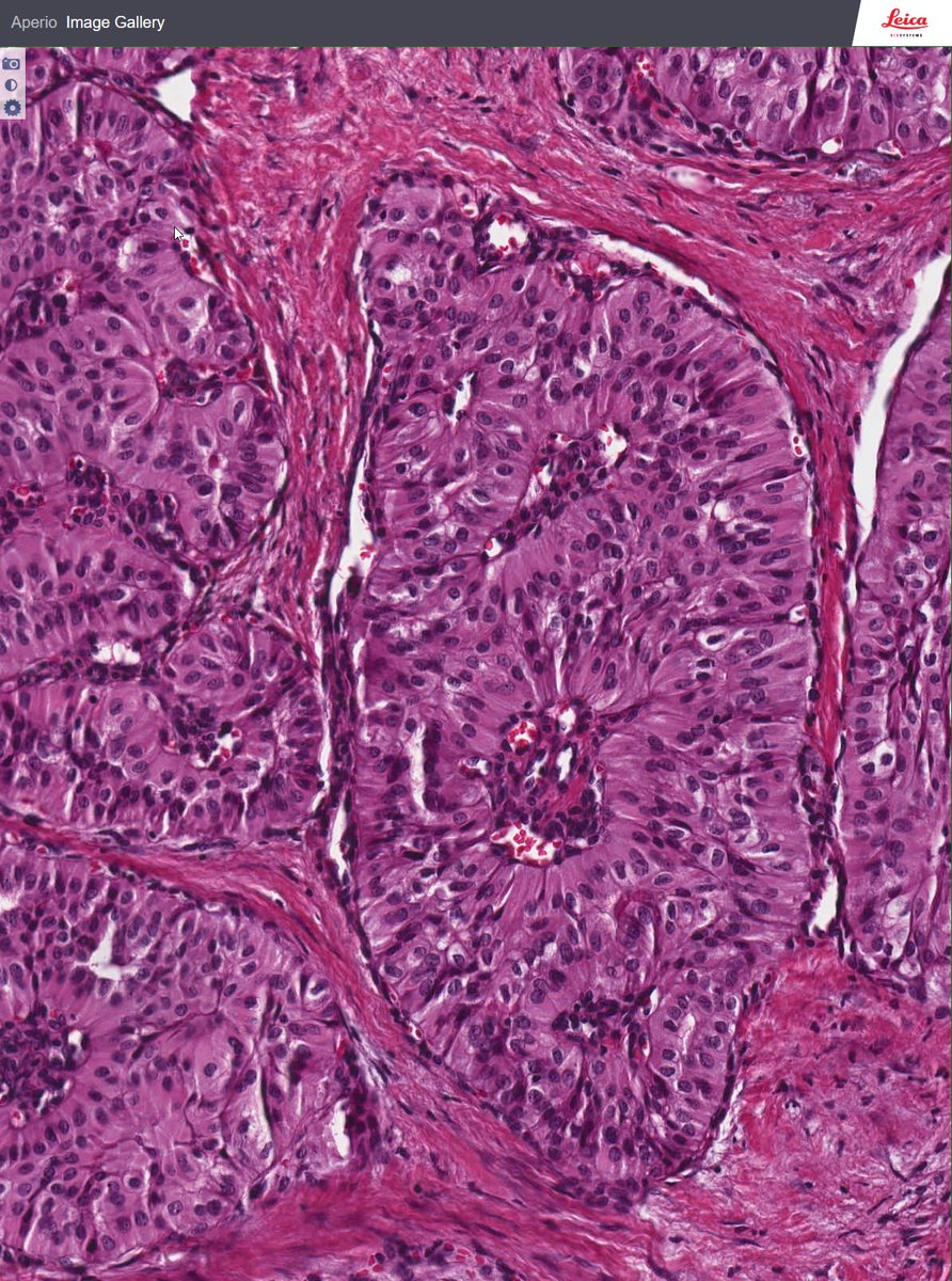
Nice case from Rosai's collection
Seminar number 1106, case 6
Breast tumour
What do you think ?

rosai.secondslide.com/view/sem1106/s…

Nice case from Rosai's collection Seminar number 1106, case 6 Breast tumour What do you think ? #BreastPath rosai.secondslide.com/view/sem1106/s…
account_circle
Craig Horbinski(@CraigHorbinski) 's Twitter Profile Photo

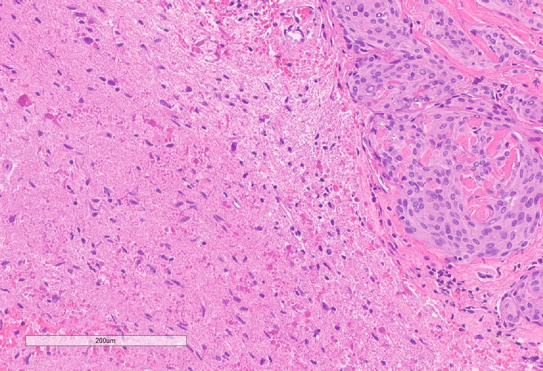
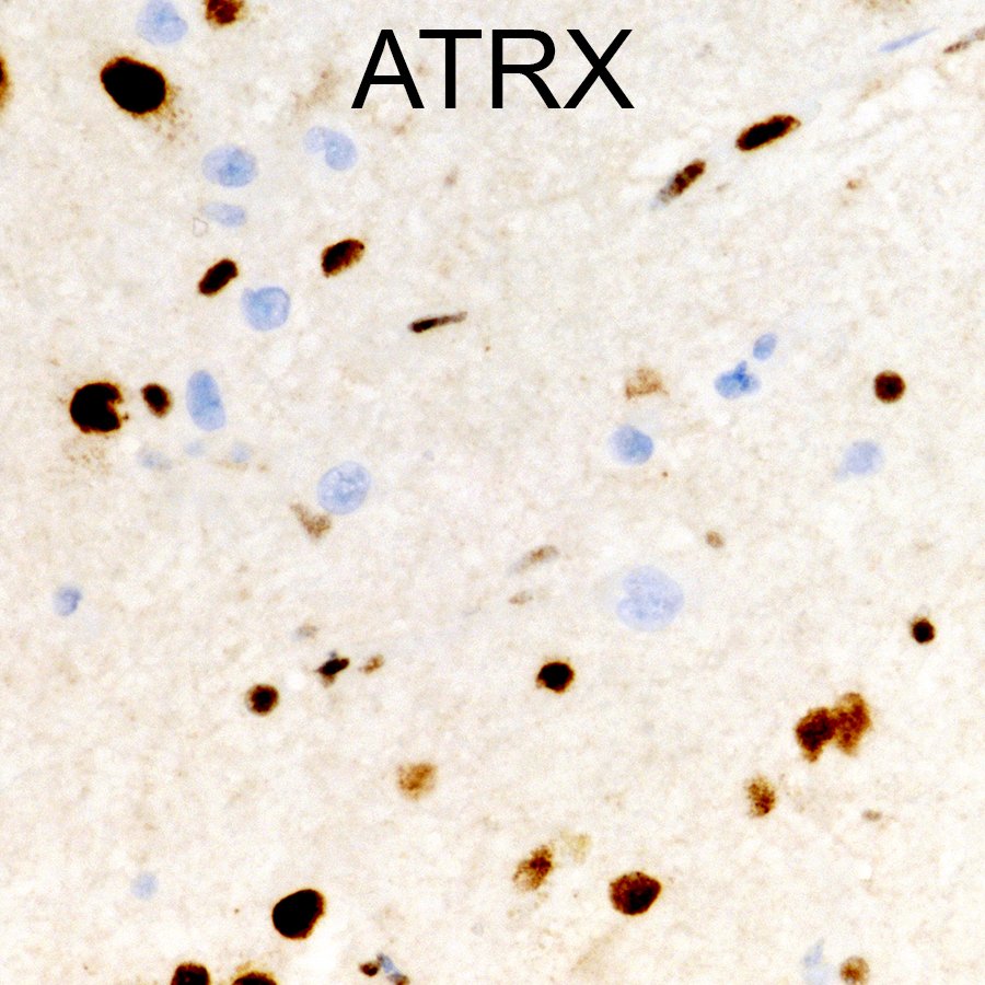
Thirtysomething man with frontal lobe tumor. What's high in your differential? (warning: this may be a Hidden GAP in your knowledge base)

Thirtysomething man with frontal lobe tumor. What's high in your differential? (warning: this may be a Hidden GAP in your knowledge base) #pathology #neuropath #PathTwitter
account_circle
Craig Horbinski(@CraigHorbinski) 's Twitter Profile Photo

SNO USCAP AANP Northwestern Pathology Neurosurgery at NM As the hint 'Hidden GAP' suggested, this is an HGAP, a high grade astrocytoma with piloid features. A PXA-like tumor with loss of ATRX raises the possibility of HGAP, which was confirmed by methylation profiling.

account_circle
William Lam(@William28538949) 's Twitter Profile Photo

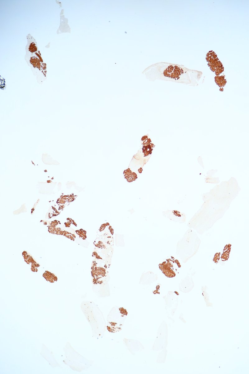
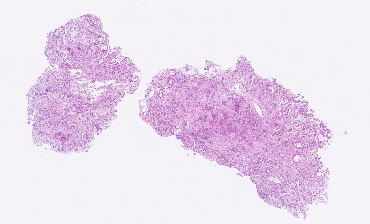
F/50 History of Ca breast pT1pN0
4 yrs later CT show 2.4cm suprasternal mass
Core biopsy
H&E and AE1/AE3
Old slides with fading.

F/50 History of Ca breast pT1pN0 4 yrs later CT show 2.4cm suprasternal mass Core biopsy H&E and AE1/AE3 Old slides with fading.
account_circle
William Lam(@William28538949) 's Twitter Profile Photo

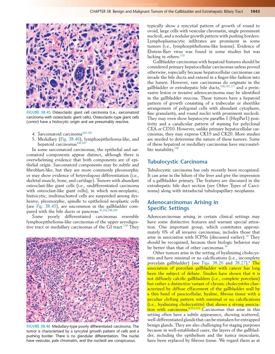
Elderly.
Gall bladder of a patient with Klatskin tumour.
Hyalinizing cholecystitis
'incomplete porcelain gallbladder' - not much calcification.
Screencap from Odze and Goldblum Surgical Pathology of the GI Tract, Liver, Biliary Tract ...

ncbi.nlm.nih.gov/pubmed/21716080

Elderly. Gall bladder of a patient with Klatskin tumour. Hyalinizing cholecystitis 'incomplete porcelain gallbladder' - not much calcification. Screencap from Odze and Goldblum Surgical Pathology of the GI Tract, Liver, Biliary Tract ... #gipath ncbi.nlm.nih.gov/pubmed/21716080
account_circle
C. Christofer Juhlin(@DrJuhlin) 's Twitter Profile Photo

Patient with unilateral hearing loss and a mass in the middle ear. Schwannoma is suspected. Biopsy is shown. Short thread follows...

Patient with unilateral hearing loss and a mass in the middle ear. Schwannoma is suspected. Biopsy is shown. Short thread follows...
account_circle