Lorand Kis(@kis_lorand) 's Twitter Profileg
Lorand Kis

@kis_lorand

pathologist; molpat, IHC, no favorite organ (yet); tweets are not medical advices, cases for educational purpose only

ID:992844732270292992

calendar_today05-05-2018 19:13:25

26,7K Tweets

6,5K Followers

745 Following

Henry YANG(@HENRYY_MD) 's Twitter Profile Photo

This sebaceous lesion on the arm of a man in his 60s ( middle aged). Mainly junctional .. quite mitotic active with high proliferation index ..but P53 is unimpressive… what you guys call this?
Dermpath_doc_trish Arjun Ramaiya Marcelasaeblima. David Terrano Tim McCalmont Gonzalo De Toro
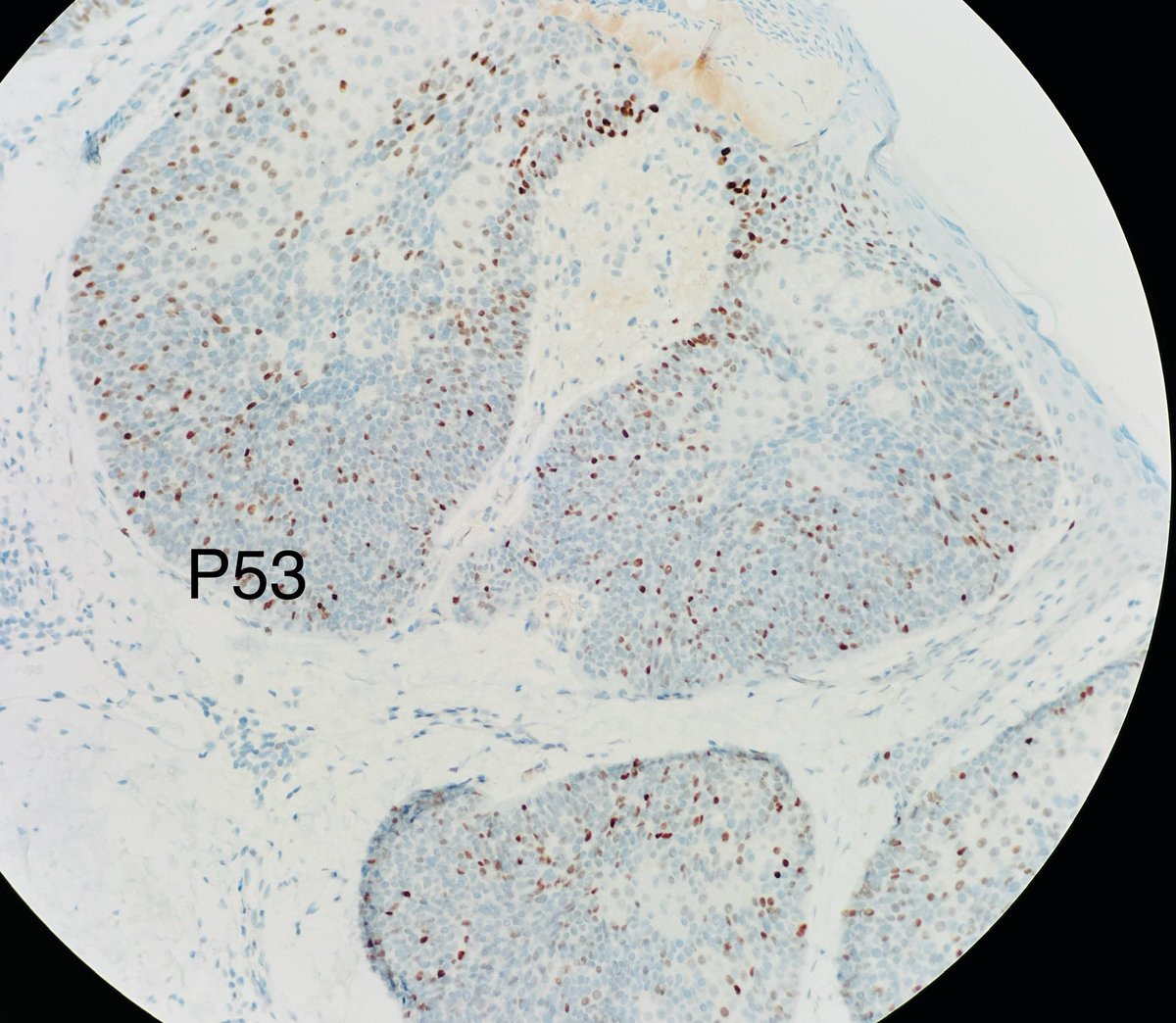
This sebaceous lesion on the arm of a man in his 60s ( middle aged). Mainly junctional .. quite mitotic active with high proliferation index ..but P53 is unimpressive… what you guys call this?#dermpath @PIorfino @ArjunRamaiya1 @MarcelaSaebL @david_terrano @mccalmo @gonzadetoro
account_circle
Sanam Loghavi, MD صنم لغوی 🔬🧬(@sanamloghavi) 's Twitter Profile Photo

The most picturesque/textbook example of AITL you will see in 2024…
What the WHO 5th now refers to as “Nodal T follicular helper cell lymphoma, angioimmunoblastic type”

why is that.. you may ask??🧐
Allow me to tell you why…🧵1/

The most picturesque/textbook example of AITL you will see in 2024… What the WHO 5th now refers to as “Nodal T follicular helper cell lymphoma, angioimmunoblastic type” why is that.. you may ask??🧐 Allow me to tell you why…🧵1/ #hemepath
account_circle
Lola Lozano(@mdlozanoe) 's Twitter Profile Photo

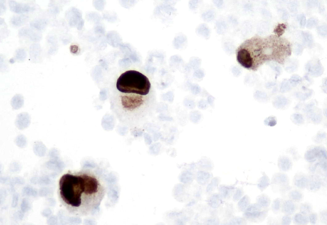
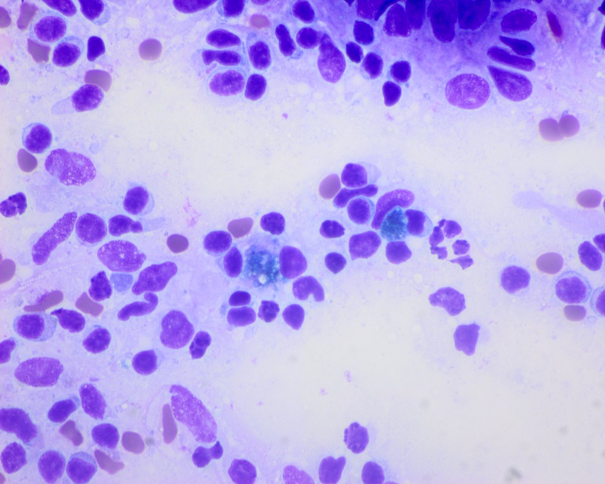
Peritoneal effusion with an unexpected guest.
ICC (on smear) leaves no doubt.....Cytomegalovirus infection. 🧐🫣🎯💡🔬
, what else?

Peritoneal effusion with an unexpected guest. ICC (on smear) leaves no doubt.....Cytomegalovirus infection. 🧐🫣🎯💡🔬 #Cytopath, what else?
account_circle
Dr Roshni Patil, MD(@Roshni141084) 's Twitter Profile Photo

Autopsy sections of brain in a clinical case of Rabies. Purkinje cell layer showing eosinophilic cytoplasmic inclusions called Negri bodies

Autopsy sections of brain in a clinical case of Rabies. Purkinje cell layer showing eosinophilic cytoplasmic inclusions called Negri bodies #neuropath #pathology #pathtwitter
account_circle
Tuçe Söylemez(@SoylemezTuce) 's Twitter Profile Photo

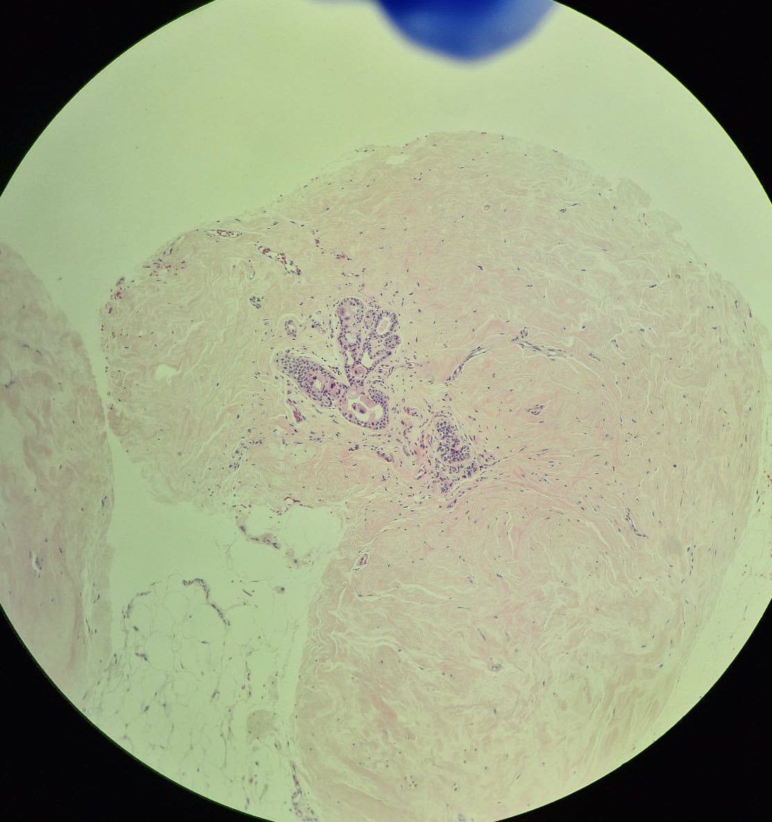
A 45-year-old female patient was initially diagnosed with a HER2-positive, invasive ductal carcinoma of the left breast in 2021. The patient was treated with lumpectomy, axillary dissection, followed by CRT. USG revealed some areas suspicious of fat necrosis.

A 45-year-old female patient was initially diagnosed with a HER2-positive, invasive ductal carcinoma of the left breast in 2021. The patient was treated with lumpectomy, axillary dissection, followed by CRT. USG revealed some areas suspicious of fat necrosis.
account_circle
Tim Bracey(@drtimbracey) 's Twitter Profile Photo

80M unusual plaque in rectum. Not clinically suspected but this is amyloid (subtype pending). Note the expansion of lamina propria by pink insoluble protein with cracks/slits picked up by Congo Red and polarising. Have a low threshold for suspecting it in unusual lesions!

80M unusual plaque in rectum. Not clinically suspected but this is amyloid (subtype pending). Note the expansion of lamina propria by pink insoluble protein with cracks/slits picked up by Congo Red and polarising. Have a low threshold for suspecting it in unusual #GIpath lesions!
account_circle
Pepe Jiménez Heffernan(@pepeheffernan) 's Twitter Profile Photo

CC325. Alone in the office. Mediastinal EBUS-TBNA with suspected sarcoidosis. Granulomas⬇️. Everything is ok. Time has taught me that in these cases it is useful to see if there is an oncologic history, whatever it is, retrieve the slides and check them. And that is what I did.

CC325. Alone in the office. Mediastinal EBUS-TBNA with suspected sarcoidosis. Granulomas⬇️. Everything is ok. Time has taught me that in these cases it is useful to see if there is an oncologic history, whatever it is, retrieve the slides and check them. And that is what I did.
account_circle
Virtual Pathology at The University of Leeds(@LeedsPathology) 's Twitter Profile Photo

🐄🔬
68M bladder biopsy. What is your diagnosis?
View the full slide and thousands of others on the Leeds website here: virtualpathology.leeds.ac.uk/slides/library…

#caseoftheweek 🐄🔬 68M bladder biopsy. What is your diagnosis? #PathTwitter #pathology View the full slide and thousands of others on the Leeds website here: virtualpathology.leeds.ac.uk/slides/library…
account_circle
Pepe Jiménez Heffernan(@pepeheffernan) 's Twitter Profile Photo

Part 3. Another case: Middle age man with psoriasis and axillary node. Large dendritic (interdigitating and Langerhans) cells will call your attention. When confluent they may vaguely resemble granulomas. Pigment is very helpful. No signs of follicular hyperplasia. Véronique Saada
Part 3. Another case: Middle age man with psoriasis and axillary node. Large dendritic (interdigitating and Langerhans) cells will call your attention. When confluent they may vaguely resemble granulomas. Pigment is very helpful. No signs of follicular hyperplasia. @saada_vsaada
account_circle
Michigan Pathology(@UMichPath) 's Twitter Profile Photo

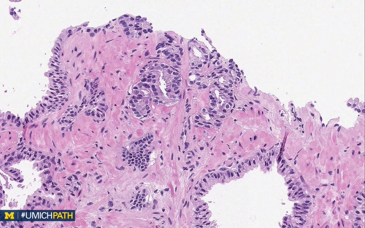
🔬 This week's theme: Prostate

➡️ One of these things is not like the other! Can you tell which image is different from the others?

➡️ We’ll tweet the answer and some quick facts tomorrow!

🔬 This week's #DailyDx theme: Prostate #Pathology ➡️ One of these things is not like the other! Can you tell which image is different from the others? ➡️ We’ll tweet the answer and some quick facts tomorrow! #UMichPath #GUPath
account_circle
Woo Cheal Cho, MD(@DrAldehyde) 's Twitter Profile Photo

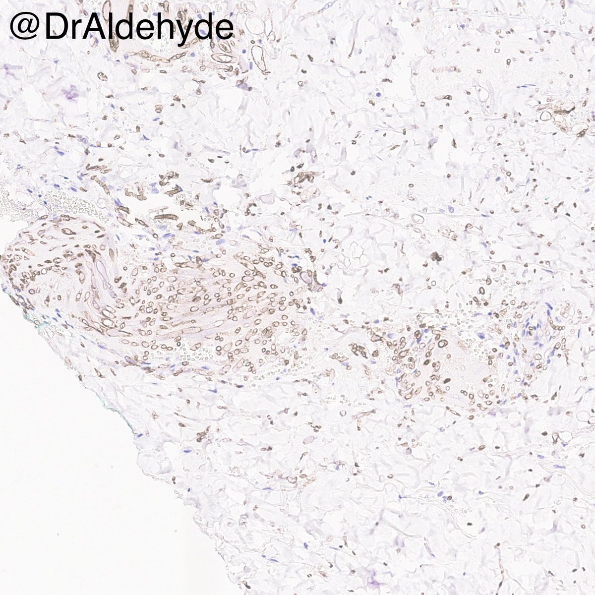
Any lovers out there routinely use fusarium ? My experience with this antibody has been great so far, and it helps me provide our treating physician team with the exact fungal species before the culture becomes available.

Any #dermpath lovers out there routinely use fusarium #IHCPath? My experience with this antibody has been great so far, and it helps me provide our treating physician team with the exact fungal species before the culture becomes available. #MDACCPath #pathology #microbiology
account_circle
Maryam Sayah, MD(@PathologySayah) 's Twitter Profile Photo

Adult granulosa cell tumor of ovary!
📍Cells show prominent longitudinal grooves. One of the characteristic pattern of this tumor is Call-Exner body (small follicle-like structures filled with acidophilic material)
📍Mutation in FOXL2
📍IHC: inhibin and calretinin

Adult granulosa cell tumor of ovary! 📍Cells show prominent longitudinal grooves. One of the characteristic pattern of this tumor is Call-Exner body (small follicle-like structures filled with acidophilic material) 📍Mutation in FOXL2 📍IHC: inhibin and calretinin #GYNpath
account_circle