Dylan_scopy
@dylanscopy
Ph.D student / Biomedical Engineering Joint @GeorgiaTech & @Emory #ShuJiaLab #microscopy 🌌🔬. Here is for ideas😉. @withBiPS
ID: 927348438039228416
https://sites.google.com/site/thejialab/ 06-11-2017 01:34:31
587 Tweet
122 Followers
197 Following


Our review with JB and Matthieu is out! CNRS Aquitaine Bordeaux Neurocampus Daniel Choquet . Advanced imaging and labelling methods to decipher brain cell organization and function | Nature Reviews Neuroscience nature.com/articles/s4158…


Excited to share an updated preprint on our new hybrid open-top light-sheet microscope. It has been a blast collecting data over the last year with our most powerful and versatile system to date. Big thank you to Jonathan Liu and all of our co-authors! biorxiv.org/content/10.110…

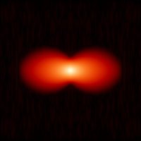


Virus walk on cells. Real-time 3D Tracking and Imaging Microscopy (3D-TrIm) of interactions between GFP+VSV lentiviruses and live cells (2D and 3D). Virions make transient contacts & their diffusion differ in actin-rich domains. Johnson et al. Welsher Lab @ Duke biorxiv.org/cgi/content/sh…
. Keio Global scientists used #organoid screening to reveal epigenetic vulnerabilities in human colorectal cancer. Nature Chemical Biology 👇 go.nature.com/3IAjg0x



Excited to share the great work of Kripa Patel demonstrating #MediSCAPE for high-speed 3D in-vivo histology. All images were acquired on fresh, uncleared tissues. Below shows human tongue imaged by just licking the probe! Just published in Nature Biomedical Engineering see it here: rdcu.be/cJ0uZ

🚨❤️🔬 #eSRRF is here! Next evolution of #SRRF. Full 3D, guides on optimal settings, and better rendering quality! An epic journey for Romain F. Laine, @Hannah_SuperRes and colleagues, with the help of fantastic labs including Bassam HAJJ Christophe Leterrier biorxiv.org/cgi/content/sh…


Computational Miniature Mesoscope V2: >7mm FOV, < 10um lateral and < 25um axial resolution; a fast & efficient 3D Shift-Variant model + a fully simulation-trained DNN generalizes to real experiments. Another impressive effort led by Yujia Xue, Ph.D.. arxiv.org/abs/2205.00123


Field of view extension in oblique plane microscopy: biorxiv.org/content/10.110… We leverage the native field of view of a NA 1.1 /25X lens by funneling it through the tertiary imaging system via optical tiling. In collaboration with Andrew York Alfred Millett-Sikking

Reto Fiolka’s (Reto Fiolka) Janelia+EMBL BioImaging seminar has just been posted on HHMI’s YouTube channel - Please watch his talk: “Improving the Spatio-temporal resolution in oblique plane microscopy.” #OPSIM #lightsheet Mark Kittisopikul Prevedel Lab 🔬 youtu.be/125wIEAUsEk


