Dan Graham(@DanGrahamMD) 's Twitter Profileg
Dan Graham

@DanGrahamMD

General Pathologist at Clinical Labs of Hawaii / Interest in BST, H&N, GU, GYN, Heme

ID:45696800

linkhttps://kikoxp.com/daniel_graham calendar_today08-06-2009 23:01:42

3,3K Tweets

1,0K Followers

806 Following

Reem Aljohani(@Aljohani_RR) 's Twitter Profile Photo

Dan Graham Under the microscope, a sight to behold,
A stroma so myxoid, cells young and old.
With vessels interspersed, and cells that float free,
A landscape so diverse, in this cardiac sea.

account_circle
Megan Smith(@megothelioma) 's Twitter Profile Photo

This spinal mass was a tough case (at least for me). The first two pics show a poorly diff component adjacent to a more mod diff area (this area stained positive with brachyury on the biopsy).

Signed out as dedifferentiated chordoma. Very cool.

This spinal mass was a tough case (at least for me). The first two pics show a poorly diff component adjacent to a more mod diff area (this area stained positive with brachyury on the biopsy). Signed out as dedifferentiated chordoma. Very cool. #PathTwitter #BSTPath
account_circle
Katrina Collins, MD(@katcollmd) 's Twitter Profile Photo

clear cell adenocarcinoma of urinary bladder

🔬solid, tubulocystic, and papillary growth patterns; hobnail cells with eosinophilic or clear cytoplasm
➕ CK7, PAX8, HNF1β, p53, Napsin
âž– GATA3, p63, ER, PR, WT1

#slidearchiveseries clear cell adenocarcinoma of urinary bladder #gupath #pathtwitter 🔬solid, tubulocystic, and papillary growth patterns; hobnail cells with eosinophilic or clear cytoplasm ➕ CK7, PAX8, HNF1β, p53, Napsin ➖ GATA3, p63, ER, PR, WT1
account_circle
Dan Graham(@DanGrahamMD) 's Twitter Profile Photo

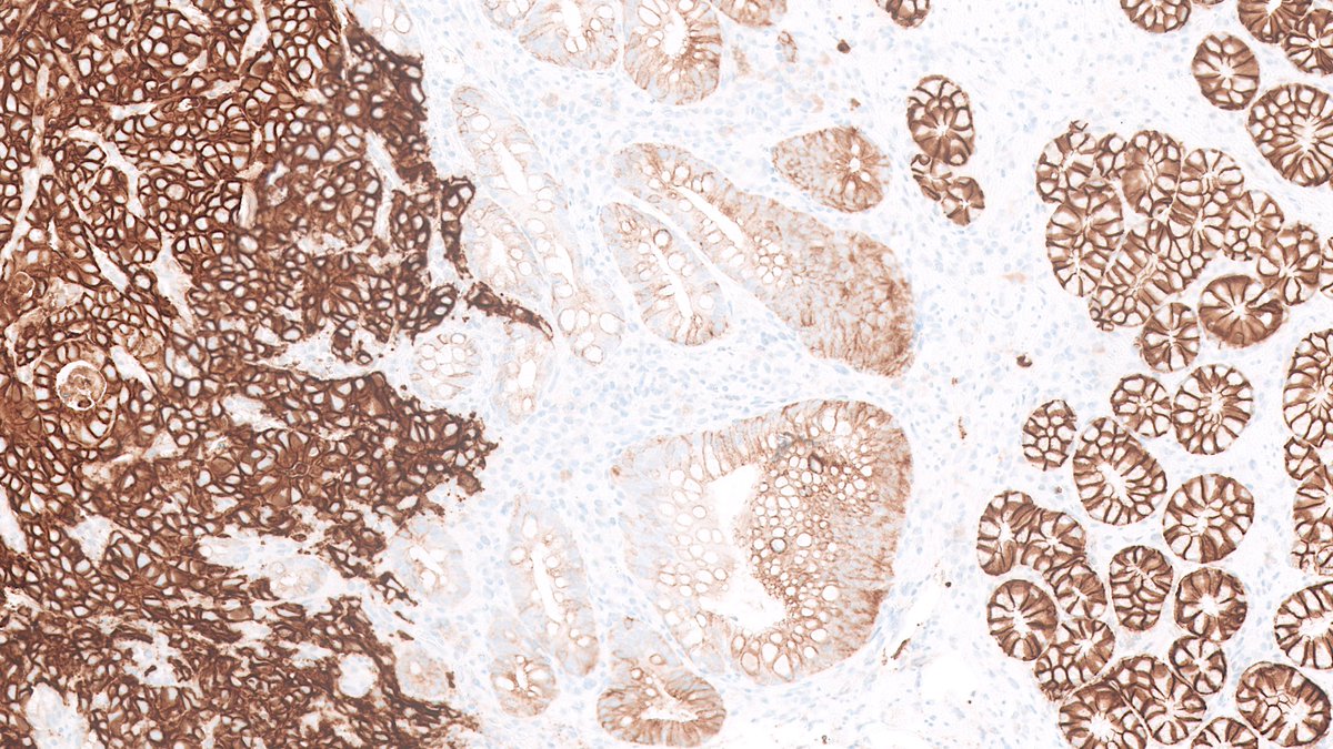
CD117 strong diffuse +
CK7 scattered single cells
GATA3 neg

Oncocytoma it is! Great job everyone, you nailed it.

account_circle
Dan Graham(@DanGrahamMD) 's Twitter Profile Photo

IHC panel:
CKAE1/3: positive
calretinin: positive
WT1: weak focal positive
TTF1: negative
desmin: negative
claudin4: negative

Enough to call mesothelioma, or need more meso markers?

IHC panel: CKAE1/3: positive calretinin: positive WT1: weak focal positive TTF1: negative desmin: negative claudin4: negative Enough to call mesothelioma, or need more meso markers?
account_circle
Dan Graham(@DanGrahamMD) 's Twitter Profile Photo

60s M 6.5 cm renal mass extending into perinephric fat. What do you think the diagnosis is? Would you get an IHC panel to help resolve any ddx you have?

60s M 6.5 cm renal mass extending into perinephric fat. What do you think the diagnosis is? Would you get an IHC panel to help resolve any ddx you have? #GUPath #PathX #PathTwitter
account_circle
Dan Graham(@DanGrahamMD) 's Twitter Profile Photo

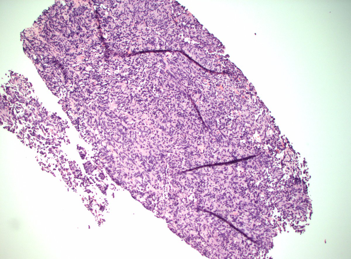
70s F with pleural and pericardial masses/studding as well as innumerable parenchymal lung masses. Bx of pleural mass.

Ddx? Dx? Immunopanel?

70s F with pleural and pericardial masses/studding as well as innumerable parenchymal lung masses. Bx of pleural mass. Ddx? Dx? Immunopanel? #ThoracicPath #PathX #PathTwitter
account_circle
Jeffrey Cloutier, MD, PhD(@JCloutierMD) 's Twitter Profile Photo

Challenging conventional belief, we found that atypical spindle cell/pleomorphic lipomatous tumor (ASPLT) can rarely undergo sarcomatous transformation. Excited to share insights on this phenomenon!

modernpathology.org/article/S0893-…

account_circle
Dan Graham(@DanGrahamMD) 's Twitter Profile Photo

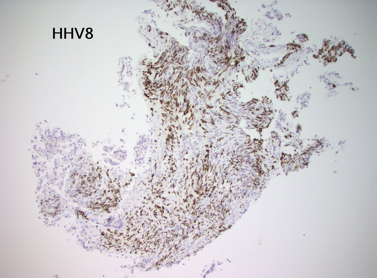
Thanks for all the responses. Indeed, the question here is: 'Is it an angiofibroma of soft tissue, or just a vocal cord polyp?' I don't think we'll ever know for sure, as it's not worth doing fusion analysis etc. A beautiful benign case nonetheless!

account_circle
Greg Charville(@Greg_Charville) 's Twitter Profile Photo

Cool new applications of GRM1 immunohistochemistry to diagnose chondromyxoid fibroma!

Subcutaneous CMF:
pubmed.ncbi.nlm.nih.gov/36810795/

CMF with FGF23 expression mimicking PMT:
pubmed.ncbi.nlm.nih.gov/38303543/

Cytopathology diagnosis of CMF:
pubmed.ncbi.nlm.nih.gov/38281480/
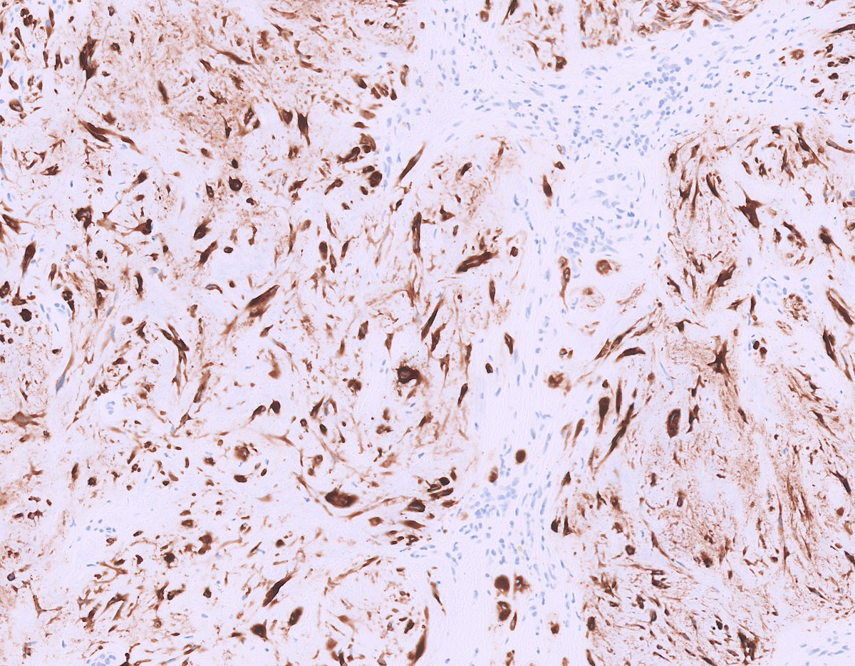
Cool new applications of GRM1 immunohistochemistry to diagnose chondromyxoid fibroma! #BSTpath #IHCpath #Cytopath Subcutaneous CMF: pubmed.ncbi.nlm.nih.gov/36810795/ CMF with FGF23 expression mimicking PMT: pubmed.ncbi.nlm.nih.gov/38303543/ Cytopathology diagnosis of CMF: pubmed.ncbi.nlm.nih.gov/38281480/
account_circle
Mark Ong(@DrMarkOng) 's Twitter Profile Photo

Mediastinal mass in young adult male.

Mixed inflammatory background and Hodgkin/Reed-Sternberg cells.

IHC in next tweets...

Mediastinal mass in young adult male. Mixed inflammatory background and Hodgkin/Reed-Sternberg cells. IHC in next tweets... #hemepath #pathology
account_circle
Greg Charville(@Greg_Charville) 's Twitter Profile Photo

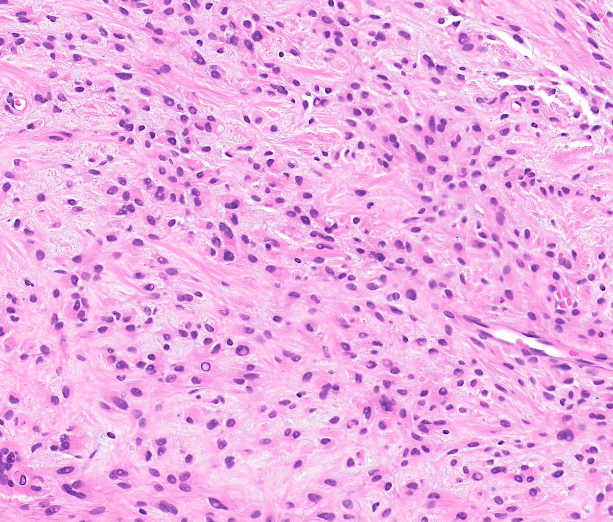
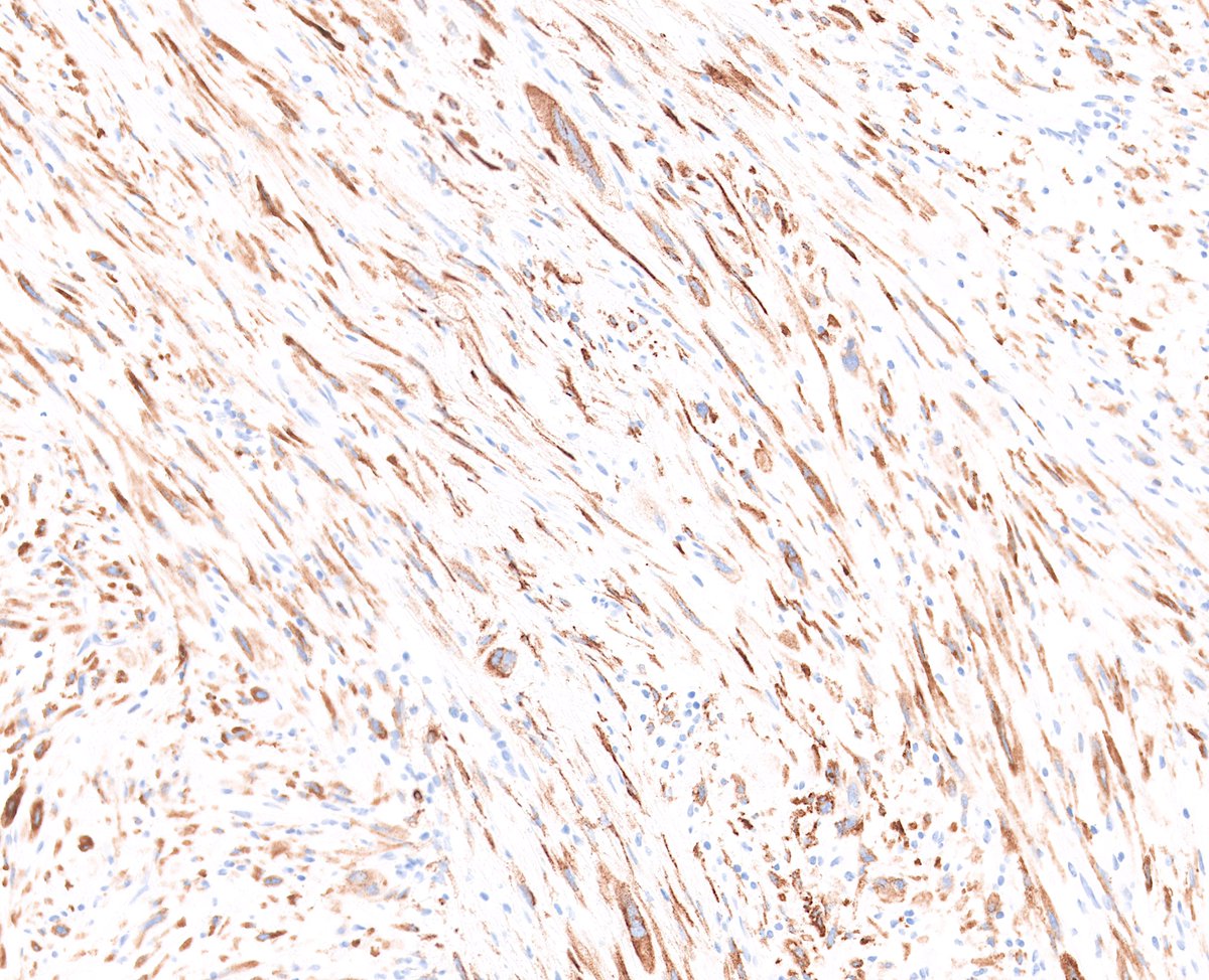
Look out - it's the myxoid, MUC4-positive variant of superficial CD34-positive fibroblastic tumor (PRDM10-rearranged soft tissue tumor)

Look out - it's the myxoid, MUC4-positive variant of superficial CD34-positive fibroblastic tumor (PRDM10-rearranged soft tissue tumor) #BSTpath
account_circle
Sanam Loghavi, MD صنم لغوی 🔬🧬(@sanamloghavi) 's Twitter Profile Photo

The fabulous Dr. Miles and her 🔥 poster and data on BRAF- mutated AML at
Come talk to her if you’re here.
Linde Miles AK Eisfeld
And a esp. thanks and shoutout to Bobby Bowman for his awesome analysis pipeline.
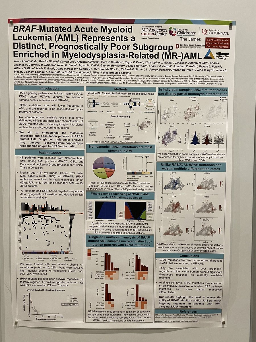
The fabulous Dr. Miles and her 🔥 poster and data on BRAF- mutated AML at #SCT24 Come talk to her if you’re here. @lindemilesphd @AkEisfeld And a esp. thanks and shoutout to @bowman_rl for his awesome analysis pipeline.
account_circle