Vanda Torous MD (@vandatorousmd) 's Twitter Profile
Vanda Torous MD

@vandatorousmd

Cytopathologist, Associate Professor @MGHpath @HarvardMed | Director of Cytopath QA/QI | Interests #CytoPath #GynPath #BreastPath #PatientSafety #QI #MedEd

ID: 839276757249323009

linkhttp://www.ezpath.org/ calendar_today08-03-2017 00:49:06

3,3K Tweet

5,5K Followers

749 Following

Vanda Torous MD (@vandatorousmd) 's Twitter Profile Photo

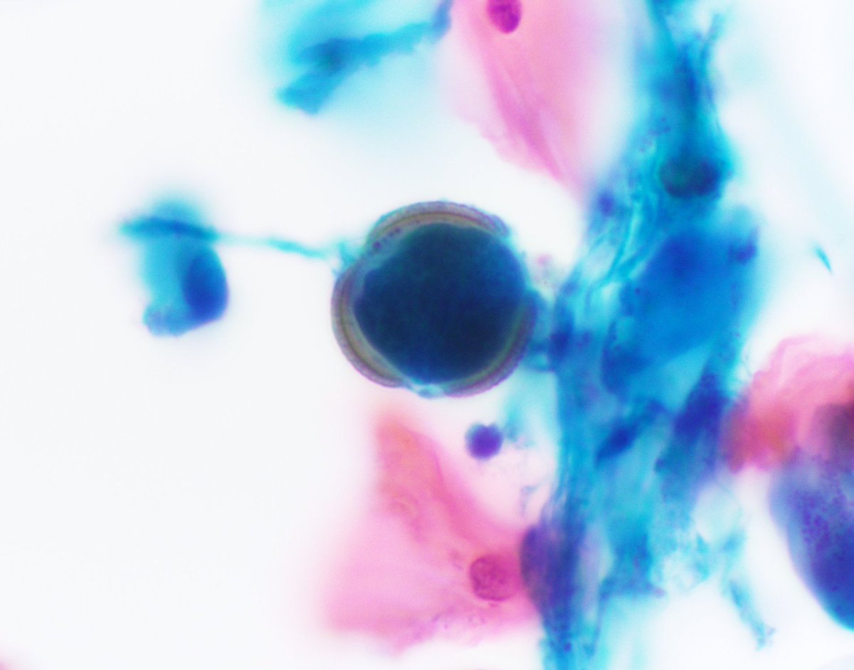
Interesting finding in this ileal loop urine sample... Vegetable matter (guar bean) consistent with Coloplast ostomy device contaminant 🫛 👀Don't mistake this for parasite eggs!👀 #CytoPath #PathTwitter

Interesting finding in this ileal loop urine sample... 

Vegetable matter (guar bean) consistent with Coloplast ostomy device contaminant 🫛

👀Don't mistake this for parasite eggs!👀

#CytoPath #PathTwitter
Vanda Torous MD (@vandatorousmd) 's Twitter Profile Photo

A case that read the book! Cervical pap with classic low grade squamous intraepithelial lesion (LSIL) If only they all came like this 🙃 #CytoPath #PathTwitter

A case that read the book! 
Cervical pap with classic low grade squamous intraepithelial lesion (LSIL)
If only they all came like this 🙃

#CytoPath #PathTwitter
Syed Z. Ali (@sza_jhcyto) 's Twitter Profile Photo

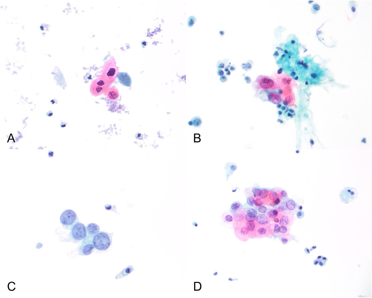
Without the intranuclear inclusions, these "oncocytoid" cells would have been a bit difficult to be called "Papillary Thyroid Carcinoma" on FNA. Follow-up, "PTC, Tall Cell Subtype".

Without the intranuclear inclusions, these "oncocytoid" cells would have been a bit difficult to be called "Papillary Thyroid Carcinoma" on FNA. Follow-up, "PTC, Tall Cell Subtype".
Cancer Cytopathology (@cancercytopath) 's Twitter Profile Photo

#OpenAccess! | HPV-positive UTCa can be identified reliably as HGUC by using TPS criteria; however, these cases may not be recognized as HPV-associated. acsjournals.onlinelibrary.wiley.com/doi/full/10.10… Poonam Vohra MD UCSF Pathology Esther Diana Rossi #CytoPath #ParisSystem

#OpenAccess! | HPV-positive UTCa can be identified reliably as HGUC by using TPS criteria; however, these cases may not be recognized as HPV-associated.

acsjournals.onlinelibrary.wiley.com/doi/full/10.10…

<a href="/PoonamVohra3/">Poonam Vohra MD</a> <a href="/UCSFPath/">UCSF Pathology</a> <a href="/DianaEstherossi/">Esther Diana Rossi</a> #CytoPath #ParisSystem
Syed Z. Ali (@sza_jhcyto) 's Twitter Profile Photo

Adenocarcinoma, presenting a sharp contrast with the juxtaposed benign squamous cells in a lower esophagus brushing. Prominent solitary macronucleoli, lack of adhered PMNs and a clean slide background with absence of inflammatory debris rules out reactive condition. (ThinPrep)

Adenocarcinoma, presenting a sharp contrast with the juxtaposed benign squamous cells in a lower esophagus brushing. Prominent solitary macronucleoli, lack of adhered PMNs and a clean slide background with absence of inflammatory debris rules out reactive condition. (ThinPrep)
American Board of Pathology (@theabpath) 's Twitter Profile Photo

Our volunteers make a difference! Thank you to our Cytopathology Test Development & Advisory Committee (TDAC) for a great meeting last week and for all you do throughout the year. A special thank you to Dr. Mehrotra for her years of service on the committee.

Our volunteers make a difference! Thank you to our Cytopathology Test Development &amp; Advisory Committee (TDAC) for a great meeting last week and for all you do throughout the year. A special thank you to Dr. Mehrotra for her years of service on the committee.
Vanda Torous MD (@vandatorousmd) 's Twitter Profile Photo

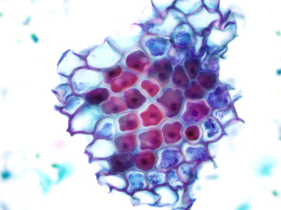
Some eye-catching 👀 vegetable matter on an anal pap Usually just glossed over, but does have the potential to be mistaken for squamous cells esp if found in smaller groups / singly #CytoPath #PathTwitter

Some eye-catching 👀 vegetable matter on an anal pap 

Usually just glossed over, but does have the potential to be mistaken for squamous cells esp if found in smaller groups / singly

#CytoPath #PathTwitter
Syed Z. Ali (@sza_jhcyto) 's Twitter Profile Photo

"Post-BCG granulomas" in voided urinary specimens may appear "atypical" to inexperienced eyes. Patient had a history of non-muscle invasive high-grade urothelial carcinoma. (SurePath)

"Post-BCG granulomas" in voided urinary specimens may appear "atypical" to inexperienced eyes. Patient had a history of non-muscle invasive high-grade urothelial carcinoma. (SurePath)
Syed Z. Ali (@sza_jhcyto) 's Twitter Profile Photo

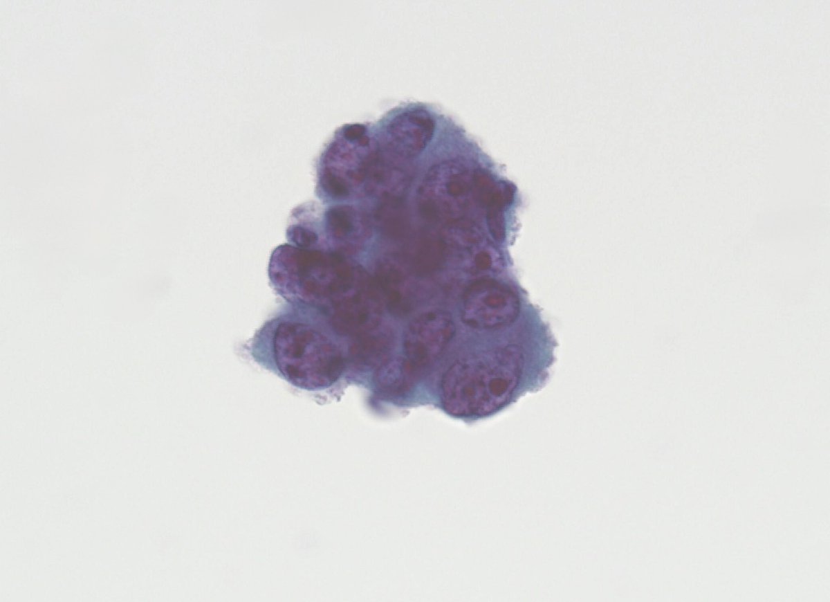
The often, notoriously difficult to diagnose, "Intestinal-type of "Endocervical Adenocarcinoma in-situ (AIS)". Also admixed is more conventional type of AIS. (SurePath, PAP Test)

The often, notoriously difficult to diagnose, "Intestinal-type of "Endocervical Adenocarcinoma in-situ (AIS)".  Also admixed is more conventional type of AIS. (SurePath, PAP Test)
Syed Z. Ali (@sza_jhcyto) 's Twitter Profile Photo

Just a great example - Cervical PAP test in a patient with history of “Colorectal Adenocarcinoma”. Complete LACK of background tumor diathesis is consistent with metastasis from the patient’s known primary over local invasion or a new gynecological primary. (SurePath)

Just a great example - Cervical PAP test in a patient with history of “Colorectal Adenocarcinoma”. Complete LACK of background tumor diathesis is consistent with metastasis from the patient’s known primary over local invasion or a new gynecological primary. (SurePath)
Syed Z. Ali (@sza_jhcyto) 's Twitter Profile Photo

A simple Papanicolaou stain may often work as good as a mucicarmine - Metastatic breast carcinoma in pleural effusion. (Cytospin)

A simple Papanicolaou stain may often work as good as a mucicarmine - Metastatic breast carcinoma in pleural effusion. (Cytospin)
Lola Lozano (@mdlozanoe) 's Twitter Profile Photo

Cytological features of fat necrosis of the breast: 🔬Foamy macrophages 🔬Necrotic debris: dirty background with cellular and fatty remnants. 🔬Adipocyte ghost cells (shadows) 🔬Absence of significant epithelial atypia International Academy Cytology Young EFCS Sociedad Española de Citología Cytopathology.org
Cytological features of fat necrosis of the breast:
🔬Foamy macrophages
🔬Necrotic debris: dirty background with cellular and fatty remnants.
🔬Adipocyte ghost cells (shadows)
🔬Absence of significant epithelial atypia 
<a href="/IACytology/">International Academy Cytology</a> <a href="/EfcsYoung/">Young EFCS</a> <a href="/SECitologia/">Sociedad Española de Citología</a> <a href="/cytopathology/">Cytopathology.org</a>