ZEISS Microscopy(@zeiss_micro) 's Twitter Profileg
ZEISS Microscopy

@zeiss_micro

We are #Microscopy. All about microscopes from #ZEISS.
LinkedIn: https://t.co/4jXTmJoUMF Facebook: https://t.co/ivuXIt7d1y | Imprint & Data Privacy: https://t.co/bvddlc6GZ2

ID:46128549

linkhttps://www.zeiss.com/microscopy calendar_today10-06-2009 14:49:22

10,6K Tweets

23,4K Followers

3,8K Following

Follow People
ZEISS Microscopy(@zeiss_micro) 's Twitter Profile Photo

Today, we introduce the new ZEISS Celldiscoverer 7 – your microscope with adaptable automation for advanced imaging workflows!

ZEISS Celldiscoverer 7 now features higher acquisition speed, and supports more versatile experiments and new applications. Here is what's new:

Today, we introduce the new ZEISS Celldiscoverer 7 – your microscope with adaptable automation for advanced imaging workflows! ZEISS Celldiscoverer 7 now features higher acquisition speed, and supports more versatile experiments and new applications. Here is what's new:
account_circle
EXCITE(@EXCITE_network) 's Twitter Profile Photo

3D views of a sillimanite-biotite-garnet migmatitic showing the peculiar, flattened shape of garnet 🏔️

📸Images by Dr. Yuntao Ji Yuntao Ji for TNA user Fabiola Caso Fabiola Caso, Uni. of Milan Università degli Studi di Milano

🔬ZEISS Microscopy Versa 610 , UU Utrecht University

account_circle
Aberdeen Microscopy(@Abdn_Micro) 's Twitter Profile Photo

We've had lots of fun being trained on our new ZEISS Microscopy Lightsheet 7 this week!
Thanks to Morgan from Tara Sutherland's lab for supplying the lung slice for us to practice on 🫁😃🔬

We've had lots of fun being trained on our new @zeiss_micro Lightsheet 7 this week! Thanks to Morgan from @DrTESutherland's lab for supplying the lung slice for us to practice on 🫁😃🔬 #FluorescenceFriday #Lightsheet #Microscopy
account_circle
ZEISS Group(@ZEISS_Group) 's Twitter Profile Photo

🌎 Protecting our environment starts with understanding the impact we have on it. Discover how researchers use ZEISS Microscopy in a project by EMBL to study the effects of human activity on Europe's ecosystems: zeiss.ly/embl-zeiss

account_circle
EXCITE(@EXCITE_network) 's Twitter Profile Photo

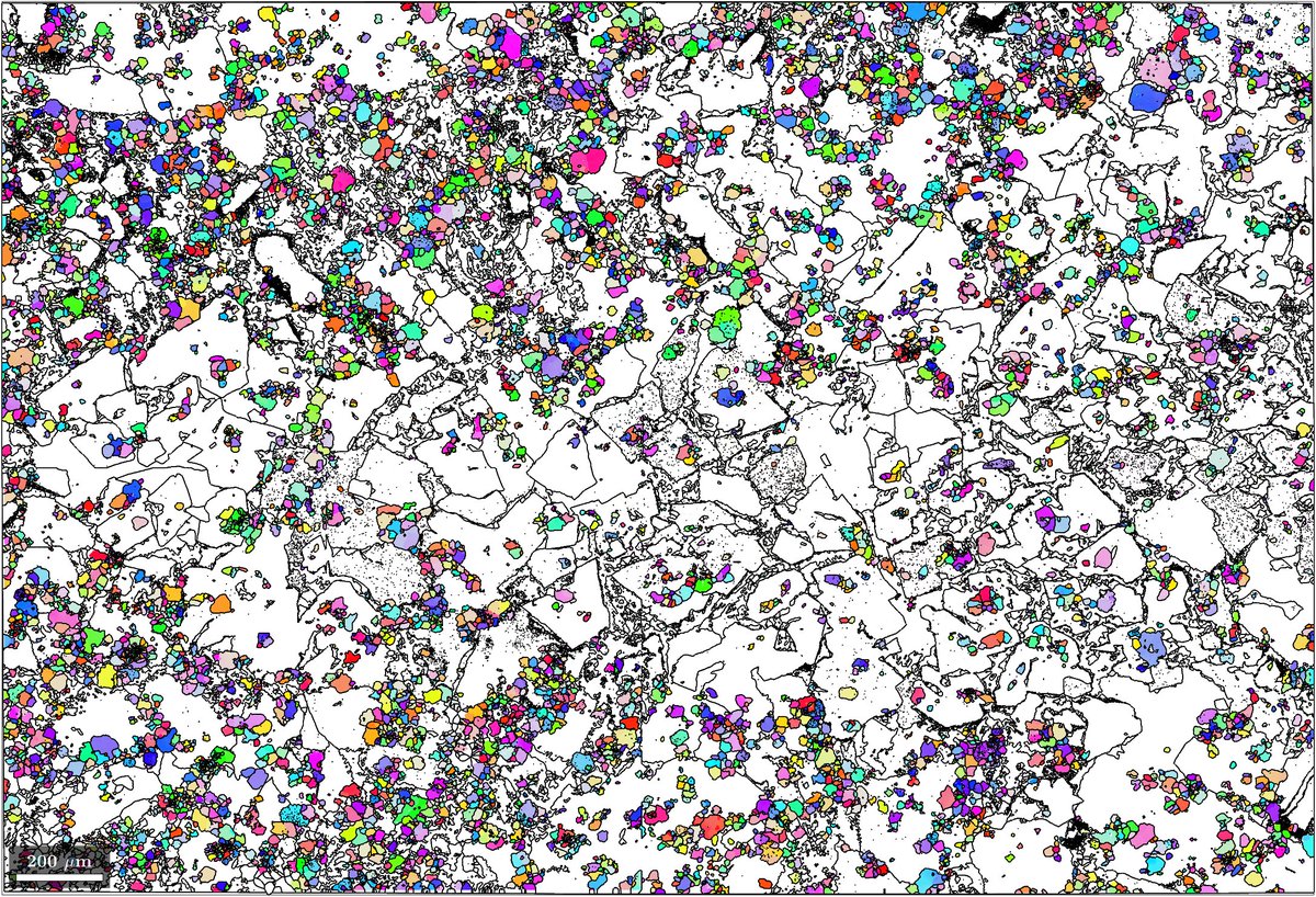
Happy ! map of a synthetic lithium slag showing lithium aluminate (LiAlO2) crystal orientation.

🔬 Imaged with a ZEISS Gemini 450 (ZEISS Microscopy) at Utrecht University by Maartje Hamers for EXCITE TNA user Cindytami Rachmawati, TU Bergakademie Freiberg

Happy #ThinSectionThursday! #EBSD map of a synthetic lithium slag showing lithium aluminate (LiAlO2) crystal orientation. 🔬 Imaged with a ZEISS Gemini 450 #SEM (@zeiss_micro) at @UniUtrecht by Maartje Hamers for @EXCITE_network TNA user Cindytami Rachmawati, @TUBergakademie
account_circle
ZEISS Microscopy(@zeiss_micro) 's Twitter Profile Photo

Currently the ZEISS Natural Resources User Group Meeting 2024 takes place in China! 🔬🌏⛰

Since yesterday, participants have the opportunity to present their research, establish new contacts and share experiences about microscopy workflows, successes and challenges.

account_circle
Dr Richard Kirby(@PlanktonPundit) 's Twitter Profile Photo

Astonishing marine life cycles. Many seabed creatures start life in the plankton. This late-stage planktonic crab larva, called a megalopa, is about to leave the plankton to begin its adult life on the seabed. (You can see the remnants of the zoea's dorsal spine.)
ZEISS Microscopy

account_circle
Danielle Beckman(@DaniBeckman) 's Twitter Profile Photo

Miss posting some photos here, so here it goes! 😁
I like taking photos of weird things happening in the brain, but homeostasis (stable equilibrium) is also beautiful! The image shows how microglia (🔵iba1) and neurons (🔴smi32 maker) are interacting. 📸Taken with ZEISS Microscopy

Miss posting some photos here, so here it goes! 😁 I like taking photos of weird things happening in the brain, but homeostasis (stable equilibrium) is also beautiful! The image shows how microglia (🔵iba1) and neurons (🔴smi32 maker) are interacting. 📸Taken with @zeiss_micro
account_circle
ibidi GmbH(@ibidiCells) 's Twitter Profile Photo

🦋Enchanted Wings: Arrival of Spring🌺

🔬Murine Colonic Organoid acquired using a ZEISS Microscopy LSM 700 Airyscan.

👩‍🔬 Scientific artwork by Jaclyn Alexis Rivas from The University of New Mexico Univ. of New Mexico, USA

🦋Enchanted Wings: Arrival of Spring🌺 🔬Murine Colonic Organoid acquired using a @zeiss_micro LSM 700 Airyscan. 👩‍🔬 Scientific artwork by Jaclyn Alexis Rivas from The University of New Mexico @UNM, USA #Butterfly #spring #organoid #sciart #microscopy #imaging #bioart #wings
account_circle
CSHL Microscopy Shared Resource(@CSHLmicroscopy) 's Twitter Profile Photo

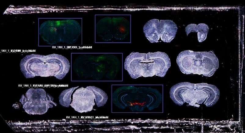
Thanks to Leo Ramirez Sanchez from Bo Li lab, this is a perfect sample and example how we can take advantage of AI sample finder, Airyscan 4Y with 10x/0.45 lens, and Zen connect to for faster scanning, whole slide + fine neuron structure’s visualization and navigation. 🔬🧠🤩

Thanks to Leo Ramirez Sanchez from Bo Li lab, this is a perfect sample and example how we can take advantage of AI sample finder, Airyscan 4Y with 10x/0.45 lens, and Zen connect to for faster scanning, whole slide + fine neuron structure’s visualization and navigation. 🔬🧠🤩
account_circle
ZEISS Microscopy(@zeiss_micro) 's Twitter Profile Photo

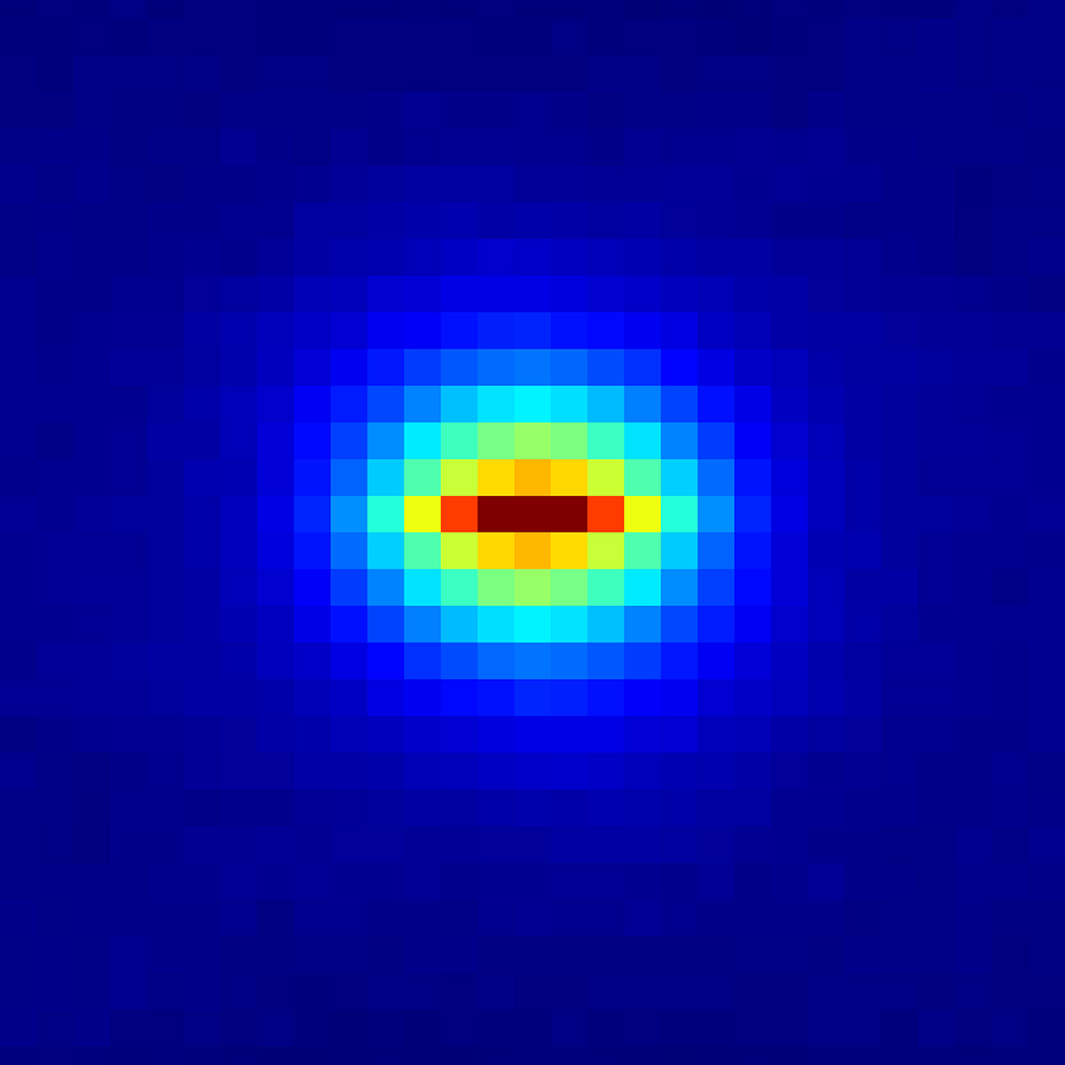
With the software module ZEISS Spectral RICS, we are releasing a new solution for understanding molecular dynamics of your living samples.

In addition to obtaining structural image information of your sample, unveil how proteins of interest move and interact in relation to their

With the software module ZEISS Spectral RICS, we are releasing a new solution for understanding molecular dynamics of your living samples. In addition to obtaining structural image information of your sample, unveil how proteins of interest move and interact in relation to their
account_circle
Dr Richard Kirby(@PlanktonPundit) 's Twitter Profile Photo

Huge spring plankton biodiversity in my net sample collected off Plymouth UK yesterday. At least: Dinoflagellata, Bacillariophyta, Haptophyta, Arthropoda, Annelida, Phoronida, Cnidaria, and Echinodermata. But what species can you see? ZEISS Microscopy

account_circle
NEMI(@ProjectNemi) 's Twitter Profile Photo

🏜️ 'Desert Rose'🌹by Ewelina Kluza from Amsterdam UMC, Amsterdam UMC

An immune cell on the endothelial surface of a mouse aorta! 🐁❤️

🔬 Imaged with a ZEISS Sigma 300 Scanning Electron Microscope ( ) ZEISS Microscopy at Amsterdam UMC

🏜️ 'Desert Rose'🌹by Ewelina Kluza from Amsterdam UMC, @amsterdamumc An immune cell on the endothelial surface of a mouse aorta! 🐁❤️ 🔬 Imaged with a ZEISS Sigma 300 Scanning Electron Microscope (#SEM) @zeiss_micro at Amsterdam UMC
account_circle
Josh Gibson, Ph.D.(@DrStrangeAnt) 's Twitter Profile Photo

MicroCT scan of a fruit fly stained with iodine, taken with Beckman Institute's brand new Zeiss Xradia ZEISS Microscopy's Xradia Versa 630 scanner! Renderings done in Amira.

account_circle
Vanderbilt Cell Imaging Shared Resource(@VUCellImaging) 's Twitter Profile Photo

Spring has sprung in Nashville! It's chilly, but the flowers are blooming, and everything is green. We found this 'helicopter seed' on campus and couldn't resist checking its autofluorescence for . ZEISS Microscopy confocal, 20x/0.8. Max. Intensity Projection.

Spring has sprung in Nashville! It's chilly, but the flowers are blooming, and everything is green. We found this 'helicopter seed' on campus and couldn't resist checking its autofluorescence for #FluorescenceFriday. @zeiss_micro confocal, 20x/0.8. Max. Intensity Projection.
account_circle
Danielle Beckman(@DaniBeckman) 's Twitter Profile Photo

Astrogliosis (🟡) in the human brain 🧠🔬😨. One of the processes occurring in neuroinflammation. These neurons (🔵) are condemned to die...
Image taken from the frontal cortex of a patient with advanced Alzheimer's disease (Braak Stage VI)
ZEISS Microscopy

Astrogliosis (🟡) in the human brain 🧠🔬😨. One of the processes occurring in neuroinflammation. These neurons (🔵) are condemned to die... Image taken from the frontal cortex of a patient with advanced Alzheimer's disease (Braak Stage VI) @zeiss_micro #neuroinflammation
account_circle
EXCITE(@EXCITE_network) 's Twitter Profile Photo

Happy ! map of a synthetic lithium slag showing crystal orientation.

🔬 Imaged with a ZEISS Gemini 450 (ZEISS Microscopy) at Utrecht University (@UniUtrecht) by Maartje Hamers for EXCITE TNA user Cindytami Rachmawati, TU Bergakademie Freiberg

Happy #ThinSectionThursday! #EBSD map of a synthetic lithium slag showing #spinel crystal orientation. 🔬 Imaged with a ZEISS Gemini 450 #SEM (@zeiss_micro) at Utrecht University (@UniUtrecht) by Maartje Hamers for @EXCITE_network TNA user Cindytami Rachmawati, @TUBergakademie
account_circle