Vanderbilt Cell Imaging Shared Resource(@VUCellImaging) 's Twitter Profileg
Vanderbilt Cell Imaging Shared Resource

@VUCellImaging

Vanderbilt University Cell Imaging Shared Resource (CISR). All things microscopy for @VanderbiltU and @VUMChealth. Confocal, Super-Res, Live Cell, TEM, SEM.

ID:1068602338779308032

linkhttps://my.vanderbilt.edu/cisr/ calendar_today30-11-2018 20:27:06

1,4K Tweets

2,3K Followers

1,0K Following

Nikon Instruments(@NikonInst) 's Twitter Profile Photo

๐Ÿ”ฌ ๐Ÿ“… Only a few weeks left to submit entries to the competitions!

Upload your best images and videos by April 30th for a chance to win up to $5000: enter.nikonsmallworld.com

account_circle
Vanderbilt Cell Imaging Shared Resource(@VUCellImaging) 's Twitter Profile Photo

Thanks to Dr. Hinton Antentor Othrell Hinton, Jr PhD (A.J. Hinton) for inviting Dr. Jenny Schafer and Dr. Evan Krystofiak from the CISR on this podcast. It was really fun, the questions are great, and we loved sharing about our professions as core scientists. Have a listen! ๐Ÿ”ฌ๐ŸŽ™๏ธ

account_circle
VUSM Basic Sciences(@VUBasicSciences) 's Twitter Profile Photo

The lab of Antentor Othrell Hinton, Jr PhD (A.J. Hinton) came together with collaborators from across Vanderbilt University, Vanderbilt Health, the U.S., and Brazil to determine the differences in the 3D morphology of mitochondria and their cristae in brown adipose tissue.

Read on ๐Ÿ‘‡

medschool.vanderbilt.edu/basic-sciencesโ€ฆ

account_circle
Vanderbilt Cell Imaging Shared Resource(@VUCellImaging) 's Twitter Profile Photo

๐Ÿ˜๐Ÿคฏ๐Ÿ˜ฎ Beautiful! CISR's Scientific Director Dr. Tyska shows us how to truly image across scales! โ˜€๏ธ๐ŸŒ˜

account_circle
Vanderbilt Cell Imaging Shared Resource(@VUCellImaging) 's Twitter Profile Photo

With a FIRST PLACE ๐Ÿฅ‡finish for Kari Seedle, we are claiming a CISR victory for ๐Ÿ€ in the VUSM Basic Sciences MBB bracket competition! Kudos to Kari for being #1 out of 92! And #1 of the 30 who picked UConn to win it all. Other CISR showings were ๐Ÿ‘Ž.

With a FIRST PLACE ๐Ÿฅ‡finish for Kari Seedle, we are claiming a CISR victory for #MarchMadness ๐Ÿ€ in the @VUBasicSciences MBB bracket competition! Kudos to Kari for being #1 out of 92! And #1 of the 30 who picked UConn to win it all. Other CISR showings were ๐Ÿ‘Ž.
account_circle
Vanderbilt Cell Imaging Shared Resource(@VUCellImaging) 's Twitter Profile Photo

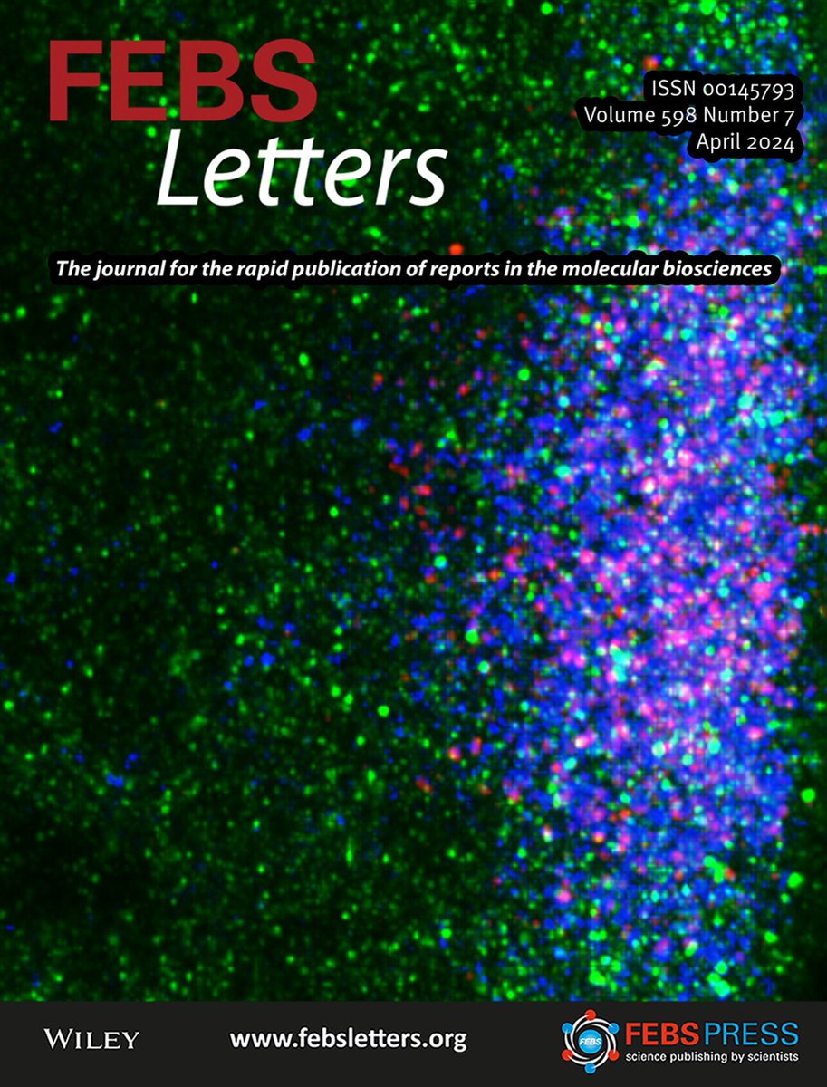
Cover alert! We always enjoy working with Dr. Alexey Arkov and his group from Murray State U. when they visit to use the ZEISS Microscopy LSM880 Airyscan. Check out their new publication and cover image. FEBS_Letters febs.onlinelibrary.wiley.com/doi/10.1002/18โ€ฆ

Cover alert! We always enjoy working with Dr. Alexey Arkov and his group from Murray State U. when they visit to use the @zeiss_micro LSM880 Airyscan. Check out their new publication and cover image. @FEBS_Letters #MicroscopyMonday febs.onlinelibrary.wiley.com/doi/10.1002/18โ€ฆ
account_circle
Vanderbilt Cell Imaging Shared Resource(@VUCellImaging) 's Twitter Profile Photo

Spring has sprung in Nashville! It's chilly, but the flowers are blooming, and everything is green. We found this 'helicopter seed' on campus and couldn't resist checking its autofluorescence for . ZEISS Microscopy confocal, 20x/0.8. Max. Intensity Projection.

Spring has sprung in Nashville! It's chilly, but the flowers are blooming, and everything is green. We found this 'helicopter seed' on campus and couldn't resist checking its autofluorescence for #FluorescenceFriday. @zeiss_micro confocal, 20x/0.8. Max. Intensity Projection.
account_circle
Vanderbilt Cell Imaging Shared Resource(@VUCellImaging) 's Twitter Profile Photo

ATTN: Graduate students! The Southeastern Microscopy Society meeting is in Chattanooga, TN, May 8-10. Students who submit an abstract and present are eligible for the Ruska competition with prize of $300.
Instructions:
southeasternmicroscopy.org/ruska-award-guโ€ฆ
Meeting:
southeasternmicroscopy.org/2024-meeting/
account_circle
Vanderbilt Biophotonics Center(@VU_Biophotonics) 's Twitter Profile Photo

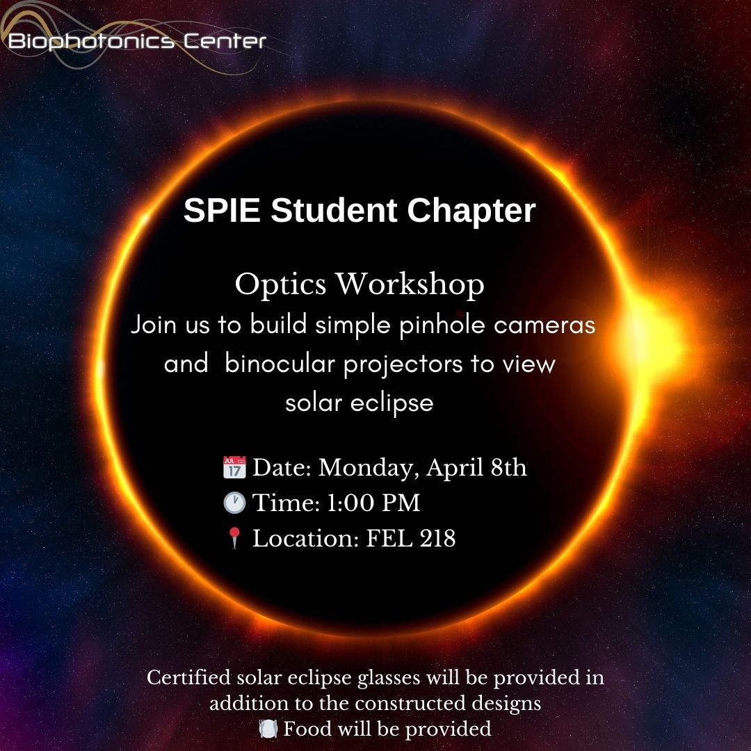
Don't miss this unique opportunity to learn, create, and witness the magic of the eclipse firsthand! Mark your calendars and see you there!

Vanderbilt SPIE student chapter organizes Optics workshop
๐Ÿš€๐Ÿ”ญ

Don't miss this unique opportunity to learn, create, and witness the magic of the eclipse firsthand! Mark your calendars and see you there! Vanderbilt SPIE student chapter organizes Optics workshop #OpticsWorkshop #EclipseViewing #STEMeducation ๐Ÿš€๐Ÿ”ญ
account_circle
VI4Research(@VI4Research) 's Twitter Profile Photo

๐ŸŽฅ Vanderbilt Cell Imaging Shared Resource provides researchers with access to state-of-the-art imaging equipment and expert technical support for sophisticated and analysis of tissue and cellular and .
๐ŸŒ my.vanderbilt.edu/cisr
loom.ly/WqEtbNY

account_circle
Vanderbilt Cell Imaging Shared Resource(@VUCellImaging) 's Twitter Profile Photo

Great Beth Cimini Lab ๐Ÿ”ฌ๐Ÿ’ป ๐Ÿ“Š paper for imagingโžก๏ธanalysis. 'every aspect of the exp. (which samples are generated...collected...processed together...how sample processing proceeds...imaging technique...scope parameters) impacts which conclusions can be reliably drawn...final analysis is done'

account_circle
Vanderbilt Cell Imaging Shared Resource(@VUCellImaging) 's Twitter Profile Photo

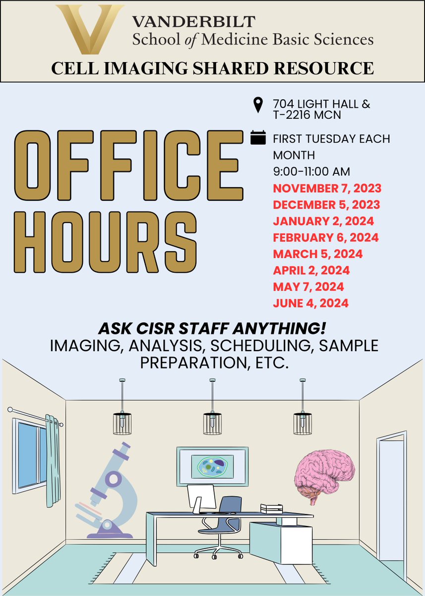
It's that time again! Join us on Tuesday, April 2 from 9:00-11:00 AM for our first Tuesday of the month CISR Office Hours. Ask us anything!

It's that time again! Join us on Tuesday, April 2 from 9:00-11:00 AM for our first Tuesday of the month CISR Office Hours. Ask us anything!
account_circle
Vanderbilt Cell Imaging Shared Resource(@VUCellImaging) 's Twitter Profile Photo

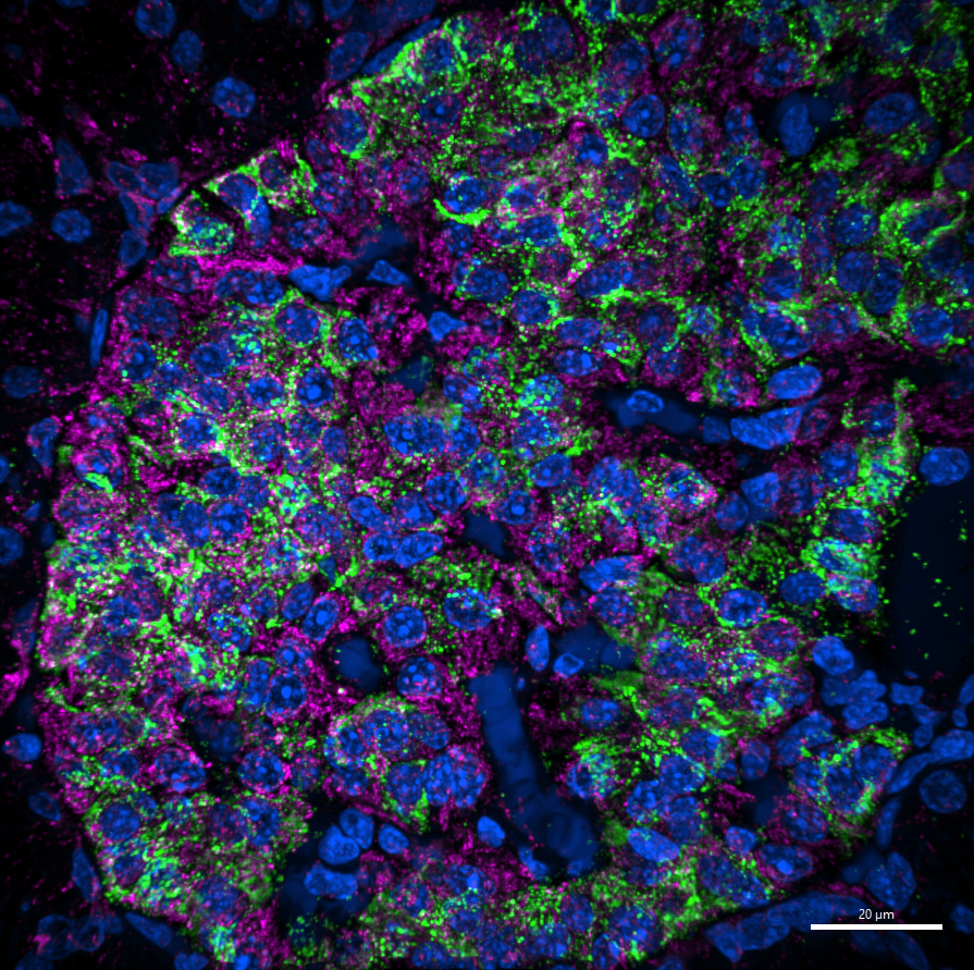
Happy ! We love this image from Darian Carroll Future Dr. Darian Carroll in the GannonLab taken on the CISR's new Nikon Instruments SDC with SoRa! ๐ŸคฉNHP islet - insulin in green, mitochondria in magenta, & DAPI in blue. Check out the resolution on those insulin granules! ๐Ÿ”ฌ๐Ÿ’š

Happy #FluorescenceFriday! We love this image from Darian Carroll @BetaCellBelle in the @GannonLab taken on the CISR's new @NikonInst SDC with SoRa! ๐ŸคฉNHP islet - insulin in green, mitochondria in magenta, & DAPI in blue. Check out the resolution on those insulin granules! ๐Ÿ”ฌ๐Ÿ’š
account_circle
ABRF(@MyABRF) 's Twitter Profile Photo

Have you registered for our Annual Meeting in Minneapolis? It's Apri 21-24! Registration rates increase April 5! buff.ly/4crmaoa

Have you registered for our Annual Meeting in Minneapolis? It's Apri 21-24! Registration rates increase April 5! #ABRF2024 buff.ly/4crmaoa
account_circle
Vanderbilt Cell Imaging Shared Resource(@VUCellImaging) 's Twitter Profile Photo

Must read when it begins... 'This research leveraged advanced light & electron microscopy to perform quantitative morphometry of the intestinal tuft cell cytoskeleton.' Great work from Matt Tyska & first author Jen Silverman. Thanks for using & acknowledging the CISR!๐Ÿ”ฌ๐Ÿ‘Š

account_circle
BioImagingUK(@BioImagingUK) 's Twitter Profile Photo

After a great first run, it's coming back!

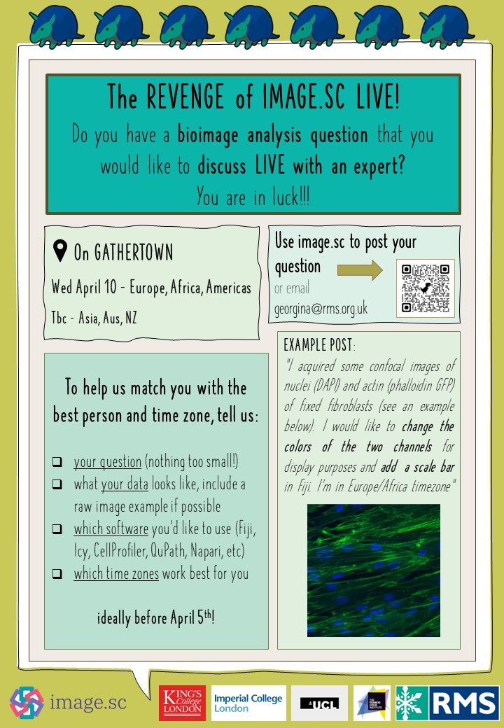
The Revenge of Image.sc LIVE! returns on Wed April 10 from 1-4pm BST!

If you want to discuss your images with an expert live, post your question here before April 5 (ideally) forum.image.sc/t/image-sc-livโ€ฆ

After a great first run, it's coming back! The Revenge of Image.sc LIVE! returns on Wed April 10 from 1-4pm BST! If you want to discuss your images with an expert live, post your question here before April 5 (ideally) forum.image.sc/t/image-sc-livโ€ฆ
account_circle