Raza Hoda MD (@razahoda) 's Twitter Profile
Raza Hoda MD

@razahoda

Pathological breast pathologist. Ask your healthcare provider if these tweets are right for you. These are just, like, my opinions, man.

ID: 820063953879519234

linkhttps://shop.lww.com/Rosen-s-Diagnosis-of-Breast-Pathology-by-Needle-Core-Biopsy/p/9781975198367 calendar_today14-01-2017 00:24:17

1,1K Tweet

1,1K Takipçi

438 Takip Edilen

Raza Hoda MD (@razahoda) 's Twitter Profile Photo

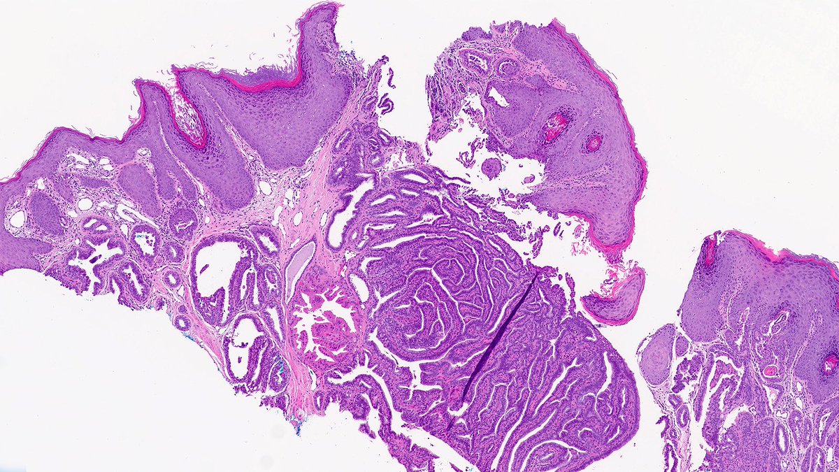
Case of Nipple Adenoma 🔬 Nipple adenoma is a benign proliferative epithelial process of the superficial lactiferous ducts that may erode the overlying squamous epithelium. #PathX #PathTwitter #breastpath

Case of Nipple Adenoma 🔬

Nipple adenoma is a benign proliferative epithelial process of the superficial lactiferous ducts that may erode the overlying squamous epithelium. 

#PathX #PathTwitter #breastpath
Raza Hoda MD (@razahoda) 's Twitter Profile Photo

That was tough! The answer was pleomorphic adenoma (PA). PA shows a biphasic tumor of epithelial and myoepithelial cells set amidst a chrondromyxoid matrix. Unlike in the major salivary glands, PA of the breast is extremely rare. #PathX #PathTwitter #breastpath #PathQuiz

Raza Hoda MD (@razahoda) 's Twitter Profile Photo

Let’s review challenging artifacts in breast specimens this week! Case of Thermocautery Artifact 🔬 The heated tip of electrosurgical unit (Bovie) can drastically distort tissue with a smudgy appearance and make margin assessment difficult. #PathX #PathTwitter #breastpath

Let’s review challenging artifacts in breast specimens this week!

Case of Thermocautery Artifact 🔬

The heated tip of electrosurgical unit (Bovie) can drastically distort tissue with a smudgy appearance and make margin assessment difficult.

#PathX #PathTwitter #breastpath
Raza Hoda MD (@razahoda) 's Twitter Profile Photo

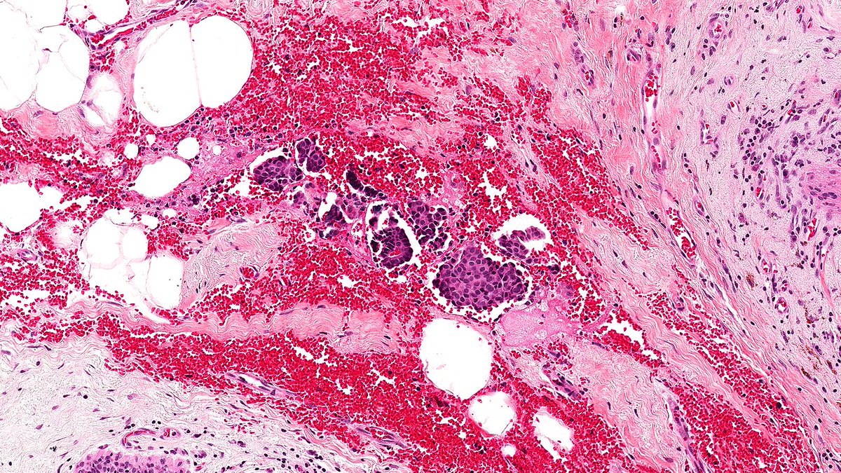
Case of Tissue Float 🔬 This isn’t benign metastasizing duodenum to breast—it’s a tissue float! Preanalytic errors are the bane of a pathologist’s existence. Tissue floats represent tissue from one patient appearing on another patient’s slide. #PathX #PathTwitter #breastpath

Case of Tissue Float 🔬

This isn’t benign metastasizing duodenum to breast—it’s a tissue float! Preanalytic errors are the bane of a pathologist’s existence. Tissue floats represent tissue from one patient appearing on another patient’s slide. 

#PathX #PathTwitter #breastpath
Raza Hoda MD (@razahoda) 's Twitter Profile Photo

Case of Running Ink 🔬 Half of breast pathology is assessment of margins, which are designated with various inks. Some, like yellow ink shown below, are notorious for seeping through and running into the specimen and can complicate measurements. #PathX #PathTwitter #breastpath

Case of Running Ink 🔬

Half of breast pathology is assessment of margins, which are designated with various inks. Some, like yellow ink shown below, are notorious for seeping through and running into the specimen and can complicate measurements.

#PathX #PathTwitter #breastpath
Raza Hoda MD (@razahoda) 's Twitter Profile Photo

Case of Blade Drag 🔬 Grossing blades can dislodge and drag tumor cells across the cut surface, creating artifacts that mimic invasion (👇) or falsely involve margins—especially in papillary tumors. #PathX #PathTwitter #breastpath

Case of Blade Drag 🔬

Grossing blades can dislodge and drag tumor cells across the cut surface, creating artifacts that mimic invasion (👇) or falsely involve margins—especially in papillary tumors.

#PathX #PathTwitter #breastpath
Raza Hoda MD (@razahoda) 's Twitter Profile Photo

Oh no—the answer was biopsy site changes! Skin can be transported into the breast through a core biopsy. As DR.Raju noted, the epithelium is deep in the breast and surrounded by features like organizing fat necrosis. #PathX #PathTwitter #breastpath

Raza Hoda MD (@razahoda) 's Twitter Profile Photo

Let’s review special morphologic patterns of invasive breast carcinoma (IBC) this week! Case of Medullary Pattern 🔬 Medullary IBC shows well-circumscribed high-grade carcinoma with syncytial growth and prominent tumor-associated lymphocytes. #PathX #PathTwitter #breastpath

Let’s review special morphologic patterns of invasive breast carcinoma (IBC) this week!

Case of Medullary Pattern 🔬

Medullary IBC shows well-circumscribed high-grade carcinoma with syncytial growth and prominent tumor-associated lymphocytes.

#PathX #PathTwitter #breastpath
Raza Hoda MD (@razahoda) 's Twitter Profile Photo

Case of Sebaceous Pattern 🔬 This rare pattern of invasive ductal carcinoma displays prominent sebaceous differentiation. The tumor must arise within the breast proper rather than cutaneous adnexal structures to be appropriately diagnosed. #PathX #PathTwitter #breastpath

Case of Sebaceous Pattern 🔬

This rare pattern of invasive ductal carcinoma displays prominent sebaceous differentiation. The tumor must arise within the breast proper rather than cutaneous adnexal structures to be appropriately diagnosed.

#PathX #PathTwitter #breastpath
Raza Hoda MD (@razahoda) 's Twitter Profile Photo

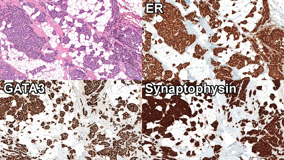
Case of Neuroendocrine Tumor (NET) of Breast 🔬 Neuroendocrine pattern shows some features of neuroendocrine differentiation and is challenging to diagnose, as up to 30% of IDC show some degree of neuroendocrine differentiation—so here’s an NET. #PathX #PathTwitter #breastpath

Case of Neuroendocrine Tumor (NET) of Breast 🔬

Neuroendocrine pattern shows some features of neuroendocrine differentiation and is challenging to diagnose, as up to 30% of IDC show some degree of neuroendocrine differentiation—so here’s an NET.

#PathX #PathTwitter #breastpath
Raza Hoda MD (@razahoda) 's Twitter Profile Photo

This January in Vegas: a pathology symposium that’ll make you want to go all in! Multiple subspecialties, high-yield topics, and packed with powerhouse faculty from Cleveland Clinic. #PathX #PathTwitter #breastpath #gipath #gynpath

Raza Hoda MD (@razahoda) 's Twitter Profile Photo

Case of Pleomorphic Pattern 🔬 This rare pattern of invasive ductal carcinoma demonstrates pleomorphic and bizarre tumor giant cells and may represent the extreme end of “dedifferentiated” breast carcinoma or perhaps even metaplastic carcinoma. #PathX #PathTwitter #breastpath

Case of Pleomorphic Pattern 🔬

This rare pattern of invasive ductal carcinoma demonstrates pleomorphic and bizarre tumor giant cells and may represent the extreme end of “dedifferentiated” breast carcinoma or perhaps even metaplastic carcinoma.

#PathX #PathTwitter #breastpath
Raza Hoda MD (@razahoda) 's Twitter Profile Photo

Close one! The answer was carcinoma with osteoclast-like stromal giant cells! These well-to-moderately differentiated tumors have hypervascular stroma filled with extravasated red blood cells, lymphocytes, and—of course—osteoclast-like giant cells. #PathX #PathTwitter

Raza Hoda MD (@razahoda) 's Twitter Profile Photo

Let’s review axillary lymph node findings this week! Case of Dermatopathic Lymphadenopathy 🔬 This reactive lymphadenopathy is characterized by paracortical expansion with dendritic cells, various immune cells, and melanophages. #PathX #PathTwitter #breastpath

Let’s review axillary lymph node findings this week!

Case of Dermatopathic Lymphadenopathy 🔬

This reactive lymphadenopathy is characterized by paracortical expansion with dendritic cells, various immune cells, and melanophages.

#PathX #PathTwitter #breastpath