Marcos Mestas Nuñez(@marcosmestas) 's Twitter Profileg
Marcos Mestas Nuñez

@marcosmestas

Chestrad

ID:609655546

calendar_today16-06-2012 03:30:36

2,6K Tweets

1,6K Followers

452 Following

Adrià Roset Altadill(@ARosetAltadill) 's Twitter Profile Photo

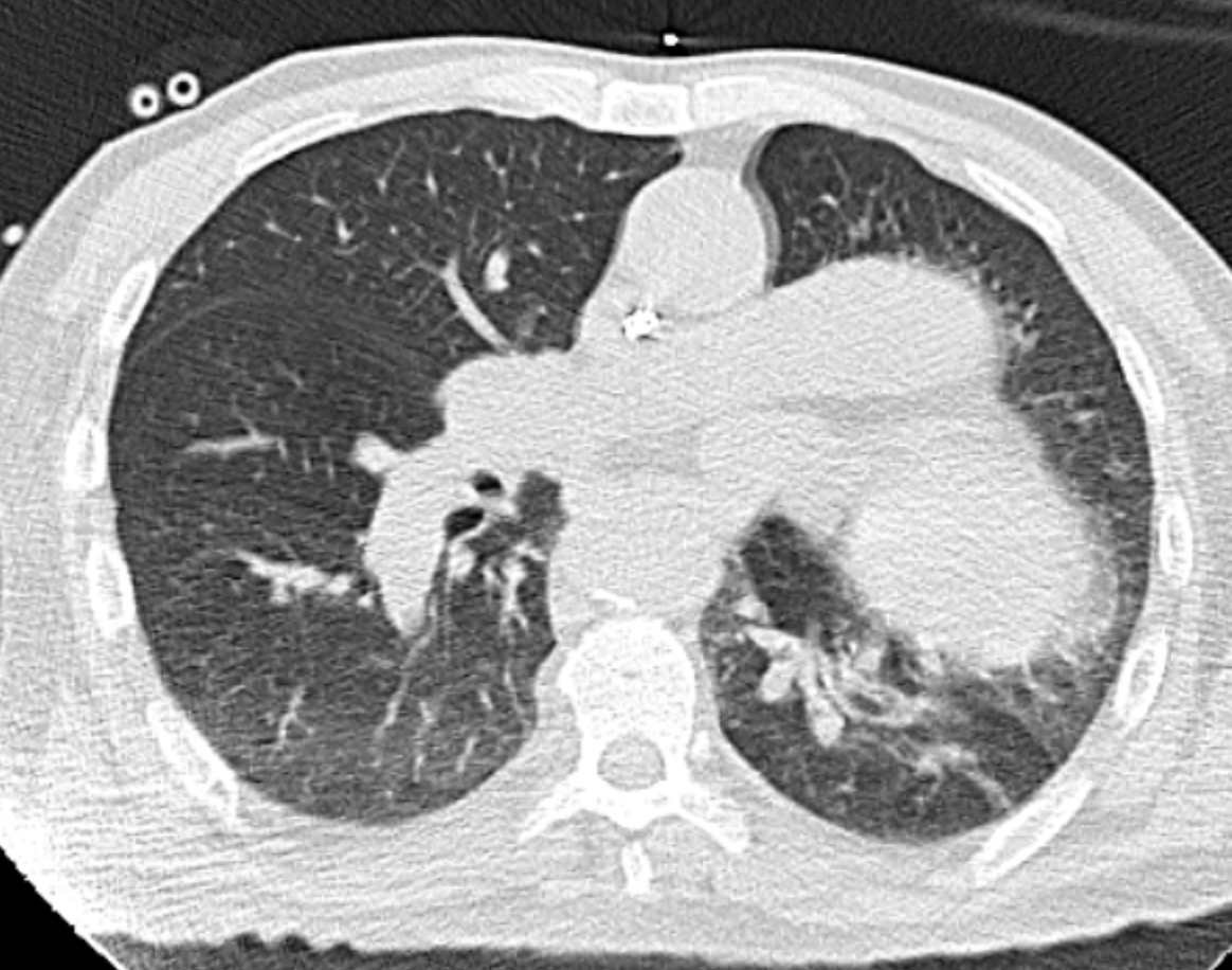
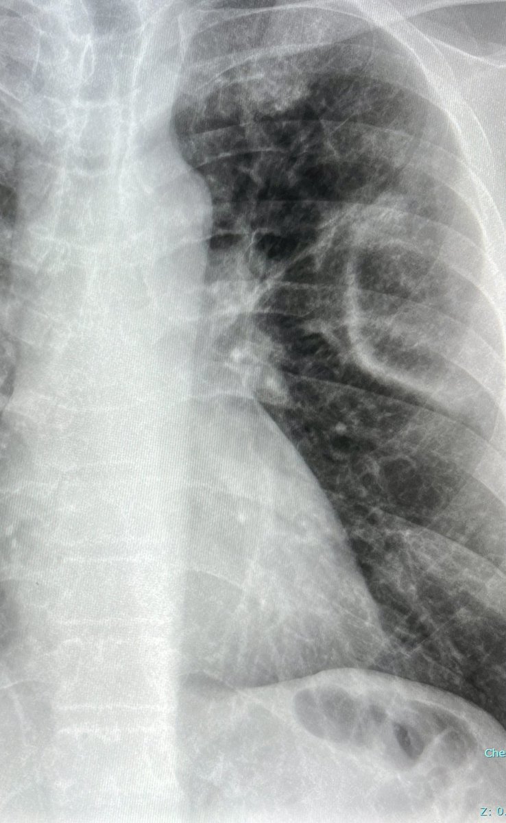
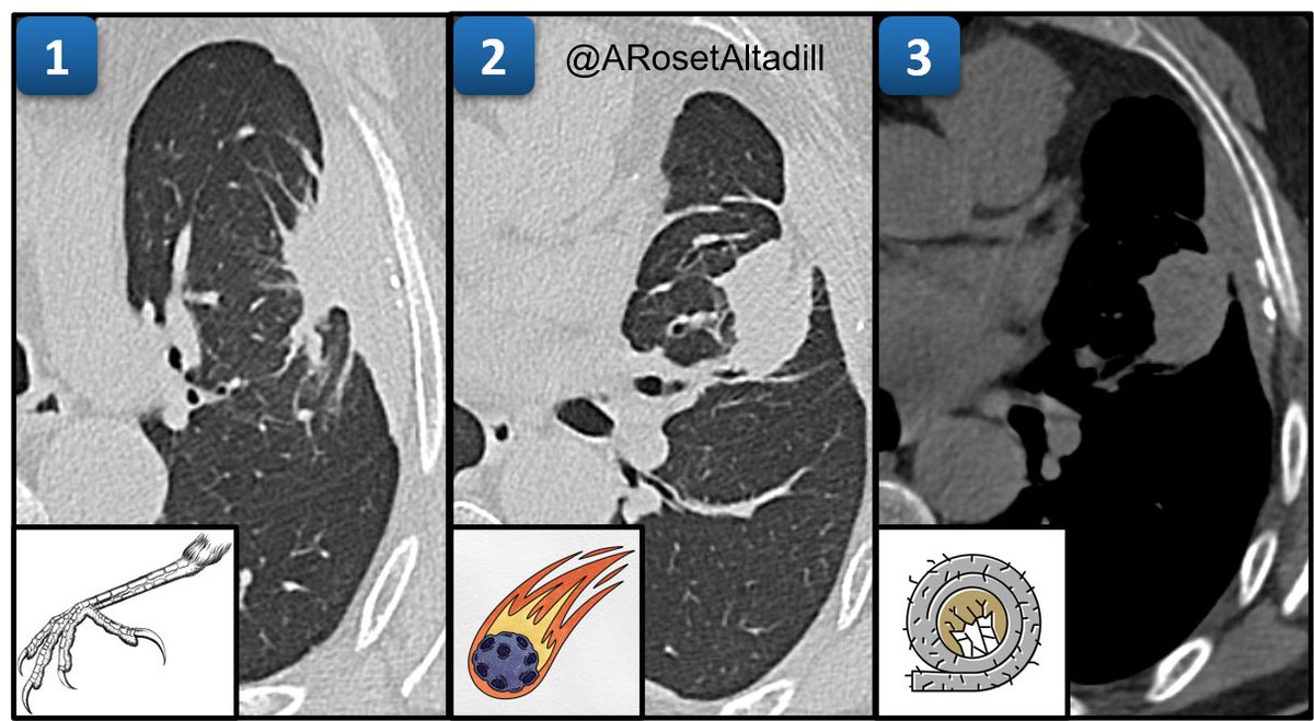
#17 Round Atelectasis

1⃣ Crow feet sign 🦅
2⃣ Comet tail sign ☄️
3⃣ Pleural plaque from asbestos exposure 🪨

Additional features: round morphology, contact with pleura & volume loss

❗️Not to be mistaken with malignancy

#17 Round Atelectasis 1⃣ Crow feet sign 🦅 2⃣ Comet tail sign ☄️ 3⃣ Pleural plaque from asbestos exposure 🪨 Additional features: round morphology, contact with pleura & volume loss ❗️Not to be mistaken with malignancy #Radiology #chestrad #radres #FOAMrad #Rad2B #MedTwitter
account_circle
Daniel Vargas, MD(@DanielVargasMD) 's Twitter Profile Photo

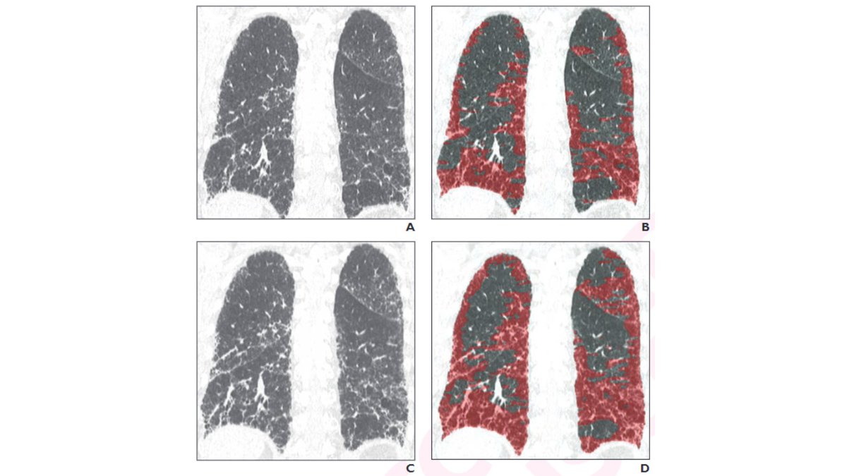
So nice to see the syllabus for the ARRS meeting categorical course on HRCT 🫁published!

👏👏 Marcos Mestas Nuñez and Gonzalo Dulcich for the great work on our chapter on Diffuse Cystic Lung Diseases!

Looking forward to Boston this week!

So nice to see the syllabus for the @ARRS_Radiology #ARRS24 meeting categorical course on HRCT 🫁published! 👏👏 @marcosmestas and @DulcichGonzalo for the great work on our chapter on Diffuse Cystic Lung Diseases! Looking forward to Boston this week! #chestrads #Radiology
account_circle
Daniel Vargas, MD(@DanielVargasMD) 's Twitter Profile Photo

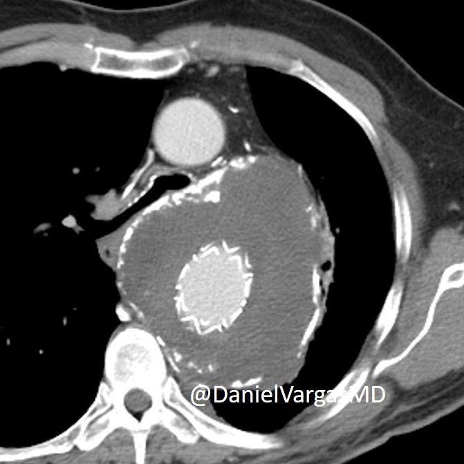
CT and answer to this case:

Post endovascular repair for massive aortic aneurysm with an 👀aorto-bronchial fistula 👀 (involving the excluded aortic aneurysm)

CT showing air filling the aneurysmal sac.

Earlier CT w large aneurysm post endovascular repair.

CT and answer to this case: Post endovascular repair for massive aortic aneurysm with an 👀aorto-bronchial fistula 👀 (involving the excluded aortic aneurysm) CT showing air filling the aneurysmal sac. Earlier CT w large aneurysm post endovascular repair.
account_circle
Sameer Raniga(@samrad77) 's Twitter Profile Photo

“Photographic negative of pulmonary edema”- a classical radiographic pattern of chronic eosinophilic pneumonia

—classic cases on-call

“Photographic negative of pulmonary edema”- a classical radiographic pattern of chronic eosinophilic pneumonia —classic cases on-call
account_circle
Andrew Rosenkrantz(@arosenkrantzmd) 's Twitter Profile Photo

New AJR Accepted Manuscript:

'Progressive Pulmonary Fibrosis and Interstitial Lung Abnormalities: AJR Expert Panel Narrative Review'

By Dr Kanne & team

ajronline.org/doi/10.2214/AJ…

New @AJR_Radiology Accepted Manuscript: 'Progressive Pulmonary Fibrosis and Interstitial Lung Abnormalities: AJR Expert Panel Narrative Review' By Dr Kanne & team ajronline.org/doi/10.2214/AJ…
account_circle
Bruno Hochhegger(@hochhegger) 's Twitter Profile Photo

Diagnostic Performance of MRI for the Detection of Pulmonary Nodules: A Systematic Review and Meta-Analysis | Radiology: Cardiothoracic Imaging pubs.rsna.org/doi/10.1148/ry…

account_circle
@RevistaRADIOLOGIA(@RevistaRADIOLO2) 's Twitter Profile Photo

¡No te pierdas el primer suplemento del año con las últimas novedades en Radiología torácica! SEICAT
elsevier.es/es-revista-rad…

¡No te pierdas el primer suplemento del año con las últimas novedades en Radiología torácica! @SEICAT_RAD elsevier.es/es-revista-rad…
account_circle
RadioGraphics(@RadioGraphics) 's Twitter Profile Photo

Various diseases can demonstrate pleural thickening, such as asbestos-related diseases, neoplasms, and systemic diseases. Click here for a comprehensive review of the clinical and multimodality imaging findings of pleural diseases. bit.ly/3U36C32

Various diseases can demonstrate pleural thickening, such as asbestos-related diseases, neoplasms, and systemic diseases. Click here for a comprehensive review of the clinical and multimodality imaging findings of pleural diseases. bit.ly/3U36C32
account_circle
RadioGraphics(@RadioGraphics) 's Twitter Profile Photo

The authors review the spectrum of intestinal and extraintestinal findings of celiac disease at CT and MR enterography and review associated complications. bit.ly/3PPkU4B @diagnosticoHI Andrea Penizzotto
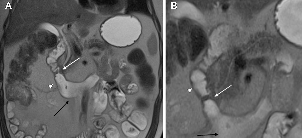
The authors review the spectrum of intestinal and extraintestinal findings of celiac disease at CT and MR enterography and review associated complications. bit.ly/3PPkU4B @diagnosticoHI @andrepenizzotto
account_circle
Maximiliano Klug(@MaxiKlugMD) 's Twitter Profile Photo

Happy to share my first full-lenght manuscript in RadioGraphics 🔥🤩 Thanks to my mentor Edith Marom for all her guidance and support. Thanks to the editor Cooky Menias and all the great reviewers who made of this article a great educational resource 🙏🙏
pubs.rsna.org/doi/10.1148/rg…
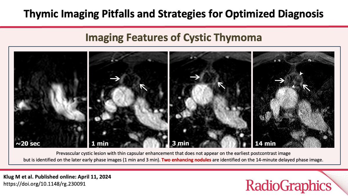
Happy to share my first full-lenght manuscript in @RadioGraphics 🔥🤩 Thanks to my mentor @MaromEdith for all her guidance and support. Thanks to the editor @cookyscan1 and all the great reviewers who made of this article a great educational resource 🙏🙏 pubs.rsna.org/doi/10.1148/rg…
account_circle
Vincent Cottin(@OrphaLung) 's Twitter Profile Photo

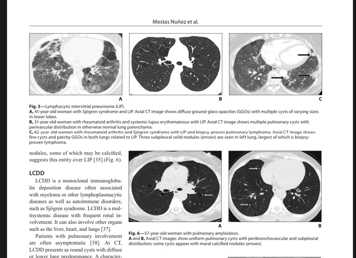
Have you heard about non-amyloid light chain deposition disease in the lung ? A rare form of cystic/bronchiectatic lung disease. See the study by the OrphaLung network
rdcu.be/dEpjk

Have you heard about non-amyloid light chain deposition disease in the lung ? A rare form of cystic/bronchiectatic lung disease. See the study by the OrphaLung network rdcu.be/dEpjk
account_circle
Tan-Lucien Mohammed, MD, FACR(@TLHM_MD) 's Twitter Profile Photo

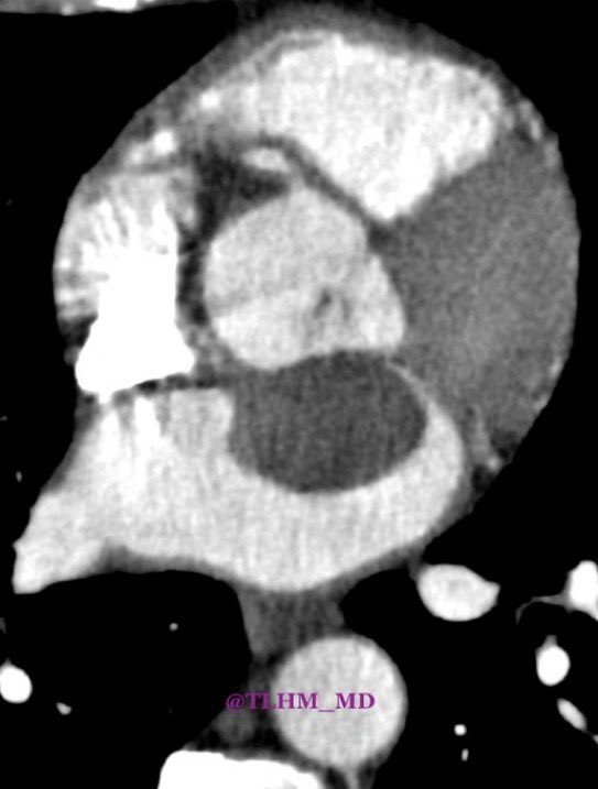
- left atrial myxoma

- MC primary cardiac neoplasm
- intracardiac masses, attached to the interatrial septum (75% in LA)
- can cause valvular obstruction, embolic phenomena
Tx: surgery

#Chestrad - left atrial myxoma - MC primary cardiac neoplasm - intracardiac masses, attached to the interatrial septum (75% in LA) - can cause valvular obstruction, embolic phenomena Tx: surgery #radiology #FOAMRad
account_circle
Daniel Vargas, MD(@DanielVargasMD) 's Twitter Profile Photo

Here's the quarterly reminder to look 👀 at the heart 🫀 on Chest CT/CTA!

Man in his 60s with acute chest pain. Large LAD distribution myocardial infarction, proximal LAD occlusion and LV apical thrombus - on Chest CTA

NASCI RSNA

Here's the quarterly reminder to look 👀 at the heart 🫀 on Chest CT/CTA! Man in his 60s with acute chest pain. Large LAD distribution myocardial infarction, proximal LAD occlusion and LV apical thrombus - on Chest CTA #FOAMed #radres @NASCISociety @RSNA
account_circle
Stacey Verzosa Weisman, MD(@svweisman) 's Twitter Profile Photo

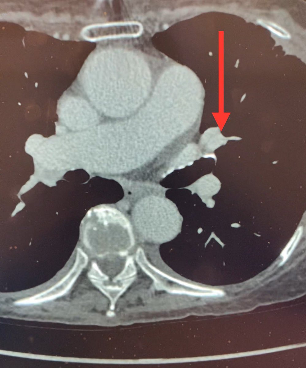
Abnormal lung can result in increased vascular resistance. So ddx is thrombus or slow flow/delayed perfusion. Latter was suspected so study was repeated with a delay. See pic - defect filled in. No PE, but rather slow flow. Have a great week! Be kind! ❤️

Abnormal lung can result in increased vascular resistance. So ddx is thrombus or slow flow/delayed perfusion. Latter was suspected so study was repeated with a delay. See pic - defect filled in. No PE, but rather slow flow. Have a great week! Be kind! ❤️
account_circle