Tengfei Wang, MD (@tengfeiwang9652) 's Twitter Profile
Tengfei Wang, MD

@tengfeiwang9652

Pathology resident at Baylor Scott and White Health-Temple

ID: 1591539637864091650

calendar_today12-11-2022 21:13:28

617 Tweet

736 Takipçi

988 Takip Edilen

Lisa Rooper (@lisaroopermd) 's Twitter Profile Photo

Does this Warthin tumor have a few too many lymphocytes? Involvement by chronic lymphocytic leukemia/small lymphocytic lymphoma has expanded its stroma in a striking phyllodal pattern. Atypical lymphs CD5+, CD23 weak for confirmation. #ENTPath #HemePath #SurgPath

Does this Warthin tumor have a few too many lymphocytes? Involvement by chronic lymphocytic leukemia/small lymphocytic lymphoma has expanded its stroma in a striking phyllodal pattern. Atypical lymphs CD5+, CD23 weak for confirmation. #ENTPath #HemePath #SurgPath
Dina Tiniakos (@dinatiniakos) 's Twitter Profile Photo

Good news! Gnomes’🔺paper on ⚡️Acute Hepatitis🔬has received over 1,800 views since publication! Read it here onlinelibrary.wiley.com/doi/10.1111/hi… #ViewsMilestone EASLnews European Society of Pathology (ESP) AASLD Hans Popper Hepatopathology Society UK Liver Pathology Group my_UEG #PathTwitter #liverTwitter David Kleiner

Charles Herndon (@drcyclopath) 's Twitter Profile Photo

✅Solitary fibrous tumor Site: anywhere Micro: patternless pattern, staghorn vessels, abundant collagen, ovoid-fusiform spindle cells, indistinct cell borders CD34+, desmin-, S100-, keratin- NAB2::STAT6 🔬tumourclassification.iarc.who.int/Viewer/Index2?… #BSTPath #PathTwitter #HNPath #Pathology

✅Solitary fibrous tumor
Site: anywhere
Micro: patternless pattern, staghorn vessels, abundant collagen, ovoid-fusiform spindle cells, indistinct cell borders
CD34+, desmin-, S100-, keratin-
NAB2::STAT6
🔬tumourclassification.iarc.who.int/Viewer/Index2?…
#BSTPath #PathTwitter #HNPath #Pathology
Chinese American Pathologists Association (@capa_comm) 's Twitter Profile Photo

📢 Join us for another #CAPA Molecular session on 11/10, 7 PM ET! Dr. Hongjie Li from Geisinger Pathology will provide an update on the molecular aspects of pancreatic, liver, and biliary duct tumors, along with pharmacogenomics. She will also cover the NCCN guidelines. Host:Qiong Zhang, MD. PhD.
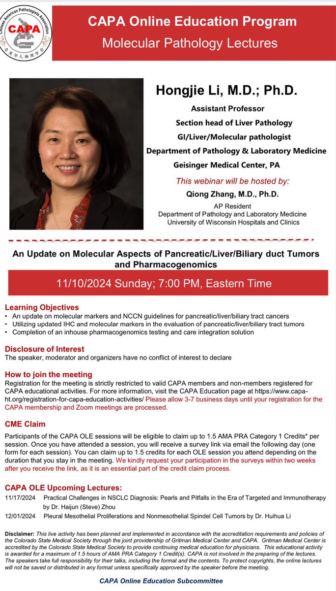
📢 Join us for another #CAPA Molecular session on 11/10, 7 PM ET! Dr. Hongjie Li from <a href="/GeisingerPath/">Geisinger Pathology</a> will provide an update on the molecular aspects of pancreatic, liver, and biliary duct tumors, along with pharmacogenomics. She will also cover the NCCN guidelines. Host:<a href="/Qiong511/">Qiong Zhang, MD. PhD.</a>
Chinese American Pathologists Association (@capa_comm) 's Twitter Profile Photo

🌟Join us 11/17, 7pm ET, for a new chapter on Thoracic session with Dr. Haijun Zhou, exploring “Practical Challenges in NSCLC Diagnosis” in the era of targeted therapy. Get valuable insights into thoracic pathology and effective molecular testing strategies. Don’t miss out!

🌟Join us 11/17, 7pm ET, for a new chapter on Thoracic session with Dr. Haijun Zhou, exploring “Practical Challenges in NSCLC Diagnosis” in the era of targeted therapy. Get valuable insights into thoracic pathology and effective molecular testing strategies. Don’t miss out!
Ankur Sangoi (@slusagar) 's Twitter Profile Photo

middle-aged M with mediastinal mass, core bx performed YOLK SAC TUMOR ✔️endodermal sinus (perivascular) pattern👉Schiller-Duval bodies ✔️microcystic/reticular pattern

middle-aged M with mediastinal mass, core bx performed

YOLK SAC TUMOR
✔️endodermal sinus (perivascular) pattern👉Schiller-Duval bodies
✔️microcystic/reticular pattern
Tengfei Wang, MD (@tengfeiwang9652) 's Twitter Profile Photo

thepathologist.com/outside-the-la… I would like to thank The Pathologist to publish my opinion piece! It is really a wonderful closure of the translational diagnostics advanced training with CAP Foundation.

Vijay Shankar S (@vijaypatho) 's Twitter Profile Photo

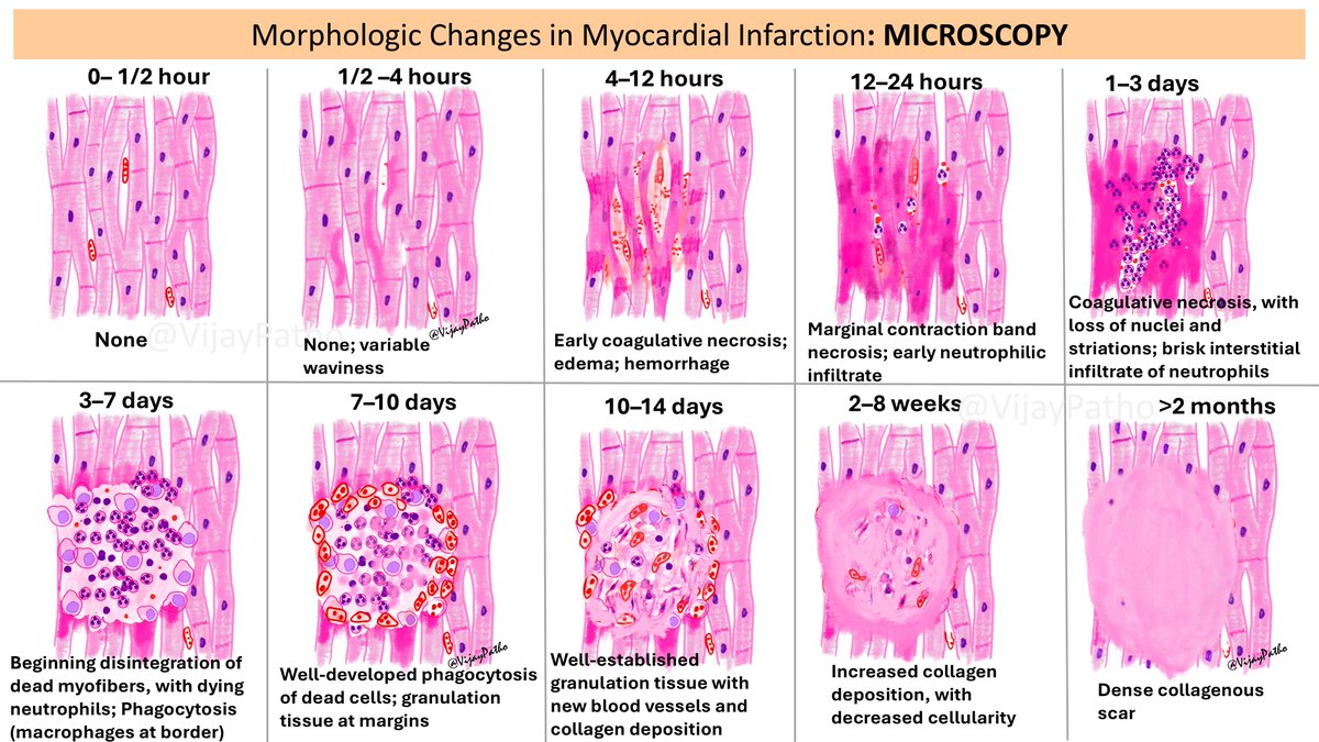
Morphologic Changes in Myocardial Infarction: A Microscopic Journey Illustrating histopathology is one of the best ways to teach and learn! If you're interested in drawing, try illustrating histological changes—it helps in retaining concepts better and makes learning fun. This

Morphologic Changes in Myocardial Infarction: A Microscopic Journey 

Illustrating histopathology is one of the best ways to teach and learn! If you're interested in drawing, try illustrating histological changes—it helps in retaining concepts better and makes learning fun.
This
Chinese American Pathologists Association (@capa_comm) 's Twitter Profile Photo

🔬 Join us for another captivating CAPA Online Education session on Pediatric Pathology this Sunday! Explore the molecular and histologic features of DICER-1 Syndrome with Dr. Yamin Ma! Mark your calendar 🗓️ 2/16, 7PM ET. Hosted by Dr. Tengfei Wang Tengfei Wang, MD #PathTwitter

🔬 Join us for another captivating CAPA Online Education session on Pediatric Pathology this Sunday! Explore the molecular and histologic features of DICER-1 Syndrome with Dr. Yamin Ma! Mark your calendar 🗓️ 2/16, 7PM ET. Hosted by Dr. Tengfei Wang <a href="/TengfeiWang9652/">Tengfei Wang, MD</a>  #PathTwitter
Charles Herndon (@drcyclopath) 's Twitter Profile Photo

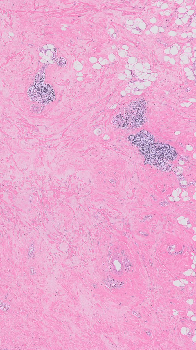
Breast biopsy shows epithelioid cells, dense collagen, and lymphocytic aggregates. What could it be?🧐🧐 A) Myofibroblastoma B) Fibroadenoma C) Lymphocytic mastopathy D) Fibromatosis #BreastPath #Pathology #PathTwitter| 🔬 learn.mghpathology.org/index.php/DZI1…

Breast biopsy shows epithelioid cells, dense collagen, and lymphocytic aggregates. 

What could it be?🧐🧐

A) Myofibroblastoma
B) Fibroadenoma
C) Lymphocytic mastopathy
D) Fibromatosis
#BreastPath #Pathology #PathTwitter| 
🔬 learn.mghpathology.org/index.php/DZI1…