Erika🔬 (@erikadidomenico) 's Twitter Profile
Erika🔬

@erikadidomenico

PhD Student @CRTDpress @tudresden_de 🧠 |
previous @CIBIO_UniTrento @CBD_VIB |
🇮🇹 Molisana in Germany 🇩🇪

ID: 1606619643531202564

linkhttps://www.linkedin.com/in/erikadidomenico/ calendar_today24-12-2022 11:56:10

53 Tweet

55 Followers

294 Following

Nicola Fattorelli (@nfattorelli) 's Twitter Profile Photo

So happy to see our latest work published today in Nature Neuroscience 🥳 terrific team effort and loads of fun with shared first authors MINDlab and Anna Martínez at De Strooper Lab. Microglia enthusiasts, let's take a short journey through the paper 🧵⬇️ go.nature.com/4av0PZd

HHMI | Janelia (@hhmijanelia) 's Twitter Profile Photo

New research led by Carolyn Ott, a senior scientist Lippincott-Schwartz Lab, provides the most detailed & comprehensive description yet of cilia in the mouse visual cortex, giving scientists new insights into the formation & function of this small & elusive organelle. 🔗doi.org/10.1016/j.cub.…

Lab of Synapse Biology (@joris_dewit_lab) 's Twitter Profile Photo

New preprint out from our lab! We uncover a novel mechanism that regulates synaptic organelle content of dendritic spines, important for the proper maturation of cortical connectivity. biorxiv.org/content/10.110…

Allen Institute (@alleninstitute) 's Twitter Profile Photo

New study in Current Biology gives the most detailed description yet of cilia in the mouse visual cortex. The study was a collaboration including researchers from the Allen Institute @hhmijanelia, University at Albany, Harvard Medical School, and others. sciencedirect.com/science/articl…

Eva-Maria Schentarra (@evaschentarra) 's Twitter Profile Photo

Happy Friday! To wrap up another exciting week of learning highly multiplexed DNA-PAINT in the Jungmann Lab, here's a nice #cellfie of a rat hippocampal neuron stained for synaptic vesicle markers (Vamp2, VGlut1 and synaptotagmin), clathrin, neurofilament and peroxisomes.

Happy Friday! To wrap up another exciting week of learning highly multiplexed DNA-PAINT in the <a href="/JungmannLab/">Jungmann Lab</a>, here's a nice #cellfie of a rat hippocampal neuron stained for synaptic vesicle markers (Vamp2, VGlut1 and synaptotagmin), clathrin, neurofilament and peroxisomes.
Schuh Lab (@schuhlab) 's Twitter Profile Photo

Women are born with all their oocytes, which need to stay viable for decades to ensure fertility. How are they maintained for that long? Our latest research in Nature Cell Biology reveals that oocyte maintenance involves exceptional protein longevity. nature.com/articles/s4155… (1/9)
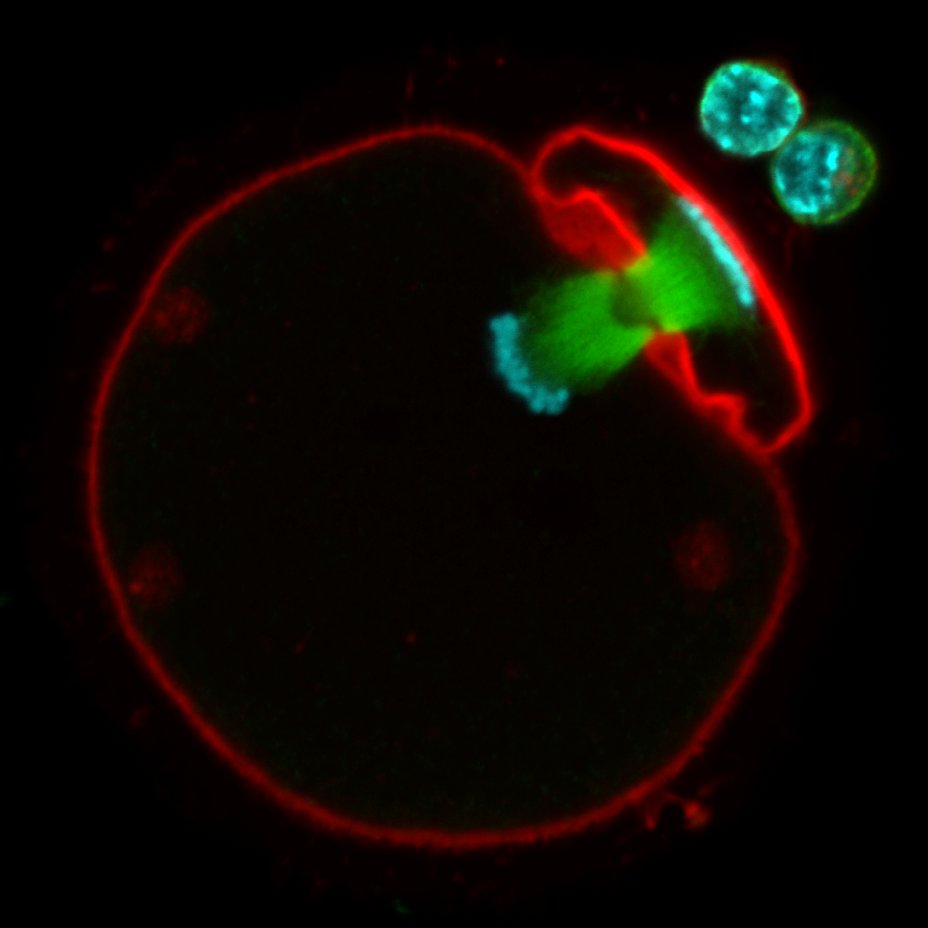
Women are born with all their oocytes, which need to stay viable for decades to ensure fertility. How are they maintained for that long?

Our latest research in <a href="/NatureCellBio/">Nature Cell Biology</a> reveals that oocyte maintenance involves exceptional protein longevity. nature.com/articles/s4155… (1/9)
Eric Vallabh Minikel (@cureffi) 's Twitter Profile Photo

Introducing CHARM: a new epigenome editor to methylate DNA at the promoter of a targeted gene. Our lab's collaboration with Jonathan Weissman's Lab's Edwin Neumann & Tessa Bertozzi shows deep silencing of brain PrP Paper: science.org/doi/10.1126/sc… Blog: cureffi.org/2024/06/27/int…
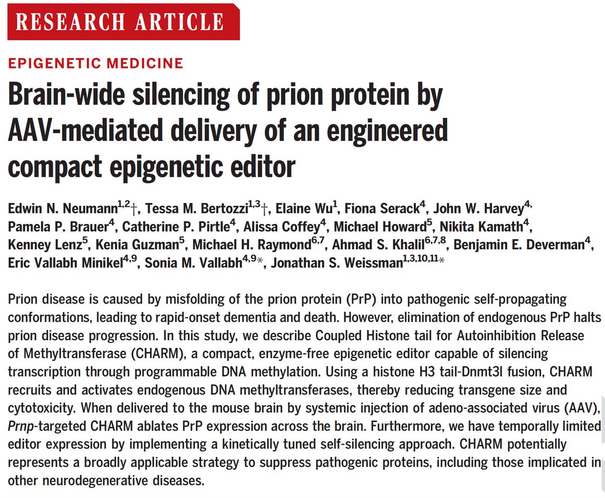
Introducing CHARM: a new epigenome editor to methylate DNA at the promoter of a targeted gene.
Our lab's collaboration with <a href="/JswLab/">Jonathan Weissman's Lab</a>'s <a href="/EdwinNNeumann/">Edwin Neumann</a> &amp; <a href="/TessaBertozzi/">Tessa Bertozzi</a> shows deep silencing of brain PrP

Paper: science.org/doi/10.1126/sc…

Blog: cureffi.org/2024/06/27/int…
Liz Chrastil (@chrastil) 's Twitter Profile Photo

A few years ago I was in a position to do something unique in neuroscience. I had been working with the phenomenal Emily G. Jacobs 🦋 @emilyjacobs.bsky.social on menopause and inspired by Laura Pritschet's menstrual study, and I was planning a pregnancy. What if we scanned my brain?? nature.com/articles/s4159…

Dylan Burnette (@mag2art) 's Twitter Profile Photo

The nuclei of the cells in the heart of a zebrafish embryo photographed through a microscope. The ventricle is on the left and the atrium is on right. #CellBiology

The nuclei of the cells in the heart of a zebrafish embryo photographed through a microscope. The ventricle is on the left and the atrium is on right. #CellBiology
FlyWire (@flywirenews) 's Twitter Profile Photo

Big news! The fly connectome is featured on the cover of a special edition of Nature. This is all possible thanks to the collaboration of 292 members of The FlyWire Consortium! nature.com/immersive/d428… Check out the thread for an overview of the 9 #flywire papers published today

Big news! The fly connectome is featured on the cover of a special edition of Nature.
This is all possible thanks to the collaboration of 292 members of The FlyWire Consortium!
nature.com/immersive/d428…
Check out the thread for an overview of the 9 #flywire papers published today