Kassaye Firde MD (@firdemd) 's Twitter Profile
Kassaye Firde MD

@firdemd

PGY2 pathology resident @templepathology
#cytology #surgicalpathology

ID: 1464440799026896906

calendar_today27-11-2021 03:48:23

27 Tweet

128 Followers

279 Following

Lola Lozano (@mdlozanoe) 's Twitter Profile Photo

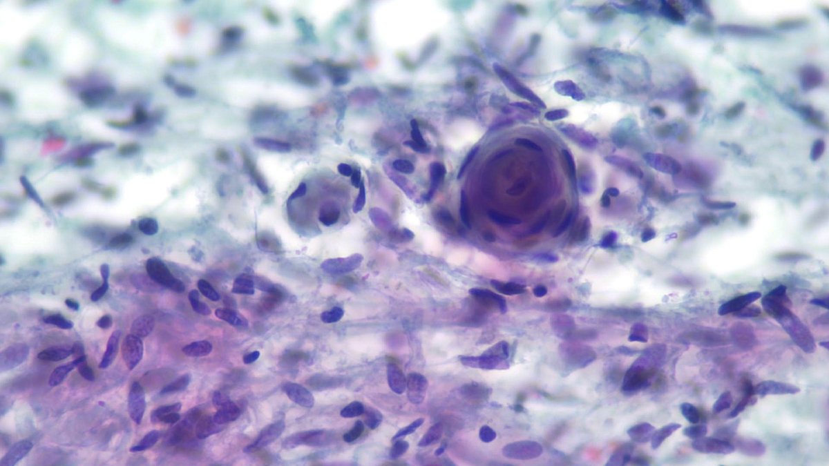
Imprints are a valuable tool for learning cytopathology 🔬💡🧠Make the most of them! #Cytopath Meningioma: -🪼clusters and sheets of monomorphic cells -🪶Wispy/feathery cytoplasm -🪹Bland nuclei (look for INCI) -🌪️🟣Meningothelial whorls and psammoma bodies are classic findings

Imprints are a valuable tool for learning cytopathology 🔬💡🧠Make the most of them! #Cytopath
Meningioma:
-🪼clusters and sheets of monomorphic cells
-🪶Wispy/feathery cytoplasm
-🪹Bland nuclei (look for INCI)
-🌪️🟣Meningothelial whorls and psammoma bodies are classic findings
Ghazal Khajir (@ghazalkhajir) 's Twitter Profile Photo

Learning from the best PAPathologists! When two legends, Syed Z. Ali and Zubair Baloch teach thyroid #pathology! Thank you Drs Darshana Jhala and nj for the wonderful opportunity and successful meeting!
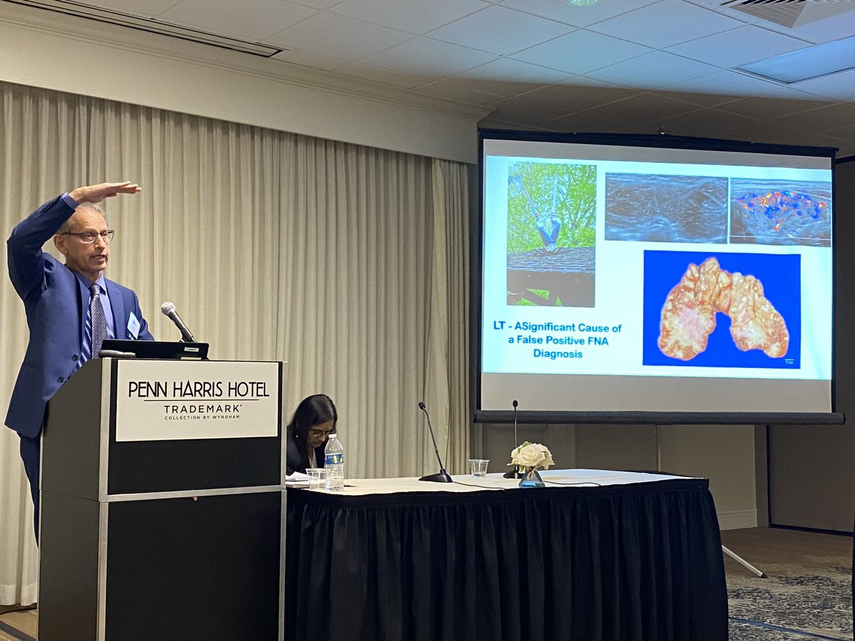
Learning from the best <a href="/PAPathologists/">PAPathologists</a>!
When two legends, <a href="/sza_jhcyto/">Syed Z. Ali</a> and <a href="/aakasharmand/">Zubair Baloch</a> teach thyroid #pathology!

Thank you Drs Darshana Jhala and <a href="/niragjhala/">nj</a> for the wonderful opportunity and successful meeting!
Dina Zenezan,MD (@dzenezan) 's Twitter Profile Photo

Such a fantastic lecture with the one and only Dr. Ali! Very nice to have you at PAP this year Syed Z. Ali ! Thanks to Dr. D Jhala and NJhala ! And thanful for Temple Pathology !
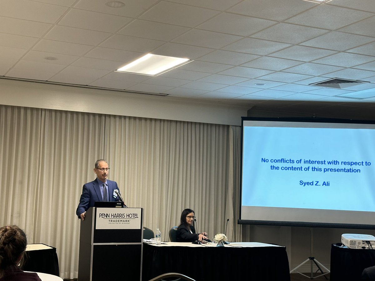
Such a fantastic lecture with the one and only Dr. Ali! Very nice to have you at PAP this year <a href="/sza_jhcyto/">Syed Z. Ali</a> ! Thanks to Dr. D Jhala and NJhala ! And thanful for <a href="/templepathology/">Temple Pathology</a> !
Dina Zenezan,MD (@dzenezan) 's Twitter Profile Photo

Shout out to my amazing program directors Dr. Dioufa and Dr. Rong , coming for full support of residents during Jeopardy and the meeting! Temple Pathology Nikolina Dioufa #PathTwitter
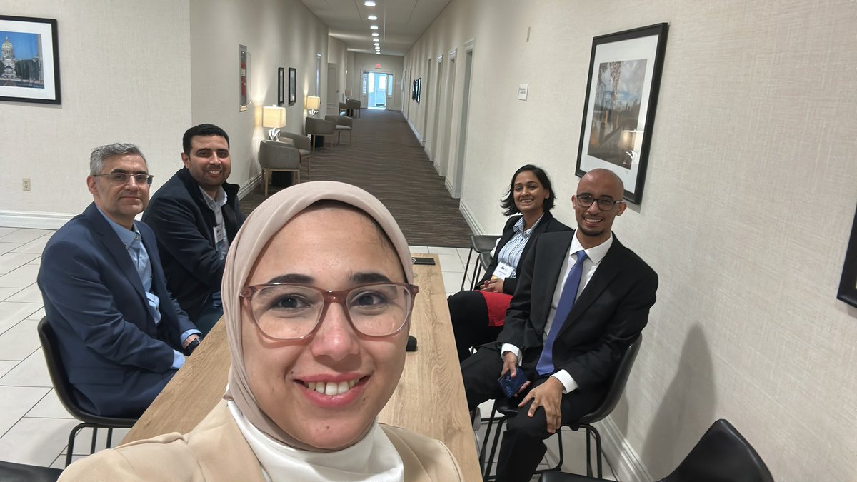
Shout out to my amazing program directors  Dr. Dioufa and Dr. Rong , coming for full support of residents during Jeopardy and the meeting!  <a href="/templepathology/">Temple Pathology</a> <a href="/DioufaNikolina/">Nikolina Dioufa</a> #PathTwitter
Fouad Boulos (@fouad_boulos) 's Twitter Profile Photo

Do not mistake peripheral palisading in invasive carcinoma for a preserved myoepithelial layer. Invasive solid papillary carcinoma with peripheral palisading (pseudo-myoepithelial layer). WashU Pathology & Immunology WashU Medicine Pathology & Immunology Education #breastpath #PathTwitter #PathX
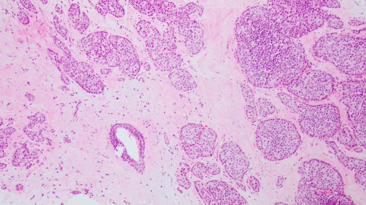
Do not mistake peripheral palisading in invasive carcinoma for a preserved myoepithelial layer.

Invasive solid papillary carcinoma with peripheral palisading (pseudo-myoepithelial layer).

<a href="/wusm_pathology/">WashU Pathology & Immunology</a> <a href="/washupathedu/">WashU Medicine Pathology & Immunology Education</a> #breastpath #PathTwitter #PathX
Israh Akhtar Khan (@israhkhan) 's Twitter Profile Photo

#cytoweek pancreatic FNA Classic definition of adenocarcinoma with drunken honeycomb in pic 2, mucin vacuoles in pic 3 and desmoplasia in pic 4 - Samir Amer Kassaye Firde MD Diff Quik stain

#cytoweek pancreatic FNA 
Classic definition of adenocarcinoma with drunken honeycomb in pic 2, mucin vacuoles in pic 3 and desmoplasia in pic 4 - <a href="/SamirAmerMD/">Samir Amer</a> <a href="/FirdeMD/">Kassaye Firde MD</a> 
Diff Quik stain
Syed Z. Ali (@sza_jhcyto) 's Twitter Profile Photo

“Basaloid” tumors can look amazingly “neuroendocrine” on FNA smears. This liver lesion is a metastatic “Basaloid Squamous Cell Carcinoma” from the patient’s known uterine cervical primary.

“Basaloid” tumors can look amazingly “neuroendocrine” on FNA smears. This liver lesion is a metastatic “Basaloid Squamous Cell Carcinoma” from the patient’s known uterine cervical primary.
Elizabeth Montgomery, MD (@lizmontgomerymd) 's Twitter Profile Photo

This example of colorectal malakoplakia presented as a mass lesion. The pale pink appearance of the histiocytes is a clue to search for Michaelis-Gutmann bodies. #umiamipath Reference: Am J Surg Pathol. 2020 Sep;44(9):1251-1258. PMID: 32301754.

This example of colorectal malakoplakia presented as a mass lesion.  The pale pink appearance of the histiocytes is a clue to search for Michaelis-Gutmann bodies. #umiamipath 

Reference:  Am J Surg Pathol. 2020 Sep;44(9):1251-1258. PMID: 32301754.
Israh Akhtar Khan (@israhkhan) 's Twitter Profile Photo

We don’t stop at making a diagnosis -we want to do more - Specimen:Pleural fluid Diagnosis:Metastatic lung adenocarcinoma (TTF1 positive)pic 3 Immunotherapy possible :PDL1 -TPS >50% (pic4) Cell block good for molecular testing All set for Rx #pathtwitter #whypathologistmatter

We don’t stop at making a diagnosis -we want to do more -
Specimen:Pleural fluid
Diagnosis:Metastatic lung adenocarcinoma (TTF1 positive)pic 3
Immunotherapy possible :PDL1 -TPS &gt;50% (pic4)
Cell block good for molecular testing 
All set for Rx
#pathtwitter #whypathologistmatter
Temple Pathology (@templepathology) 's Twitter Profile Photo

🎉 Get ready for an exciting day🎉 Join us for the TemplePath open house on 13th September—don’t miss out! Scan the QR code 👇👇👇👇 #PathMatch25 #PathX

🎉 Get ready for an exciting day🎉 Join us for the TemplePath open house on 13th September—don’t miss out! Scan the QR code 👇👇👇👇 #PathMatch25 #PathX
Kristen (@mismatchdef) 's Twitter Profile Photo

In GI luminal biopsies, amyloid deposition in the muscularis mucosae is easy to miss because the MM is often fragmented and at the tissue edge. Thankfully, the team asked me to exclude amyloid upfront otherwise I may have missed this focus. #GIpath

In GI luminal biopsies, amyloid deposition in the muscularis mucosae is easy to miss because the MM is often fragmented and at the tissue edge. Thankfully,  the team asked me to exclude amyloid upfront otherwise I may have missed this focus. #GIpath
Israh Akhtar Khan (@israhkhan) 's Twitter Profile Photo

TRPS-1: IHC pleural fluid Dx breast ca including metaplastic and nonmetaplastic triple negative breast ca(TNBC) Differentiates GATA3 +IDC from GATA3+ urothelial ca Differentiates IDC from adenoCa of nonbreast origin (eg lung, GI, pancreas, etc Differentiates TNBC from melanoma

TRPS-1: IHC pleural fluid
Dx breast ca including metaplastic and nonmetaplastic triple negative breast ca(TNBC)
Differentiates  GATA3 +IDC  from GATA3+ urothelial ca
Differentiates IDC from adenoCa of nonbreast origin (eg lung, GI, pancreas, etc
Differentiates TNBC  from melanoma