MCDB/BSCRC Microscopy Core
@coremcdb
Twitter account of the @UclaMcdb/@UCLAstemcell #microscopy core facility. Tweets by @Nat_Prunet
ID: 1045006047323119616
https://sites.lifesci.ucla.edu/mcdb-microscopy/ 26-09-2018 17:43:51
875 Tweet
694 Takipçi
445 Takip Edilen
 's Twitter Profile Photo")
We are super excited to present our newest #publication in the The EMBO Journal : "MICOS assembly controls mitochondrial inner membrane remodeling and crista junction redistribution to mediate cristae formation" embopress.org/doi/10.15252/e… #mitochondria #superresolution #EM
 on Twitter photo We are super excited to present our newest #publication in the <a href=\"/embojournal/\">The EMBO Journal</a> : \"MICOS assembly controls mitochondrial inner membrane remodeling and crista junction redistribution to mediate cristae formation\" embopress.org/doi/10.15252/e…
#mitochondria #superresolution #EM")
 's Twitter Profile Photo")
 's Twitter Profile Photo")
Out today from Dong Li's lab, a deep learning approach that uses frequency content information in the Fourier domain to improve SIM reconstruction under low-SNR conditions. And an accompanying News and Views from David Hoffman! nature.com/articles/s4159… nature.com/articles/s4159…
 on Twitter photo Out today from Dong Li's lab, a deep learning approach that uses frequency content information in the Fourier domain to improve SIM reconstruction under low-SNR conditions. And an accompanying News and Views from <a href=\"/davephoffman/\">David Hoffman</a>! nature.com/articles/s4159… nature.com/articles/s4159…")
 's Twitter Profile Photo")
 on Twitter photo The power of #UExM 😍
(Green: Tubulin, Bleu: DAPI, Magenta: NHS Ester, widefield picture)")
 's Twitter Profile Photo")
 's Twitter Profile Photo")
 's Twitter Profile Photo")
 's Twitter Profile Photo")
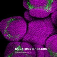
 on Twitter photo Check this 4D atlas of early #Arabidopsis #flower #development, now out in <a href=\"/Dev_Cell/\">Developmental Cell</a>. I contributed a bunch of #confocal #microscopy data from my postdoc <a href=\"/Caltech/\">Caltech</a>. Fun to collaborate with @RDPlab where I did my PhD!
doi.org/10.1016/j.devc…
#devbio #plantscience #flowers")
 's Twitter Profile Photo")
 on Twitter photo Happy #FluorescenceFriday")
 's Twitter Profile Photo")
Love seeing all of the #FluorescenceFriday posts especially from #MUSC Kelsey S. Moore, PhD🔬 Rachel Biggs Cortney Gensemer, PhD Amy Engevik #womeninstem
 on Twitter photo Love seeing all of the #FluorescenceFriday posts especially from #MUSC <a href=\"/sciencekelso/\">Kelsey S. Moore, PhD🔬</a> <a href=\"/rachinstem/\">Rachel Biggs</a> <a href=\"/CortDoesScience/\">Cortney Gensemer, PhD</a> <a href=\"/AmyEngevik/\">Amy Engevik</a> #womeninstem")
 's Twitter Profile Photo")
 's Twitter Profile Photo")
 on Twitter photo Hundreds of beautiful #microscope images were submitted to this year's Global Image of the Year Award competition. Here's a glimpse at a few of the images that received honorable mentions by our panel of jurors.
Click here to see all winning images - olympus-lifescience.com/IOTY")
 's Twitter Profile Photo")
 on Twitter photo Happy Monday! Does anyone else see balloons in this picture? These are actually individually labeled axons in an embryonic chick ciliary ganglion. #MondayFunday
qoo.ly/3bjnfk
Cred: Dr. Ryo Egawa")
 on Twitter photo Congratulations to the winners of the @OlympusLifeSci Image of the Year awards! Stunning images 😍
#microscopy #sciart #bioart #confocal #beautyinsmallthings #microscopicworld")
 's Twitter Profile Photo")
For #FluorescenceFriday, we love this #lightsheet image from our ZEISS Microscopy Z.1! Installed 2 wks before shutdown, now it's popular. Beautiful E9.5 mouse embryo with vascular perfusion from Linh Trinh in Mark Magnuson Lab. Expert imaging with CISR's Nick Mignemi. Scale=200um.
 's Twitter Profile Photo")
I am thrilled to share my first preprint from the Alex Schier about the extraordinary development of the African turquoise killifish!
 's Twitter Profile Photo")
 on Twitter photo Combining #dSTORM & #ExpansionMicroscopy to achieve molecular scale #imaging. Awesome work from <a href=\"/LabSauer/\">SauerLab</a> that I somehow missed when it was published last summer!
nature.com/articles/s4146…
#exM #microscopy #superresolution")
 's Twitter Profile Photo")
Cute little red #algae I picked in #Malibu and imaged on ZEISS Microscopy LSM880 #confocal #microscope. #Autofluorescence including #chlorophyll. #bioart #sciart #microscopy #plamtscience #botany #microscopyart #fluorescence #redalgae #beautyinsmallthings #microscopic
 on Twitter photo Cute little red #algae I picked in #Malibu and imaged on <a href=\"/zeiss_micro/\">ZEISS Microscopy</a> LSM880 #confocal #microscope. #Autofluorescence including #chlorophyll.
#bioart #sciart #microscopy #plamtscience #botany #microscopyart #fluorescence #redalgae #beautyinsmallthings #microscopic")