Viola Nähse (@violanahse) 's Twitter Profile
Viola Nähse

@violanahse

Molecular biologist working in the Stenmark lab, Oslo

ID: 1496761520923025411

calendar_today24-02-2022 08:19:19

58 Tweet

58 Followers

92 Following

Anne Carpenter, PhD (@drannecarpenter) 's Twitter Profile Photo

Delightful 😠 I've had enough of big-name scientists leveraging their fame to exploit others. Is science well served by one dude taking 30 postdoc salaries from the funding ecosystem & producing a few high-tier papers/year? Leaving human wreckage in his wake?

Delightful 😠
I've had enough of big-name scientists leveraging their fame to exploit others.

Is science well served by one dude taking 30 postdoc salaries from the funding ecosystem & producing a few high-tier papers/year?

Leaving human wreckage in his wake?
The EMBO Journal (@embojournal) 's Twitter Profile Photo

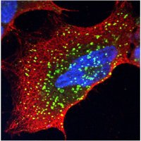
#TECPR1 is activated by damage-induced #sphingomyelin exposure to mediate noncanonical #autophagy Alf Håkon Lystad and colleagues Universitetet i Oslo embopress.org/doi/full/10.15…

#TECPR1 is activated by damage-induced #sphingomyelin exposure to mediate noncanonical #autophagy
Alf Håkon Lystad and colleagues <a href="/UniOslo/">Universitetet i Oslo</a>
embopress.org/doi/full/10.15…
Harald Stenmark (@haraldstenmark) 's Twitter Profile Photo

The roles of ER-derived "omegasomes" and their hallmark protein DFCP1 in autophagosome biogenesis has remained enigmatic. We now provide evidence that DFCP1 is an ATPase that constricts omegasomes which shape autophagosomes in selective autophagy. rdcu.be/dgmP1

Kay Schink (@kschink) 's Twitter Profile Photo

Very happy and proud that this story is finally published. We show that DFCP1 is an ATPase which constricts forming autophagosomes Fantastic work from Viola Nähse Centre for Cancer Cell Reprogramming. Many thanks to our great collaborators Terje Johansen , Veijo Salo, IkonenLab nature.com/articles/s4146…

Carlson Laboratory (@lacarlsonlab) 's Twitter Profile Photo

Autophagic membranes expand by protein-mediated lipid transfer. Want to know how? We're very happy to have played a part in this cool story from the Stjepanovic lab. ATG2 really is a mind-blowing structure!

Justin Korfhage (@jleonard_01) 's Twitter Profile Photo

I’m excited to finally share this project from the Melia Lab where we demonstrate that the lipid transport protein ATG2 is active and highly efficient on phospholipid monolayers. (1/3) biorxiv.org/content/10.110…

Kelsey Hickey (@kelsey_hickey22) 's Twitter Profile Photo

Macroautophagy prioritizes turnover of ER and Golgi proteins during nutrient stress. Check out our latest work defining this and the discovery of a selective Golgi degradation pathway. Sharan Swarup, Ian Smith, Wade Harper, Harvard Med Cell Bio nature.com/articles/s4158…

Alex van Vliet (@alex_van_vliet) 's Twitter Profile Photo

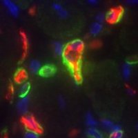
Proud to share our latest work, published in J Cell Science where we investigate the interactome of the core autophagy protein ATG9A, and characterize a new interaction with the lipid transport protein VPS13A. journals.biologists.com/jcs/article/do… #Autophagy #Lipidtransport #Membranetrafficking
Hao Yin (@haoyin20) 's Twitter Profile Photo

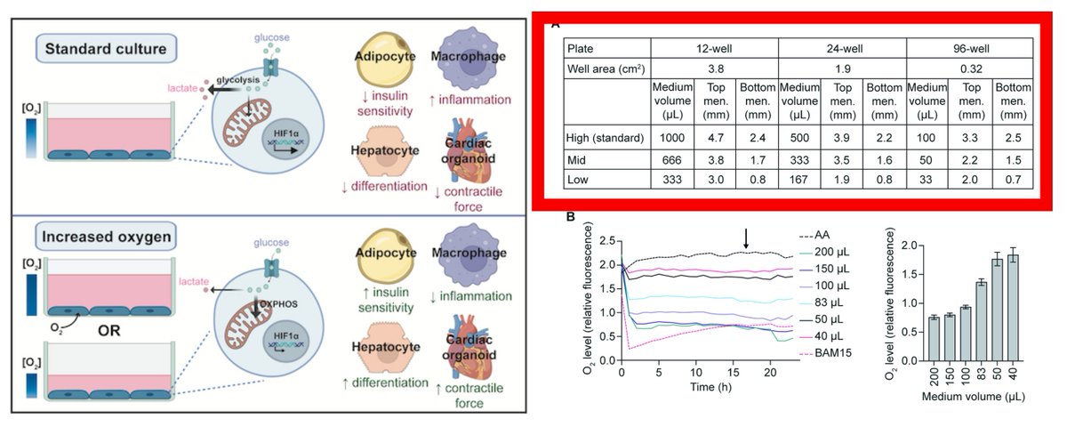
Cultured Cells in standard plastics can actually be Hypoxic & lactate-producing It matters how High the medium level is in your dish/well!🤯 The medium as liquid phase creates a steep oxygen gradient...... 33~50 µL per 96-well make your cells happily oxygenated (Have you added

Cultured Cells in standard plastics can actually be Hypoxic &amp; lactate-producing

It matters how High the medium level is in your dish/well!🤯
The medium as liquid phase creates a steep oxygen gradient......

33~50 µL per 96-well make your cells happily oxygenated (Have you added