Satrajit Chakrabarty
@satrajit_c
Senior Data Scientist @GEHealthCare #AI #Imaging #Informatics #Healthcare #DataScience
ID: 1404909273100996615
https://www.linkedin.com/in/satrajitchakrabarty 15-06-2021 21:10:58
19 Tweet
24 Followers
35 Following

The first Brain Tumor Sequence Registration (BraTS-Reg) challenge in conjunction with ISBI 2022 is now live! Details are available at: med.upenn.edu/cbica/brats-re… Looking forward to your participation! #ImageRegistration Diana Waldmannstetter Satrajit Chakrabarty Spyridon Bakas CBICA Announcements

At #5: "MRI-based identification and classification of major intracranial tumor types by using a 3D convolutional neural network" doi.org/10.1148/ryai.2… from WashUMedMIR Satrajit Chakrabarty #NeuroRad #DeepLearning #RadAIchat
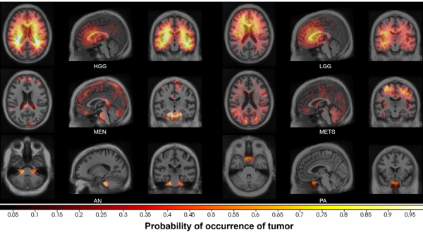

The #BRAin Tumor Sequence #REGistration #Challenge (#BraTS-Reg) is now live in #MICCAI2022Challenges . More details can be found at: med.upenn.edu/cbica/brats-re… CBICA Announcements Spyridon Bakas Penn Radiology Diana Waldmannstetter Satrajit Chakrabarty #machinelearning


“Non-invasive classification of Isocitrate Dehydrogenase mutation status of gliomas from multi-modal MRI using a 3D convolutional neural network” on 2/22 10:50am. Work by Satrajit Chakrabarty, Pamela LaMontagne, Joshua Shimony, Daniel Marcus, and Aris Sotiras.

“Deep learning-based end-to-end scan-type classification, pre-processing, and segmentation of clinical neuro-oncology studies” on 2/21 2pm. Work by Satrajit Chakrabarty, Syed Abidi, Mina Mousa, M Mokkarala, Matthew Kelsey, Pamela LaMontagne, Aris Sotiras, and Daniel Marcus.

.Satrajit Chakrabarty from CIRC and MINDS lab proposes his end-to-end framework using convolutional neural networks to segment tumor tissue subtypes from MRI scans. The framework adopts an expert-in-the-loop approach allowing for segmentation refinements. #SPIEMedicalImaging


.Satrajit Chakrabarty on his second talk at #SPIEMedicalImaging proposes a deep learning-based method to non-invasively and preoperatively determine IDH status of high- and low-grade gliomas by leveraging their phenotypical characteristics from volumetric MRI scans. WashUMedMIR
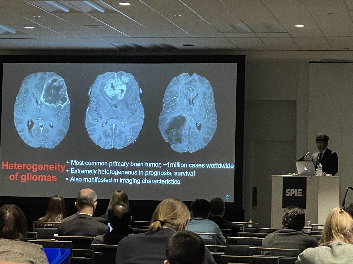

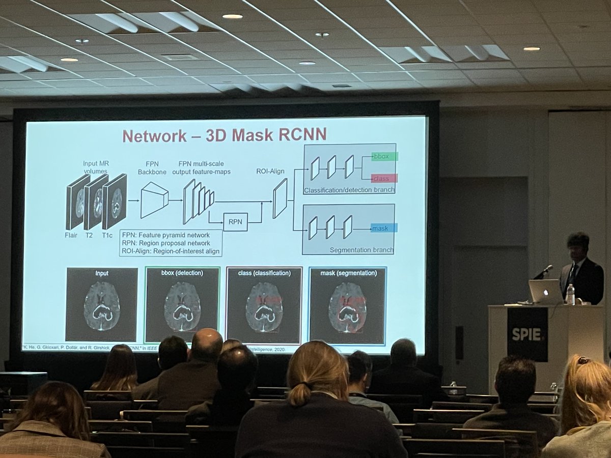
The method can be used as a pre-operative ‘virtual biopsy’ of gliomas and utilizes a 3D Mask R-CNN-based approach to simultaneously detect and segment glioma as well as classify its IDH status. Work with LaMontagne P, Simony J, Marcus D, and Aris Sotiras WashUMedMIR Test Account

Thrilled to share our #NeuroOncology Advances article on #AI-driven molecular subtyping of #gliomas using a hybrid 2.5D #CNN - tested on data from 11 centers! Paper: doi.org/10.1093/noajnl… SNO Aris Sotiras WashUMedMIR Test Account @WashUi2 WashU McKelvey Engineering #neuroradiology


👀 Check out what’s popular this week in #JCOCCI: Integrative Imaging #Informatics for #CancerResearch: Workflow Automation for Neuro-Oncology (I3CR-WANO) Satrajit Chakrabarty #AI ➡️ fal.cn/3yK0J

The 2023 BraTS on brain metastases (BraTS-METS) is live! synapse.org/#!Synapse:syn5… Arxiv paper here arxiv.org/abs/2306.00838 Mariam Aboian Spyridon Bakas Ujjwal Baid Ahmed W. Moawad Anastasia Janas
