Lacey Schrader (@ljschradermd) 's Twitter Profile
Lacey Schrader

@ljschradermd

Pathology enthusiast. #pulmpath. Alaska born. Living the good life.

ID: 896533054323179521

calendar_today13-08-2017 00:45:11

25 Tweet

232 Takipçi

171 Takip Edilen
David Gandara (@drgandara) 's Twitter Profile Photo

Important long term survival data in ALK+ NSCLC show 4 yr survival of about 50%. Equals transformation to “chronic disease”, which is our goal in all advanced cancers. And newer ALK inhibitors are adding to this advance. x.com/jco_asco/statu…

Aurélien Morini, MD, MSc 🇫🇷🇪🇺💙💛 (@a_morini_md) 's Twitter Profile Photo

Memo of WHO Histological Classification of Thymoma and Thymic Carcinoma (2 others pictures in comments). Pictures from WebPathology. Articles : J Thorac Oncol. 2014 May;9(5):596-611. #pathclues #pathboards #surgpath #thymoma Gerônimo Jr. @ritaescarvalho Ankur Sangoi Woo Cheal Cho, MD
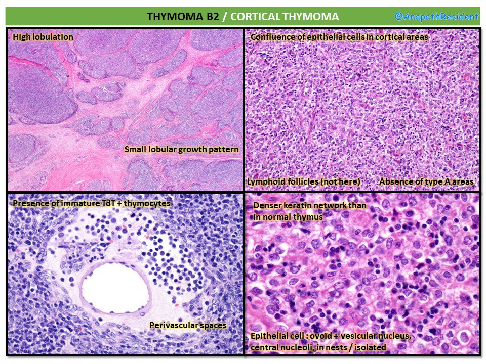
Memo of WHO Histological Classification of Thymoma and Thymic Carcinoma
(2 others pictures in comments). Pictures from WebPathology.
Articles : J Thorac Oncol. 2014 May;9(5):596-611.
#pathclues #pathboards #surgpath #thymoma
<a href="/GeronimoJrLapac/">Gerônimo Jr.</a> @ritaescarvalho <a href="/slusagar/">Ankur Sangoi</a> <a href="/DrAldehyde/">Woo Cheal Cho, MD</a>
Lacey Schrader (@ljschradermd) 's Twitter Profile Photo

My husband is going on 3 years taking the same TKI with no radiological evidence of disease. Within this time he got to deliver our second child and watch our daughter’s first graduation. It may not be the imatinib of lung ca but to our family his TKI is a #gamechanger. x.com/leciasequist/s…

Mayo Clinic Pathology (@mayoclinicpath) 's Twitter Profile Photo

1/5 #HEARTThursday w/ Melanie Bois! Where did that week go!?! But wait! For your patience, we have a two-fer bonus question! Hold on tight, and get ready for a great case comin’ at ya now! The floor is yours, Dr. Bois!
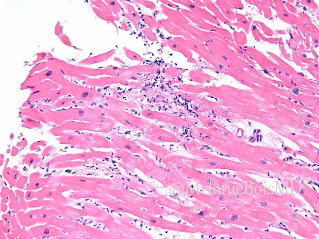
1/5
#HEARTThursday w/ <a href="/MelanieBoisMD/">Melanie Bois</a>!  Where did that week go!?!  But wait!  For your patience, we have a two-fer bonus question!  Hold on tight, and get ready for a great case comin’ at ya now!  
The floor is yours, Dr. Bois!
Lacey Schrader (@ljschradermd) 's Twitter Profile Photo

Pulmonary Blastomycosis characterized by broad based-budding and double contoured thick wall highlighted on GMS. Mayo Clinic Pathology andrewlayman #dimorphicfungi #Bforbroadbasedbudding
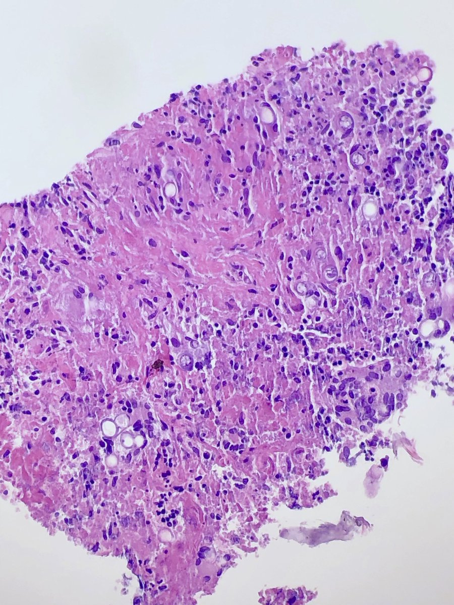
Pulmonary Blastomycosis characterized by broad based-budding and double contoured thick wall highlighted on GMS. <a href="/MayoClinicPath/">Mayo Clinic Pathology</a> <a href="/AndrewLayman/">andrewlayman</a> #dimorphicfungi #Bforbroadbasedbudding
Jerad Gardner, MD (@jmgardnermd) 's Twitter Profile Photo

How do you know if is a benign nevus or not? Learn the basic features of melanocytic nevi in my new YouTube video: buff.ly/2Hm65SV. This is part 1 of an in progress series I’m doing on melanocytic #dermpath. Part 2 will be basics of #melanoma. #Pathology #dermatology

How do you know if is a benign nevus or not? Learn the basic features of melanocytic nevi in my new YouTube video: buff.ly/2Hm65SV. This is part 1 of an in progress series I’m doing on melanocytic #dermpath. Part 2 will be basics of #melanoma. #Pathology #dermatology
Silvija Gottesman MD (@sgottesmanmd) 's Twitter Profile Photo

Gottlieb’s sandwich sign: Fungal hyphae are “sandwiched” between two zones of cornified cells: orthokeratosis on top & parakeratosis or compact orthokeratosis below. Can be used as a clue for low power dx of dermatophyte infection. #dermpath #dermatology #SilviPearls #pathology

Gottlieb’s sandwich sign: Fungal hyphae are “sandwiched” between two zones of cornified cells: orthokeratosis on top &amp; parakeratosis or compact orthokeratosis below. Can be used as a clue for low power dx of dermatophyte infection. #dermpath #dermatology #SilviPearls #pathology
Steven Billings, MD (@stevebillingsmd) 's Twitter Profile Photo

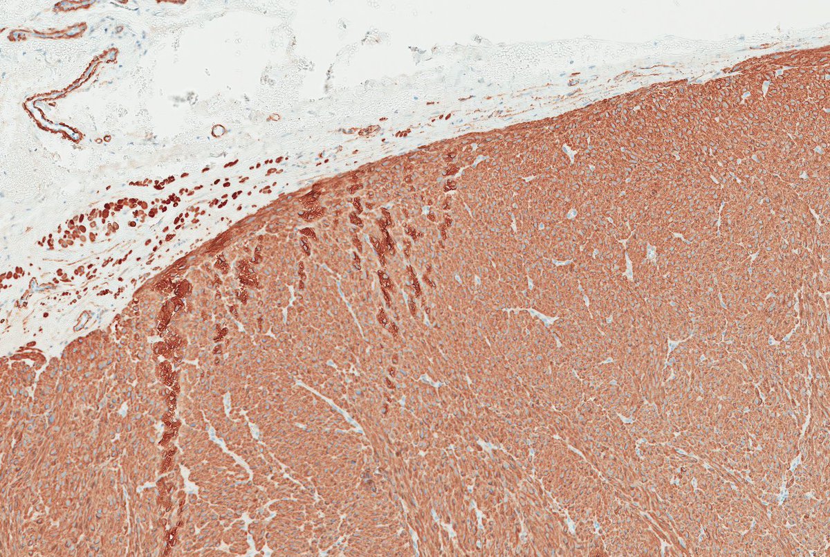
Remember leiomyosarcomas arising from vessels may be aggressive unlike leiomyosarcoma from arrector pili. This LMS arising from vein; note residual vascular wall at periphery. Residual vein top left corner on image 2 & 3. Ref: Italiano A, et al. PubMed PMID: 20167595. #dermpath

Remember leiomyosarcomas arising from vessels may be aggressive unlike leiomyosarcoma from arrector pili. This LMS arising from vein; note residual vascular wall at periphery.  Residual vein top left corner on image 2 &amp; 3. Ref: Italiano A, et al. PubMed PMID: 20167595. #dermpath
IASLC (@iaslc) 's Twitter Profile Photo

A study presented at a press briefing ahead of #ASCO18 shows that comprehensive gen. testing in adv. #lungcancer is more cost-effective than testing 1/limited number of genes at a time. Nathan A. Pennell MD, PhD, FASCO is lead author and study will be presented at ASCO. #LCSM bit.ly/2wMTqGF
Jerad Gardner, MD (@jmgardnermd) 's Twitter Profile Photo

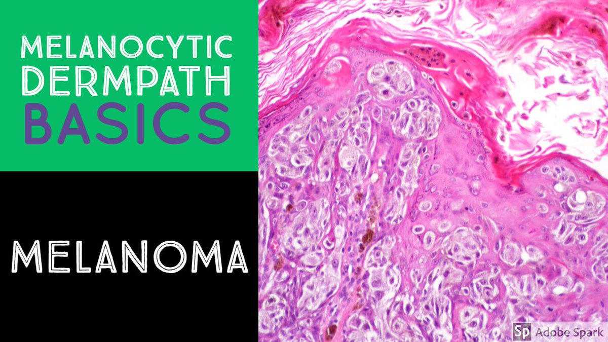
Learn the basic histologic features of #melanoma in my new YouTube video. buff.ly/2s5bczB. If you missed my video on the basics of melanocytic nevi, check that out, too: buff.ly/2xcRxmO. #pathology #dermatology #dermpath #meded #usmle

Learn the basic histologic features of #melanoma in my new YouTube video. buff.ly/2s5bczB. If you missed my video on the basics of melanocytic nevi, check that out, too: buff.ly/2xcRxmO. #pathology #dermatology #dermpath #meded #usmle
Lacey Schrader (@ljschradermd) 's Twitter Profile Photo

Palliative femur resection from an older chap with Paget’s disease. Cement lines in random orientation highlight a jigsaw puzzle pattern. #bonepathology #mosaic

Palliative femur resection from an older chap with Paget’s disease. Cement lines in random orientation highlight a jigsaw puzzle pattern.  #bonepathology #mosaic