GI James
@GIJamesMD
GI and liver pathologist @UTSW_GIpath. 🐈 dad. 🌏 traveler.
#GIpath #pathology
ID:824412489899184129
https://profiles.utsouthwestern.edu/profile/196597/james-mitchell.html 26-01-2017 00:23:49
2,6K Tweets
5,0K Followers
4,5K Following

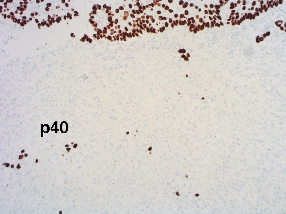
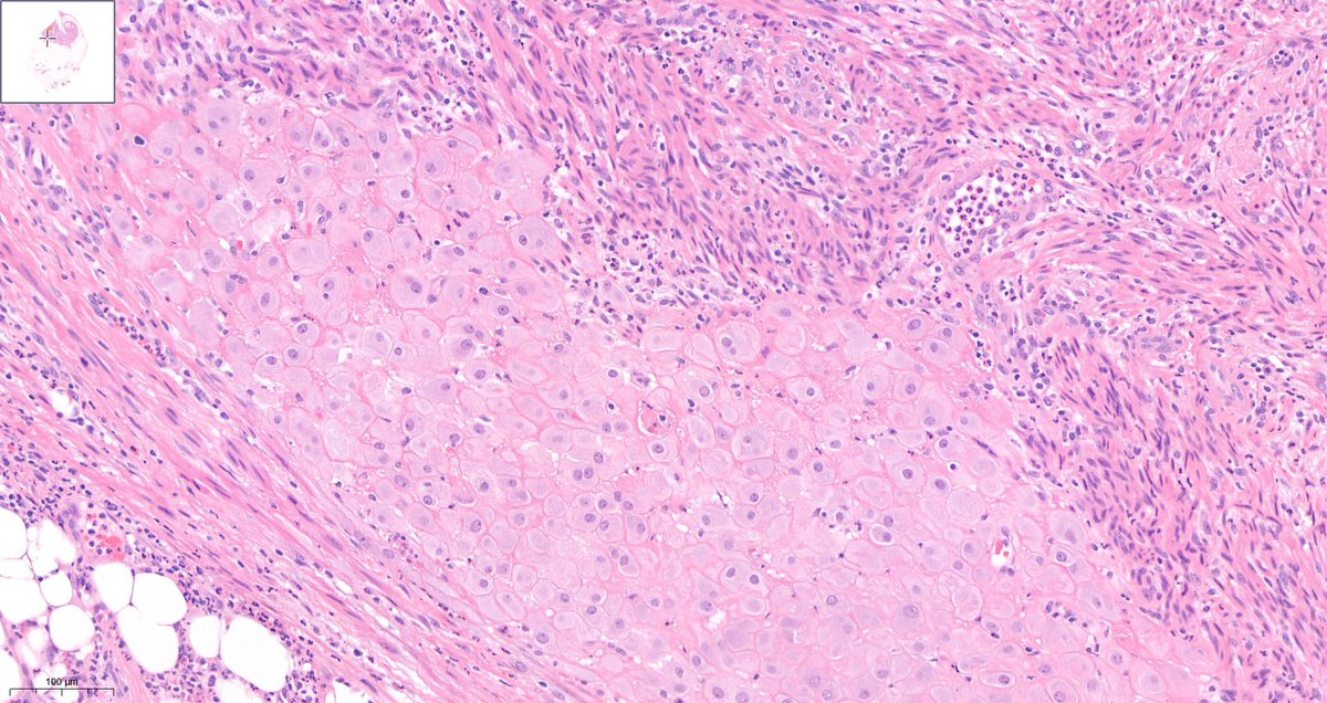
⚕️ Rectal traumatic neuroma
🔪Arising at polypectomy site (Dx was a Well differentiated NET grade 1).
➡️Residual NET in adjacent block.
#GIpath #BSTpath #Pathtwitter #PathX #Pathresidents


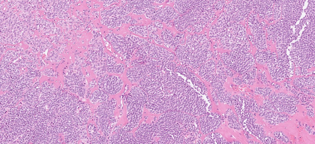
60F stomach, submucosal lesion, diagnosis?
A) Gastrointestinal stromal tumor
B) Plexiform fibromyxoma
C) Inflammatory myofibroblastic tumor
D) Inflammatory fibroid polyp
#GIPath #BSTPath #Pathology #PathTwitter #PathX


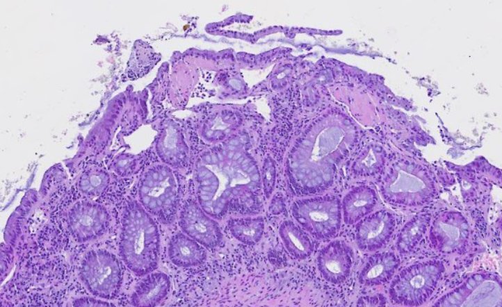
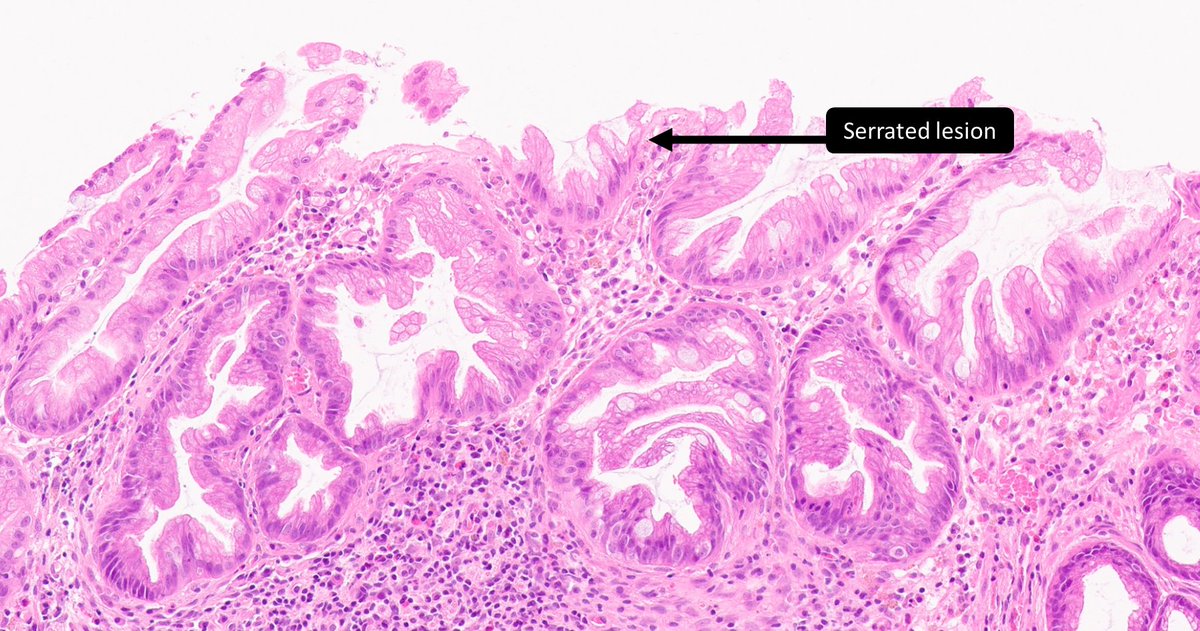
Serrated sigmoid polyp. Diagnosis?
A) Sessile serrated lesion
B) Perineurioma
C) Schwann cell hamartoma
D) Schwannoma
#GIPath #BSTPath #PathTwitter #pathology
virtualpathology.leeds.ac.uk/slides/library…


New in 2024: Each month we will share the best #PathTweetAward from the trainee category.
We will continue to announce nominees for the Open Category at the end of the year.
The March 2024 Trainee #PathTweetAward goes to Miruna Popescu, MD, congratulations 👏🏻👏🏻👏🏻

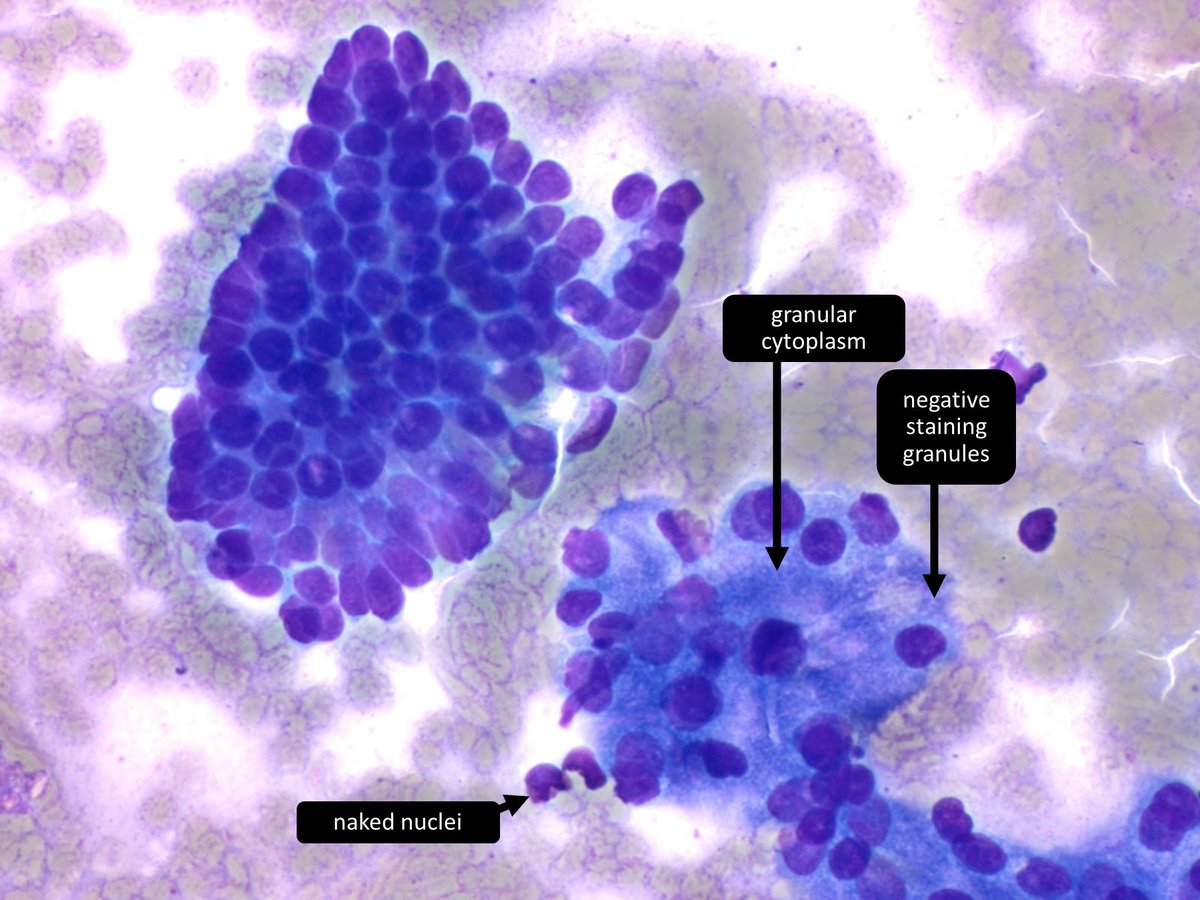
Pancreatic mass. Diagnosis?
A) Non-diagnostic
B) Negative (for malignancy)
C) Neoplastic (benign or other)
D) Malignant
#cytopath #pathology #gipath #pathtwitter


Join us at the Diagnexia Symposium, Aug 21-23, 2024, Oxford! Delighted to feature Dr. Adam L. Booth from Washington University. Don't miss his insights in Pathology & Immunology.
📆 Save the date & stay tuned for more!
➡️ Register: news.diagnexia.com/4aQpLuE
#DiagnexiaSymposium2024


What mismatch repair gene(s) have mutated?
A) MSH2
B) MSH6
C) A & B
#GIPath #DermPath #PediPath #BSTPath #PathTwitter #PathResidents


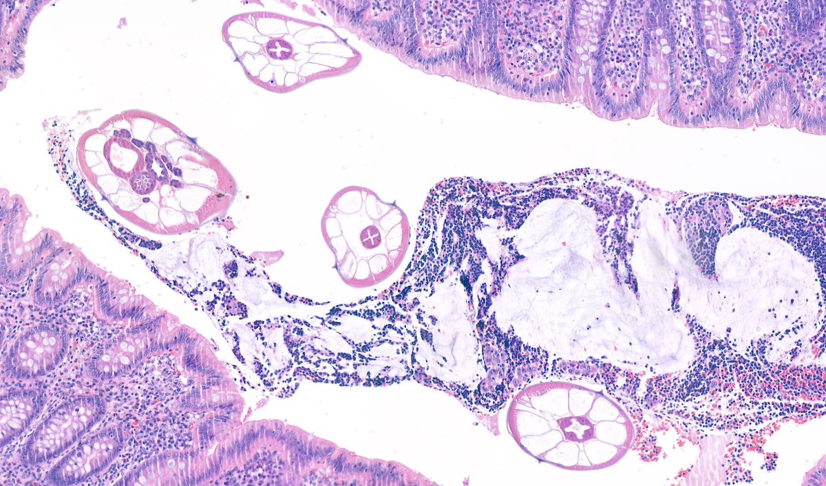
Young woman with appendicitis and these unusual changes at the tip #Pathologists what’s going on here?

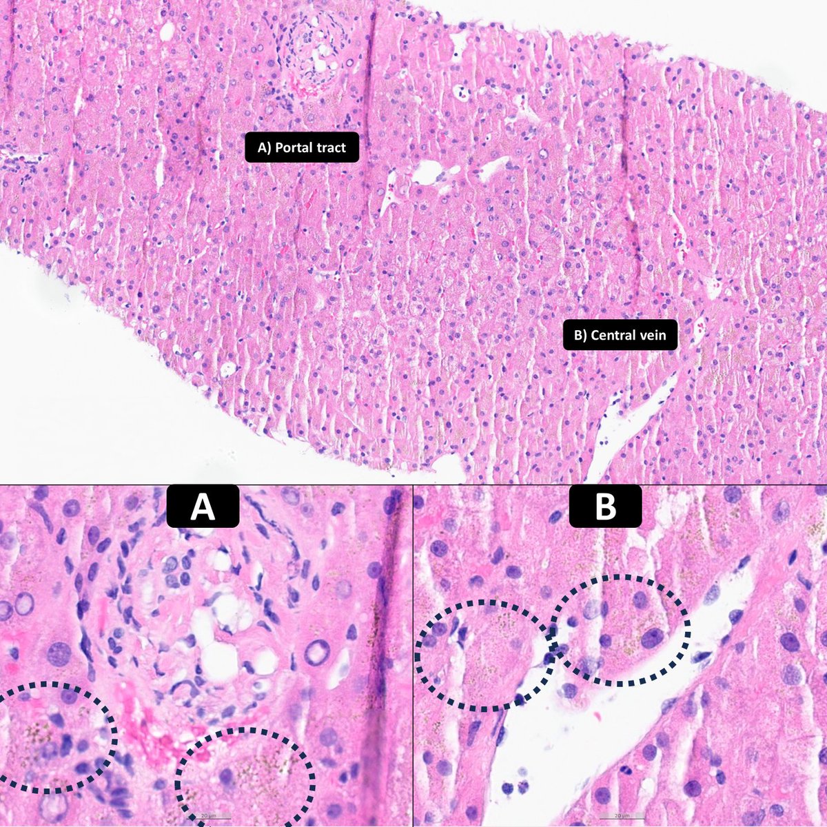
Pigment A or B = Iron?
#Hemochromatosis #LiverPath
🙏John Hart, MD at #uscap2024
#GIPath #PathTwitter #pathresidents

How about a little poll to end the week? #BreastPath Dr. Luca Olaleke Folaranmi GI James Celina Stayerman MD Nusrat Z. Bokhari Amal Asar, FRCpath. Tristan Rutland MBBS FRCPA IFCAP Kristen Anandi Lobo, MD Sumanta Das Lorand Kis Laura G. Pastrián MD Padma Priya J Carlos Miguel Ruiz Carlos Nieves Barry McGinn Pascual Meseguer


