ZEISS Microscopy (@zeiss_micro) 's Twitter Profile
ZEISS Microscopy

@zeiss_micro

We are #Microscopy. All about microscopes from #ZEISS.
LinkedIn: zeiss.ly/ln Facebook: zeiss.ly/fb | Imprint & Data Privacy: zeiss.com

ID: 46128549

linkhttps://www.zeiss.com/microscopy calendar_today10-06-2009 14:49:22

10,10K Tweet

23,23K Followers

3,3K Following

ibidi GmbH (@ibidicells) 's Twitter Profile Photo

🌌Celestial Intestine🌠 🌟Image shows a mouse intestinal organoid cultured on a µ-Dish 35mm, high Glass Bottom and stained for tight junction protein (green) and actin (red). Captured with ZEISS Microscopy LSM 980 🎨By Sarbari Saha from Uni Stuttgart #organoid #microscopy #sciart

🌌Celestial Intestine🌠

🌟Image shows a mouse intestinal organoid cultured on a µ-Dish 35mm, high Glass Bottom and stained for tight junction protein (green) and actin (red). Captured with <a href="/zeiss_micro/">ZEISS Microscopy</a> LSM 980

🎨By Sarbari Saha from <a href="/Uni_Stuttgart/">Uni Stuttgart</a>

#organoid #microscopy #sciart
ZEISS Microscopy (@zeiss_micro) 's Twitter Profile Photo

On the tip of your tongue... 👅 Check out this application image of oral bacteria, acquired with a ZEISS LSM 900 laser scanning microscope and discover the story behind it: zeiss.ly/user-story-tip… Courtesy: Dr. Tagide deCarvalho, UMBC Biology

On the tip of your tongue... 👅
Check out this application image of oral bacteria, acquired with a ZEISS LSM 900 laser scanning microscope and discover the story behind it: zeiss.ly/user-story-tip…
Courtesy: Dr. Tagide deCarvalho, <a href="/UMBCBiology/">UMBC Biology</a>
Aberdeen Microscopy (@abdn_micro) 's Twitter Profile Photo

These cells were prepared for #imaging many years ago and the slide lives on the desk next to our ZEISS Microscopy LSM880 #confocal 🔬 Despite it's age, and the fact it's been sitting out in the open for a long time, it still gives us beautiful images! 😃 #microscopy #fluorescence

These cells were prepared for #imaging many years ago and the slide lives on the desk next to our <a href="/zeiss_micro/">ZEISS Microscopy</a> LSM880 #confocal 🔬

Despite it's age, and the fact it's been sitting out in the open for a long time, it still gives us beautiful images! 😃

#microscopy #fluorescence
Instituto de Neurociencias UMH-CSIC (@neuroalc) 's Twitter Profile Photo

#SciencePhoto_IN "Of Flowers and Mitochondria" Author: Enrico Negri Enrico Negri Lab: Dr. Victor Borrell Borrell Lab "Chick embryo neural progenitors (magenta in lower half) and neurons (magenta in upper half, multipolar morphology) and their mitochondria (cyan). With ZEISS Microscopy."

#SciencePhoto_IN
"Of Flowers and Mitochondria"
Author: Enrico Negri <a href="/en_negri/">Enrico Negri</a>
Lab: Dr. Victor Borrell <a href="/BorrellLab/">Borrell Lab</a>
"Chick embryo neural progenitors (magenta in lower half) and neurons (magenta in upper half, multipolar morphology) and their mitochondria (cyan). With <a href="/zeiss_micro/">ZEISS Microscopy</a>."
Identifyn (@identifynllc) 's Twitter Profile Photo

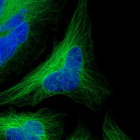
Identifyn™ SRM Alexa Fluor™ 750 Affinity Purified Donkey Anti-Mouse, IgG (H + L) visualized using our new Zeiss LSM 980 confocal with near infra-red (NIR) resolution. Image shows alpha Tubulin with the Identifyn™ 750 secondary antibody. Image by J.K. Bennett © Copyright

Identifyn™ SRM Alexa Fluor™ 750 Affinity Purified Donkey Anti-Mouse, IgG (H + L) visualized using our new Zeiss LSM 980 confocal with near infra-red (NIR) resolution. Image shows alpha Tubulin with the Identifyn™ 750 secondary antibody.

Image by J.K. Bennett

© Copyright
edward lachica (@edwardlachica3) 's Twitter Profile Photo

Hey nerds: tufted cells from the mouse olfactory bulb courtesy of Shawn Burton Lehigh University & Nathan Urban lab. Captured with #lightsheet #microscope from 3i & FusionBT from Hamamatsu. Nearly isotropic res using a 1.5X/0.37NA obj from @Zeiss_micro. Happy #FluorescenceFriday

EXCITE2 (@excite_network) 's Twitter Profile Photo

#SEM image of Mg #carbonate showing the transformation and hydration of #nesquehonite. 📸 Imaged by Dr. Xu Zhang (Shoobeedoobeedoo) & Hans van Melick Utrecht University 🔬ZEISS Microscopy Gemini 450 SEM Geosciences UU Earth Sciences - Utrecht University #EXCITE #Microscopy #Geology #MgCO3

#SEM image of Mg #carbonate showing the transformation and hydration of #nesquehonite.

📸 Imaged by Dr. Xu Zhang (<a href="/Xuuuuuu/">Shoobeedoobeedoo</a>) &amp; Hans van Melick <a href="/UniUtrecht/">Utrecht University</a> 

🔬<a href="/zeiss_micro/">ZEISS Microscopy</a> Gemini 450 SEM

<a href="/UUGeo/">Geosciences UU</a> <a href="/UUEarthSciences/">Earth Sciences - Utrecht University</a> #EXCITE #Microscopy #Geology #MgCO3
Instituto de Neurociencias UMH-CSIC (@neuroalc) 's Twitter Profile Photo

#SciencePhoto_IN "Mouse Pad with Heart" Author: Miguel A Serrano Lab: Dr. Francisco Taberner "Mouse skin section expressing GFP in Schwann Cells (green) and tdTomato in advillin fibers (red) and nuclei labelled in cyan. Image acquired with ZEISS Microscopy Axioscan."

#SciencePhoto_IN 
"Mouse Pad with Heart" 
Author: Miguel A Serrano 
Lab: Dr. Francisco Taberner 
"Mouse skin section expressing GFP in Schwann Cells (green) and tdTomato in advillin fibers (red) and nuclei labelled in cyan. Image acquired with <a href="/zeiss_micro/">ZEISS Microscopy</a> Axioscan."
Dr Richard Kirby (@planktonpundit) 's Twitter Profile Photo

Acantharia are in my Plymouth plankton tow today. I always see the appearance of these fascinating, single celled protists and their symbiotic algae as the end of summer and start of autumn. Also among them now however, is our ever present microplastic pollution. ZEISS Microscopy

ZEISS Microscopy (@zeiss_micro) 's Twitter Profile Photo

ZEISS are proud sponsors of the 6th European Cilia Meeting meeting. If you are attending come and see us for all the latest information on our range of Super-Resolution Microscopes: zeiss.com/microscopy/en/… #zeiss #microscopy #superresolution #cilia2024

ZEISS are proud sponsors of the <a href="/EuroCilia2024/">6th European Cilia Meeting</a> meeting. If you are attending come and see us for all the latest information on our range of Super-Resolution Microscopes:
zeiss.com/microscopy/en/…

#zeiss #microscopy #superresolution #cilia2024
Identifyn (@identifynllc) 's Twitter Profile Photo

SIM² image of Mitochondria visualized with Identifyn™ SRM Alexa Fluor™ 532 Affinity Purified Donkey Anti-Mouse, F(ab’)₂ (H + L) in HeLa cells. Captured with the Zeiss Elyra 5 Lattice SIM. Image by P. Raut Explore more at identifyn.com © Copyright 2023-2024

SIM² image of Mitochondria visualized with Identifyn™ SRM Alexa Fluor™ 532 Affinity Purified Donkey Anti-Mouse, F(ab’)₂ (H + L) in HeLa cells.

Captured with the Zeiss Elyra 5 Lattice SIM.

Image by P. Raut

Explore more at identifyn.com

© Copyright 2023-2024
ZEISS Microscopy (@zeiss_micro) 's Twitter Profile Photo

We’re thrilled to see ZEISS microscopy playing a key role in advancing research on Alzheimer’s Tau protein! Congratulations to the team on this important publication and your innovative use of microfluidics and AD-FCS. Exciting work! 📷📷 #ProudPartner

ibidi GmbH (@ibidicells) 's Twitter Profile Photo

The Order of the #Neural Phoenix🐦 Mouse brain coronal section showing #vasculature (magenta) and neural #network (orange). Imaged using ZEISS Microscopy Lightsheet 7🔬 🥼👩‍🎨Scientific #artwork by Lea Amina Kalayci from Rheinische Friedrich-Wilhelms-Universität Bonn, Germany #science #microscopy #sciart #bioart #brain

The Order of the #Neural Phoenix🐦

Mouse brain coronal section showing #vasculature (magenta) and neural #network (orange). Imaged using <a href="/zeiss_micro/">ZEISS Microscopy</a> Lightsheet 7🔬

🥼👩‍🎨Scientific #artwork by Lea Amina Kalayci from <a href="/UniBonn/">Rheinische Friedrich-Wilhelms-Universität Bonn</a>, Germany 

#science #microscopy #sciart #bioart #brain
ZEISS Microscopy (@zeiss_micro) 's Twitter Profile Photo

In this upcoming Bitesize Bio live webinar, learn about microscopy techniques for urine analysis, how to identify and differentiate between normal and abnormal urine particles, and how to interpret different urine particles. Join Dr. José Antonio Tesser Poloni on September 25,

In this upcoming Bitesize Bio live webinar, learn about microscopy techniques for urine analysis, how to identify and differentiate between normal and abnormal urine particles, and how to interpret different urine particles.

Join Dr. José Antonio Tesser Poloni on September 25,