UConn Advanced Light Microscopy Facility (@uconnalmf) 's Twitter Profile
UConn Advanced Light Microscopy Facility

@uconnalmf

The ALMF is a part of the Center for Open Research Resources and Equipment at UConn. We provide microscopy expertise and access to cutting edge instruments.

ID: 1569413872490270723

linkhttp://confocal.uconn.edu calendar_today12-09-2022 19:53:28

51 Tweet

77 Followers

13 Following

UConn Advanced Light Microscopy Facility (@uconnalmf) 's Twitter Profile Photo

New instrument alert! Our newest addition, the Nikon Instruments point-scanning AXR #confocal, also equipped for TIRF, is up and running! Full information about the instrument and use/trainings can be found here: confocal.uconn.edu/nikonaxr/


New instrument alert! Our newest addition, the <a href="/NikonInst/">Nikon Instruments</a> point-scanning AXR #confocal, also equipped for TIRF, is up and running! Full information about the instrument and use/trainings can be found here: confocal.uconn.edu/nikonaxr/
UConnCOR²E (@uconncore) 's Twitter Profile Photo

Hello Twitterverse - UConn COR²E is now on Twitter! We are a thriving OVPR Center located on the Storrs academic campus comprised of 11 unique core facilities that boast diverse capabilities and expertise. We exist to support the equally diverse UConn research community!

UConn Advanced Light Microscopy Facility (@uconnalmf) 's Twitter Profile Photo

Happy #FluorescenceFriday! The ALMF Core has been busy the past few weeks, so we have some repeat images collected on our Leica Microsystems SP8 with different LUT displays. We're also interested in some robust specimen slides for practice, so if anyone has recs please comment 👇

Happy #FluorescenceFriday! The ALMF Core has been busy the past few weeks, so we have some repeat images collected on our <a href="/LeicaMicro/">Leica Microsystems</a> SP8 with different LUT displays. We're also interested in some robust specimen slides for practice, so if anyone has recs please comment 👇
UConnCOR²E (@uconncore) 's Twitter Profile Photo

COR²E has many important updates to share with the UConn research community: recently completed projects, state-of-the-art instrumentation installations, and new people! Read all about them in our brand new Summer 2023 COR²E newsletter here: core.uconn.edu/wp-content/upl…

COR²E has many important updates to share with the UConn research community: recently completed projects, state-of-the-art instrumentation installations, and new people!  Read all about them in our brand new Summer 2023 COR²E newsletter here: 
core.uconn.edu/wp-content/upl…
UConn Advanced Light Microscopy Facility (@uconnalmf) 's Twitter Profile Photo

Having fun with our new Nikon Instruments AXR confocal and using the NIS Elements movie maker function. This is a 3D reconstruction of the crypts in a mouse intestine section showing goblet cells (blue), nuclei (green), and actin (red).#confocal

UConn Advanced Light Microscopy Facility (@uconnalmf) 's Twitter Profile Photo

The value of machine learning using ZEISS arivis 🔬 software for segmentation becomes clear in a sample with high dynamic range, such as this mitotic spindle (bottom). Most of the dimmer astral microtubules are lost with traditional segmentation algorithms (top).

The value of machine learning using <a href="/zeiss_arivis/">ZEISS arivis 🔬</a>   software for segmentation becomes clear in a sample with high dynamic range, such as this mitotic spindle (bottom).  Most of the dimmer astral microtubules are lost with traditional segmentation algorithms (top).
UConn Advanced Light Microscopy Facility (@uconnalmf) 's Twitter Profile Photo

Happy #FluorescenceFriday! We've been playing around with large image + z-stack using JOBS on the Nikon Instruments AXR, which is super useful! Image 1 is a 2D overview, and 2 & 3 are max projections from select regions of this kidney section at 40X.

Happy #FluorescenceFriday! We've been playing around with large image + z-stack using JOBS on the <a href="/NikonInst/">Nikon Instruments</a> AXR, which is super useful! Image 1 is a 2D overview, and 2 &amp; 3 are max projections from select regions of this kidney section at 40X.
UConn Advanced Light Microscopy Facility (@uconnalmf) 's Twitter Profile Photo

Happy #MicroscopyMonday! Here we have a fern rhizome imaged with a color camera on a Leica Microsystems Thunder without (Left) and with (Right) a DIC prism in the lightpath, creating interference that results in other colors.

Happy #MicroscopyMonday! Here we have a fern rhizome imaged with a color camera on a <a href="/LeicaMicro/">Leica Microsystems</a> Thunder without (Left) and with (Right) a DIC prism in the lightpath, creating interference that results in other colors.
UConn Advanced Light Microscopy Facility (@uconnalmf) 's Twitter Profile Photo

If you're a lab at UConn or @UConnHealth there's still a few weeks left to apply for internal funds if you want to try out one of the UConn Core facilities!

UConn Advanced Light Microscopy Facility (@uconnalmf) 's Twitter Profile Photo

This #FluorescenceFriday we bring you some fun #sciart of a "microscopic sunflower," found from a hibiscus sample! Captured using green and far red fluorescence (yellow and orange here, respectively) and DIC on our Leica Microsystems Thunder. Any #plantsci people able to ID this?
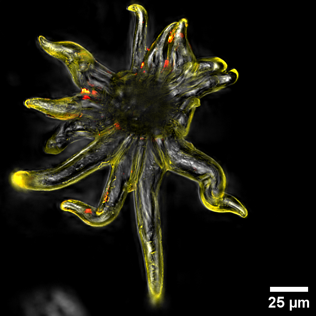
This #FluorescenceFriday we bring you some fun #sciart of a "microscopic sunflower," found from a hibiscus sample! Captured using green and far red fluorescence (yellow and orange here, respectively) and DIC on our <a href="/LeicaMicro/">Leica Microsystems</a> Thunder. Any #plantsci people able to ID this?
UConn Advanced Light Microscopy Facility (@uconnalmf) 's Twitter Profile Photo

Double dipping on this same stellate trichome (left) for #microscopymonday! This image is using the Leica Microsystems Thunder K5C color camera and applying extended depth of focus. We also imaged part of the stem itself (right) and found other interesting structures!
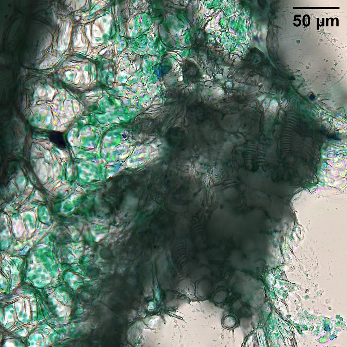
Double dipping on this same stellate trichome (left) for #microscopymonday! This image is using the <a href="/LeicaMicro/">Leica Microsystems</a> Thunder K5C color camera and applying extended depth of focus. We also imaged part of the stem itself (right) and found other interesting structures!
UConn Advanced Light Microscopy Facility (@uconnalmf) 's Twitter Profile Photo

Want to see more of the core facilities that UConn Storrs and Health have to offer? Register for the Open House! Free lunch with registration, and vendors will be here too!

UConn Advanced Light Microscopy Facility (@uconnalmf) 's Twitter Profile Photo

The Advanced Light Microscopy Facility will be holding a one day workshop on image analysis using the free program ImageJ. It will be held on January 9th and is open to all members of the UConn community. Information and registration can be found at confocal.uconn.edu/analysisworksh…

The Advanced Light Microscopy Facility will be holding a one day workshop on image analysis using the free program ImageJ.  It will be held on January 9th and is open to all members of the UConn community.  Information and registration can be found at confocal.uconn.edu/analysisworksh…
Leica Microsystems (@leicamicro) 's Twitter Profile Photo

According to Dr. Chen, “we developed a nanoparticle-based therapeutic for treating osteoarthritis. Our nanoparticles have a unique nano-rod shape, enabling better penetration into articular cartilage compared to traditional spherical nanoparticles.