Cody Raul Cardenas(@nepbaphomet) 's Twitter Profile Photo

Finally back from Prague and backing up all this MicroCT scan data. I'll be excited to start learning about proper segmentation and how to do some 3D morphological analyses! The short video is a rotating 3D microCT scan of a Calosoma Callisthenes reticulatum.

account_circle
Jimmy Bernot, PhD 🦐🔬🧬🏳️‍🌈(@JimmyBernot) 's Twitter Profile Photo

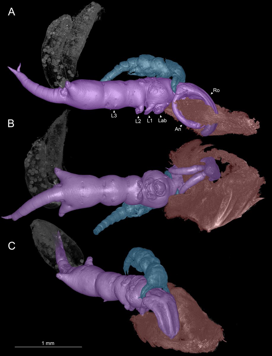
Our new paper is out today using microCT to understand the family Shiinoidae, some of the most unusual parasitic copepods 🧵
peerj.com/articles/16966/

Our new paper is out today using microCT to understand the family Shiinoidae, some of the most unusual parasitic copepods 🧵
peerj.com/articles/16966/
account_circle
National Museums of Kenya(@museumsofkenya) 's Twitter Profile Photo

For over 25 years National Museums of Kenya has made medical CT scans of our fossil humans available for scientific research. A new large addition of available microCT scans can now be viewed at human-fossil-record.org

For over 25 years National Museums of Kenya has made medical CT scans of our fossil humans available for scientific research. A new large addition of available microCT scans can now be viewed at human-fossil-record.org
account_circle
Neuralink(@neuralink) 's Twitter Profile Photo

At Neuralink, we have imaging capabilities that let us see exactly what’s going on in fully sealed devices. For implant debugging, we often capture high-resolution 2D and 3D images with microCT. It’s what’s on the inside that counts ♥️

account_circle
The Anatomical Record(@AnatRecord) 's Twitter Profile Photo

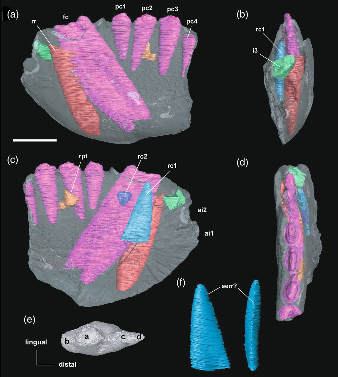
MicroCT scans reveal insights into the tooth replacement pattern of the enigmatic Charruodon tetracuspidatus, a . Early ontogenetic stage hints at potential taxonomic mysteries! New research by Carolina Hoffmann et al.:
doi.org/10.1002/ar.253…

MicroCT scans reveal insights into the tooth replacement pattern of the enigmatic Charruodon tetracuspidatus, a #Triassic #cynodont. Early ontogenetic stage hints at potential taxonomic mysteries! New research by Carolina Hoffmann et al.:
doi.org/10.1002/ar.253…
account_circle
Richard Dearden(@Euphanerops) 's Twitter Profile Photo

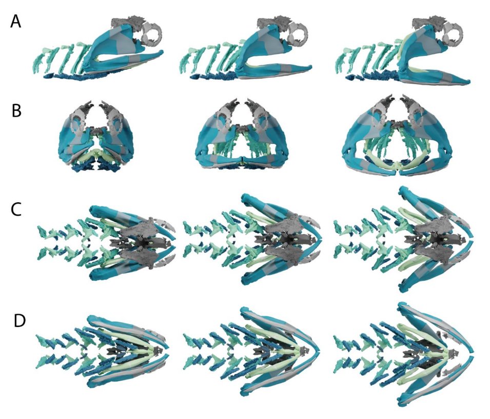
Bamboozled by the all the different reconstructions of the gill arch anatomy of the Permian acanthodian Acanthodes? Us too! Check out our preprint, in which we use non acid-prepped specimens in the MNHN to redescribe it with microCT. All feedback welcome!

Bamboozled by the all the different reconstructions of the gill arch anatomy of the Permian acanthodian Acanthodes? Us too! Check out our preprint, in which we use non acid-prepped specimens in the MNHN to redescribe it with microCT. All feedback welcome!
account_circle
Emma R Schachner PhD(@paleofox) 's Twitter Profile Photo

New collaborative hybrid review paper (old data/new data) out today in The Anatomical Record with a diverse team looking at CT/microCT based segmentation of LUNGS in biology: evolution to medicine! The madness behind the methods. 🥳🫁🐊🦅 anatomypubs.onlinelibrary.wiley.com/doi/epdf/10.10…
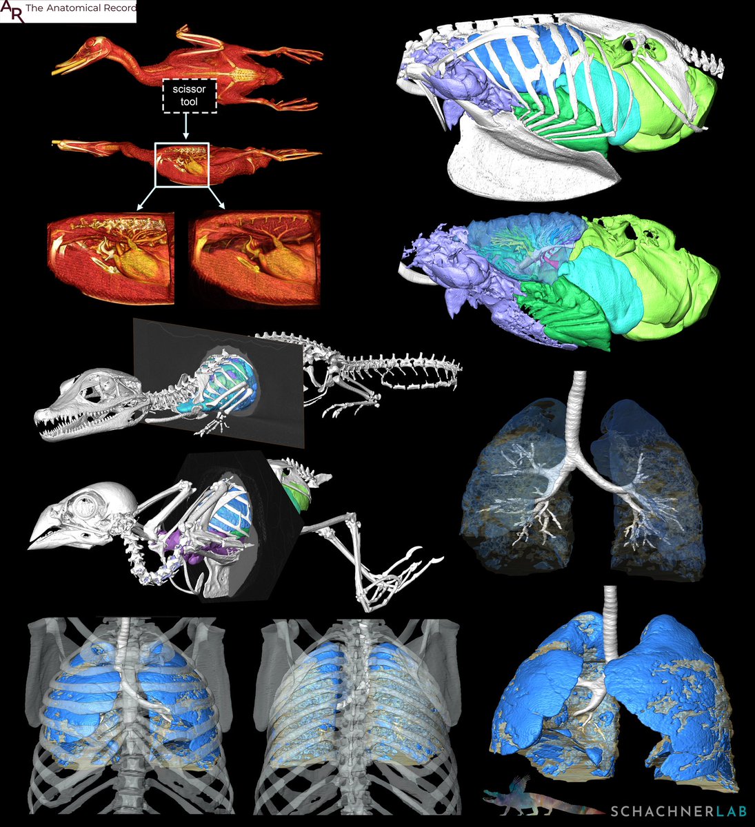
New collaborative hybrid review paper (old data/new data) out today in @AnatRecord with a diverse team looking at CT/microCT based segmentation of LUNGS in biology: evolution to medicine! The madness behind the methods. 🥳🫁🐊🦅 anatomypubs.onlinelibrary.wiley.com/doi/epdf/10.10…
account_circle
Masato Hirose(@moostiere) 's Twitter Profile Photo

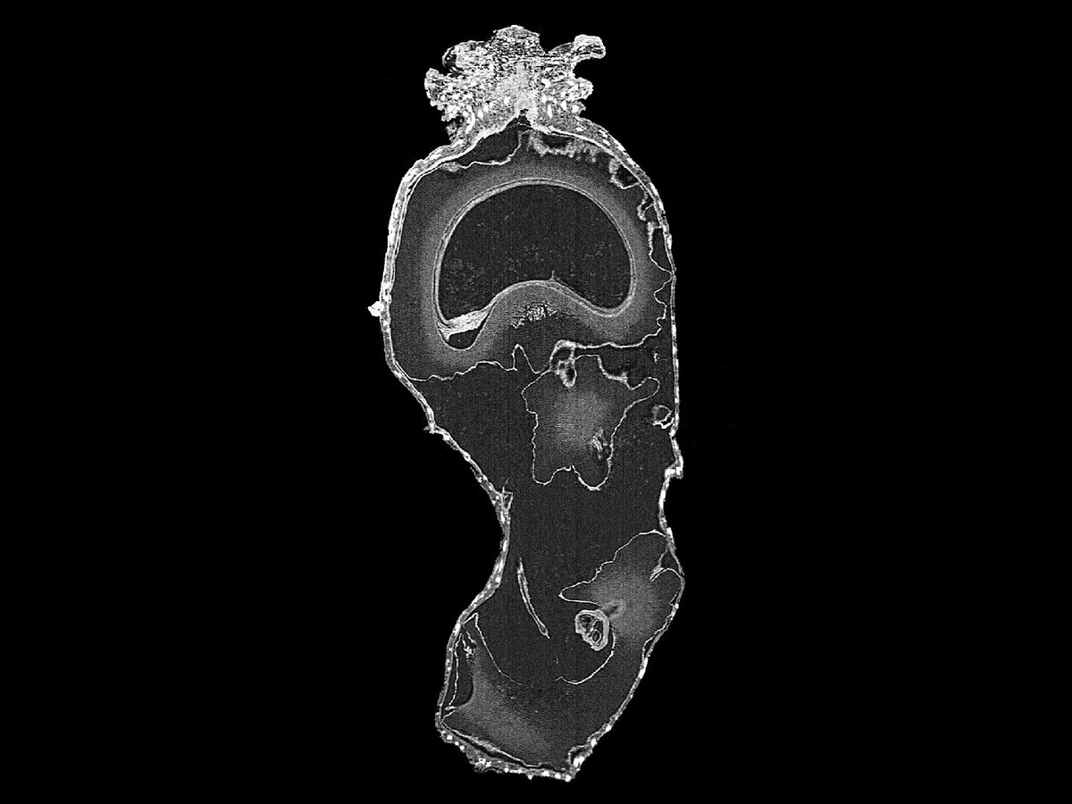
コブコケムシ科の一種の樹状群体をmicroCTで観察すると、このように年輪のような成長線がくっきりと見えます。

account_circle
Jerónimo Morales Toledo(@JMT_MHL) 's Twitter Profile Photo

Look at the wonders of seeds and their shapes! 🌱🔍 Take a look at this micro CT scan of Cyrtosperma senegalese seeds – they're campylotropous (bending), with intricate cristate seed coats. The stylar region is full of oxalate crystals!

Look at the wonders of seeds and their shapes! 🌱🔍 Take a look at this micro CT scan of Cyrtosperma senegalese seeds – they're campylotropous (bending), with intricate cristate seed coats. The stylar region is full of oxalate crystals! #Seeds #Botany #MicroCT
account_circle
Alain Chédotal(@AlainChedotal) 's Twitter Profile Photo

Taking Volumetric correlated light & EM (vCLEM) to the next level 1)Design 8 recombinant  smaller single-chain variable fragments (scFv) able to penetrate 100µm deep w/o detergent 2) confocal microscopy 3) microCT 4) serial EM & registration. Lichtman lab. biorxiv.org/cgi/content/sh…

account_circle
Zach Cofran(@ZCofran) 's Twitter Profile Photo

Your daily gibbon microCT: semitransparent view of the left half of the developing skull. If you look closely at the back of the jaws you can see the nascent cusps of the 'wisdom teeth' still buried in bony crypts, haunted by the dense specter of the bony labyrinth beyond

Your daily gibbon microCT: semitransparent view of the left half of the developing skull. If you look closely at the back of the jaws you can see the nascent cusps of the 'wisdom teeth' still buried in bony crypts, haunted by the dense specter of the bony labyrinth beyond
account_circle
🪱Ekin Tilic(@TilicEkin) 's Twitter Profile Photo

Check out our paper for stunning and 3D reconstructions of in its 'home.' It's like architectural digest but for parasites! 3/9

account_circle
Zach Cofran(@ZCofran) 's Twitter Profile Photo

This time last year I was getting ready to microCT scan more gibbons—it was a lot of time alone in a basement but well worth it for me and my students!

This time last year I was getting ready to microCT scan more gibbons—it was a lot of time alone in a basement but well worth it for me and my students!
account_circle
Dan Ridley-Ellis(@FlyingQuercus) 's Twitter Profile Photo

A microct scan of Norway spruce - turned into a cup & 3d printed in PLA. At 1:1 scale it would be about 1 mm diameter. As you can see, this size is a little leaky. You can get the model here: skfb.ly/6YzTy
For more on microCT & wood structure see doi.org/10.15126/thesi…

account_circle
The Anatomical Record(@AnatRecord) 's Twitter Profile Photo

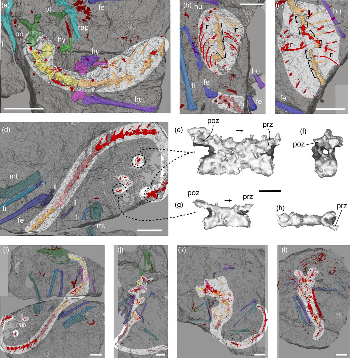
New microCT data on enigmatic Scleromochlus taylori reveal its anatomy & early diverging position within Pterosauromorpha & add important information on pterosaur origins, the 1st group of vertebrates to evolve powered flight. Davide Foffa et al
anatomypubs.onlinelibrary.wiley.com/doi/10.1002/ar…

New microCT data on enigmatic Scleromochlus taylori reveal its anatomy & early diverging position within Pterosauromorpha & add important information on pterosaur origins, the 1st group of vertebrates to evolve powered flight. @davfof et al
anatomypubs.onlinelibrary.wiley.com/doi/10.1002/ar…
#FossilFriday
account_circle