Arghadip Samaddar, MD(@dr_arghadip) 's Twitter Profile Photo

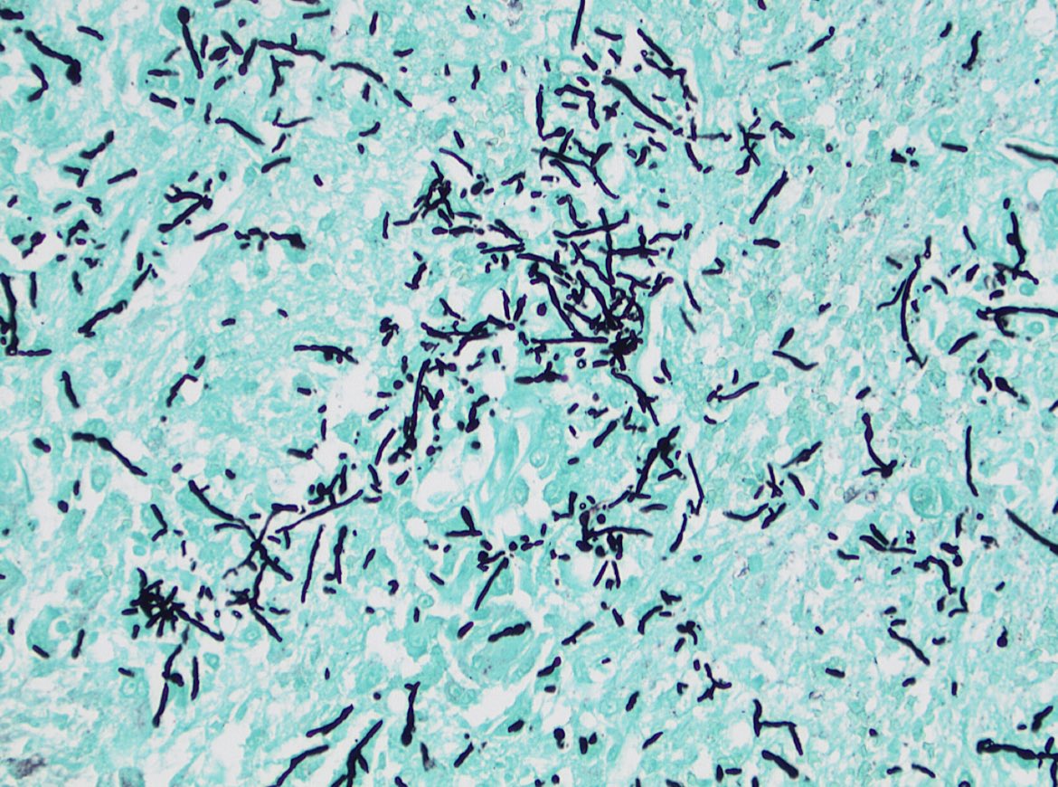
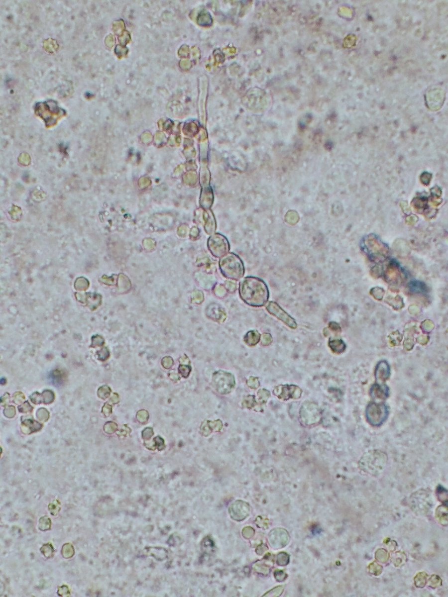
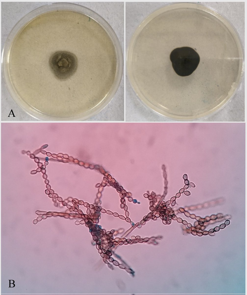
A case of cerebral phaeohyphomycosis caused by Cladosporium cladosporioides. MRI brain: infiltrative lesion in skull base with left IJV thrombophlebitis. KOH mount: pigmented septate fungal hyphae. HPE: brownish black hyphal elements. Patient recovered with Vori + Caspofungin

A case of cerebral phaeohyphomycosis caused by Cladosporium cladosporioides. MRI brain: infiltrative lesion in skull base with left IJV thrombophlebitis. KOH mount: pigmented septate fungal hyphae. HPE: brownish black hyphal elements. Patient recovered with Vori + Caspofungin
account_circle
Fernando A. Messina(@famessina) 's Twitter Profile Photo

π‘·π’‰π’Šπ’‚π’π’π’‘π’‰π’π’“π’‚ π’‚π’Žπ’†π’“π’Šπ’„π’‚π’π’‚

TEAM WORK
doi.org/10.1177/205511…

π‘·π’‰π’Šπ’‚π’π’π’‘π’‰π’π’“π’‚ π’‚π’Žπ’†π’“π’Šπ’„π’‚π’π’‚
#phaeohyphomycosis #Thinkfungus #Feline
 TEAM WORK 
doi.org/10.1177/205511…
account_circle
Arghadip Samaddar, MD(@dr_arghadip) 's Twitter Profile Photo

Cladophialophora bantiana is a neurotropic fungus & the commonest cause of cerebral phaeohyphomycosis. The condition mimics other SOL like tuberculoma & malignancy,making dx challenging.Mortality~70% despite sx & antifungal. Reported an interesting case.
germs.ro/en/Articles/Fa…

Cladophialophora bantiana is a neurotropic fungus & the commonest cause of cerebral phaeohyphomycosis. The condition mimics other SOL like tuberculoma & malignancy,making dx challenging.Mortality~70% despite sx & antifungal. Reported an interesting case.
germs.ro/en/Articles/Fa…
account_circle
Daniel D. Rhoads(@danrhoads) 's Twitter Profile Photo

Phaeohyphomycosis. 200x H&E with granulomas, 1000x GMS with moniliform hyphae, and 1000x H&E with dematiaceous fungus visible within the granuloma.

Phaeohyphomycosis. 200x H&E with granulomas, 1000x GMS with moniliform hyphae, and 1000x H&E with dematiaceous fungus visible within the granuloma.
account_circle
Sandhya Suresh(@Sandyrvsdav) 's Twitter Profile Photo

My poster of a post-transplant recurrent phaeohyphomycosis at .
Interesting to discuss the incidence of this post-Tx fungal infection with nephrologists from other parts of the country.
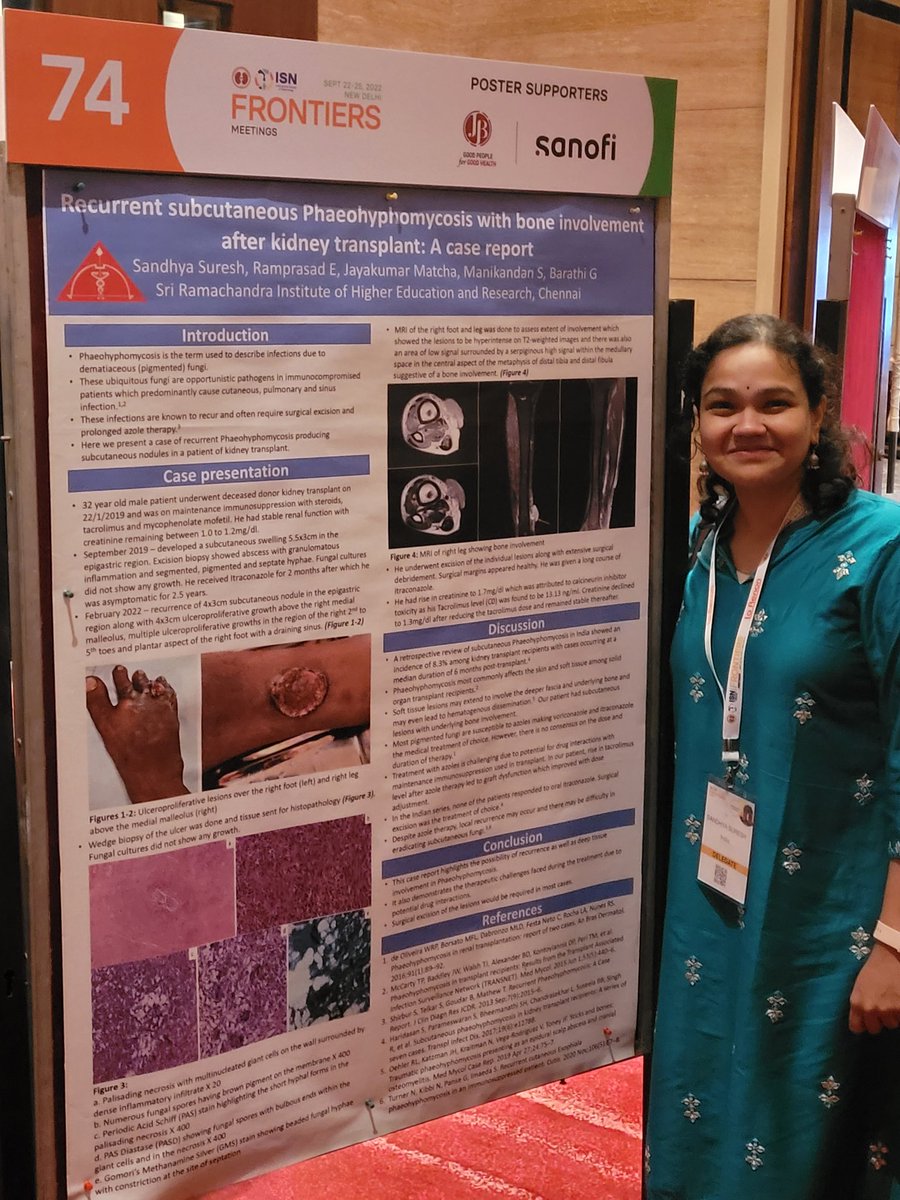
My poster of a post-transplant recurrent phaeohyphomycosis at #ISNFrontiers.
Interesting to discuss the incidence of this post-Tx fungal infection with nephrologists from other parts of the country.
account_circle
Jared T. Ahrendsen MD, PhD (AP/NP/FP)(@JaredAhrendsen) 's Twitter Profile Photo

Cerebral phaeohyphomycosis: brain infection by darkly pigmented dematiaceous fungi. Notice melanin pigment in the fungal walls. Rare, often fatal, and occurrence is unrelated to immune status. 🧠 πŸ”¬

Cerebral phaeohyphomycosis: brain infection by darkly pigmented dematiaceous fungi. Notice melanin pigment in the fungal walls. Rare, often fatal, and occurrence is unrelated to immune status. #neuropath #pathology #purplepath 🧠 πŸ”¬
account_circle
Jose Candido(@JCandidoXavier) 's Twitter Profile Photo

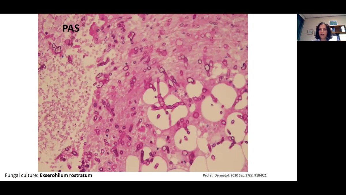
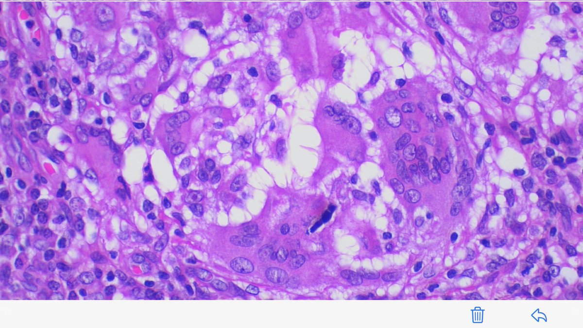
SCC? Phaeohyphomycosis? Pseudoepitheliomatous hyperplasia? Contamination? What should we do when we find an atypical epithelial proliferation with granulomas and hyphae???

SCC? Phaeohyphomycosis? Pseudoepitheliomatous hyperplasia? Contamination? What should we do when we find an atypical epithelial proliferation with granulomas and hyphae??? #pathology #dermpath #patologΓ­a #patologia #dermatopathology #histopathology #dermatology
account_circle
Michael P. Lee MD(@MPLEEMD) 's Twitter Profile Photo

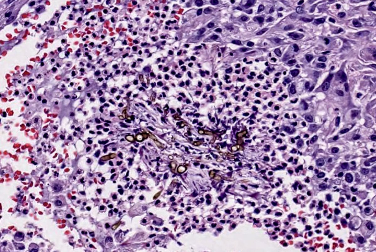
Phaeohyphomycosis diverse group of pigmented molds commonly in soil and wood and enter skin through trauma. Hyphae=phaeohypho, chromo=no hyphae. Cystic cavity - mixed inflammation with giant cells, histiocytes, and neutrophils.

Phaeohyphomycosis diverse group of pigmented molds commonly in soil and wood and enter skin through trauma. Hyphae=phaeohypho, chromo=no hyphae. Cystic cavity - mixed inflammation with giant cells, histiocytes, and neutrophils. #Dermpath #Dermatology #pathology
account_circle
fortheloveofmicrobiology(@fortheloveofmi2) 's Twitter Profile Photo

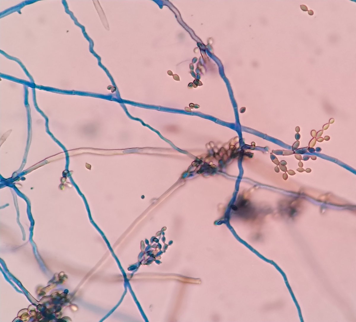
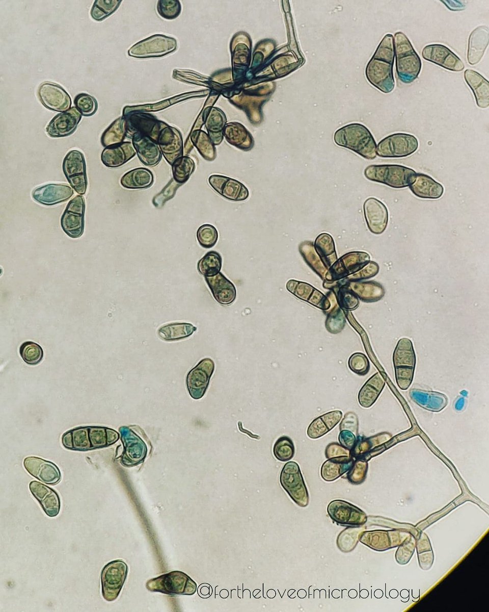
CurvulariaΒ spp on lactophenol blue prep

Etiologic agents of opportunistic infections, most commonly causing keratitis and allergic/chronic sinusitis. Also causes onychomycosis, mycetoma and phaeohyphomycosis at various sites, including subcutaneous tissue and deep tissues.

CurvulariaΒ spp on lactophenol blue prep

Etiologic agents of opportunistic infections, most commonly causing keratitis and allergic/chronic sinusitis. Also causes onychomycosis, mycetoma and phaeohyphomycosis at various sites, including subcutaneous tissue and deep tissues.
account_circle
JAAD Journals(@JAADjournals) 's Twitter Profile Photo

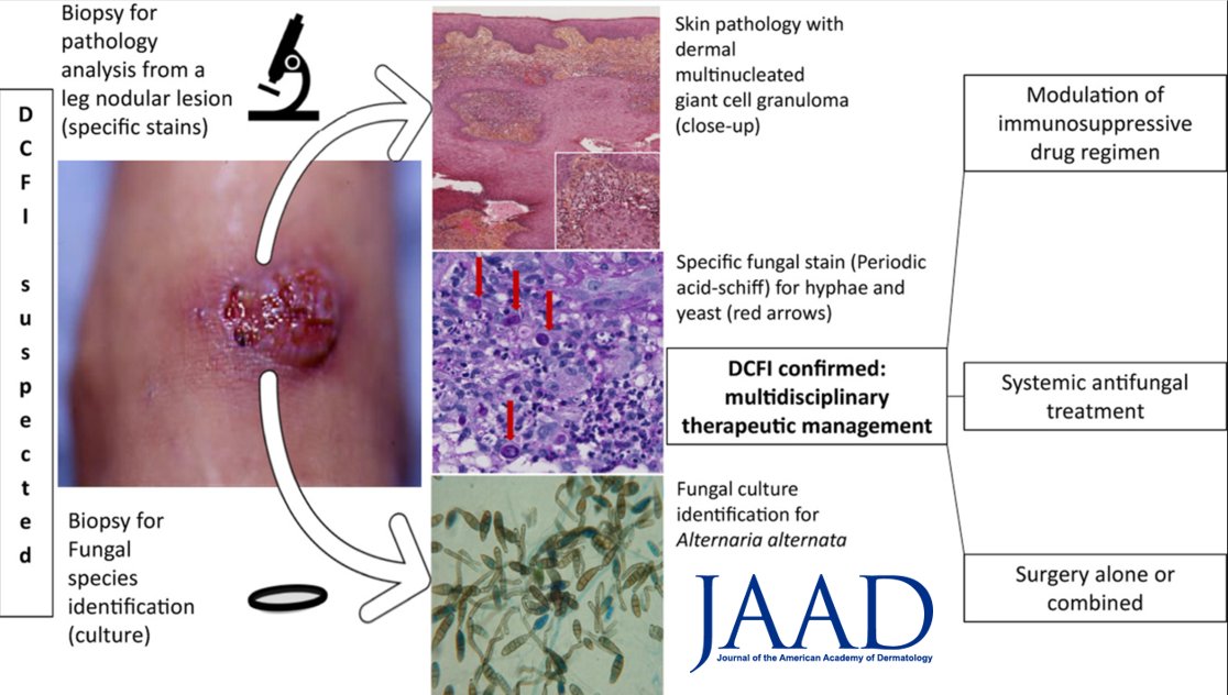
Deep fungal infections in solid-organ transplant recipients, 20-year retrospective study from France with 46 cases.
-Phaeohyphomycosis 45.7%
-Hyalohyphomycosis 17.5%
-Crypto 13%
-Histo 4.3%
Are these frequencies similar to what’s seen at your institution? bit.ly/2OXLWXR

Deep fungal infections in solid-organ transplant recipients, 20-year retrospective study from France with 46 cases.
-Phaeohyphomycosis 45.7%
-Hyalohyphomycosis 17.5%
-Crypto 13%
-Histo 4.3%
Are these frequencies similar to what’s seen at your institution? bit.ly/2OXLWXR
account_circle
Ilana Rosman, MD(@ilanarosman) 's Twitter Profile Photo

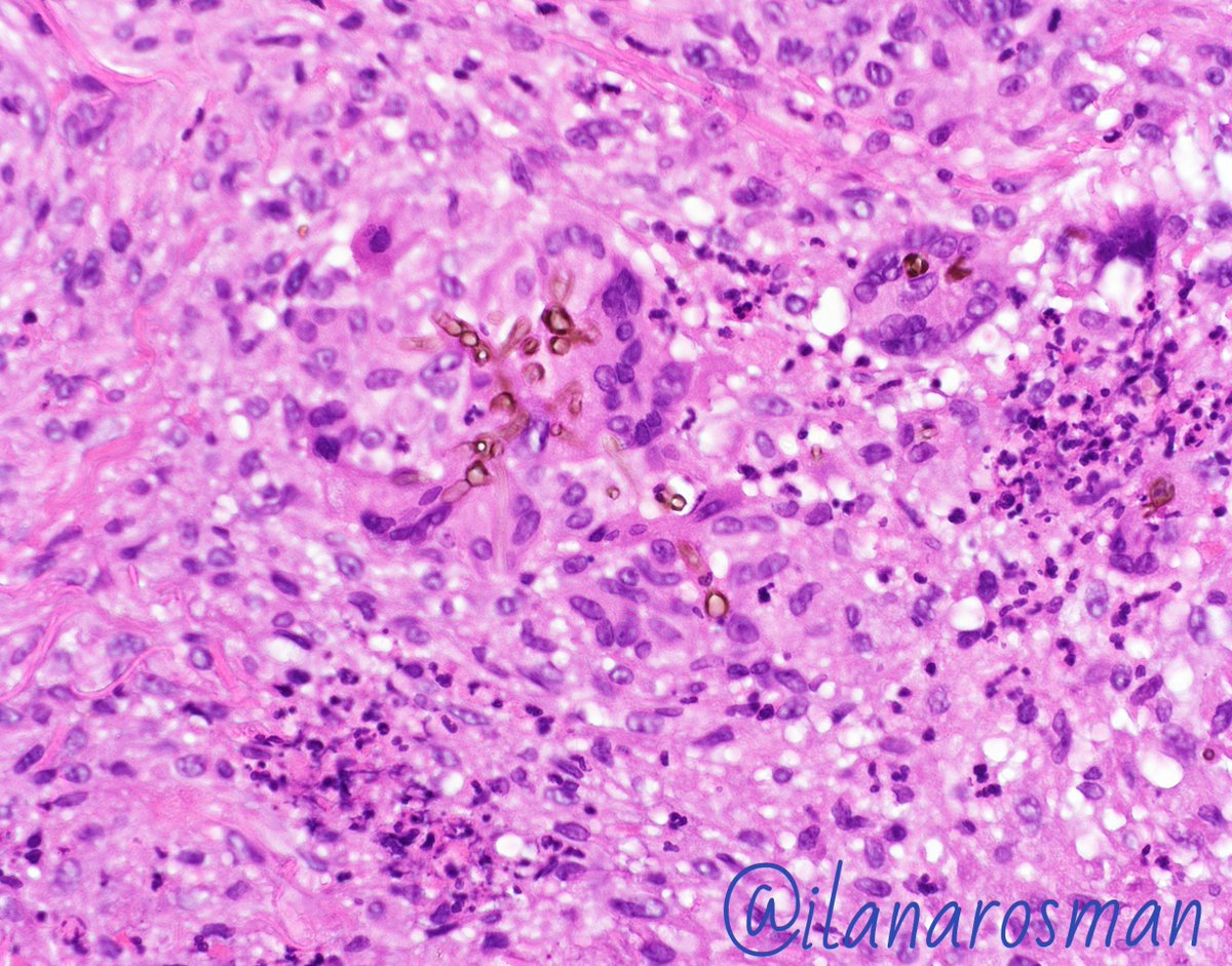
Fungus Friday: this one came in as rule out squamous cell carcinoma on the leg of a 70-something year old. Cutaneous phaeohyphomycosis caused by one of several dematiaceous fungi, typically from inoculation.

Fungus Friday: this one came in as rule out squamous cell carcinoma on the leg of a 70-something year old. Cutaneous phaeohyphomycosis caused by one of several dematiaceous fungi, typically from inoculation. #dermpath #dermatology #pathology #dermtwitter
account_circle
Diego Morales, MD(@DiegoMoralesN) 's Twitter Profile Photo

My mnemonic 2 remember phaeohyphomycosis:

*Phaeo (I think of pheomelanin bc it is pigmented [dematiaceous])

*hypho (bc hyphae predominate & lacks Medlar bodies, as opposed to Chromomycosis) and

*mycosis (bc πŸ„)

All the rest is in this brilliant πŸ§΅πŸ‘‡πŸΌ

My mnemonic 2 remember phaeohyphomycosis:

 *Phaeo (I think of pheomelanin bc it is pigmented [dematiaceous])

*hypho (bc hyphae predominate & lacks Medlar bodies, as opposed to Chromomycosis) and 

*mycosis (bc πŸ„)

All the rest is in this brilliant πŸ§΅πŸ‘‡πŸΌ

#PathTwitter #dermpath
account_circle
SPIN(@spinacademics) 's Twitter Profile Photo

🧠SPIN case of the week & SPIN 2021 case prize recipient: 'Invasive cerebral phaeohyphomycosis and CARD9 deficiency'

Congratulations Dr Leanne Chin (Leanne Chin)!!

Watch now: youtu.be/a8D0qxowxD4 and join us for more at SPIN 2021

🧠SPIN case of the week & SPIN 2021 case prize recipient: 'Invasive cerebral phaeohyphomycosis and CARD9 deficiency'

Congratulations Dr Leanne Chin (@leaaish)!!

Watch now: youtu.be/a8D0qxowxD4 and join us for more at SPIN 2021

#pedineurorad #radres #FOAMrad #MedEd
account_circle
Arghadip Samaddar, MD(@dr_arghadip) 's Twitter Profile Photo

Cerebral Phaeohyphomycosis is a fatal disease known for decades. How can we optimise the diagnostic & treatment strategies? Listen to Alexandre Alanio & Mycology in our upcoming webinar Central Nervous System Infections due to Melanized Fungi. Register here: events.teams.microsoft.com/event/de2e0390…

Cerebral Phaeohyphomycosis is a fatal disease known for decades. How can we optimise the diagnostic & treatment strategies? Listen to @a_alanio & @mycoprakash in our upcoming webinar Central Nervous System Infections due to Melanized Fungi. Register here: events.teams.microsoft.com/event/de2e0390…
account_circle
Karius(@KariusInc) 's Twitter Profile Photo

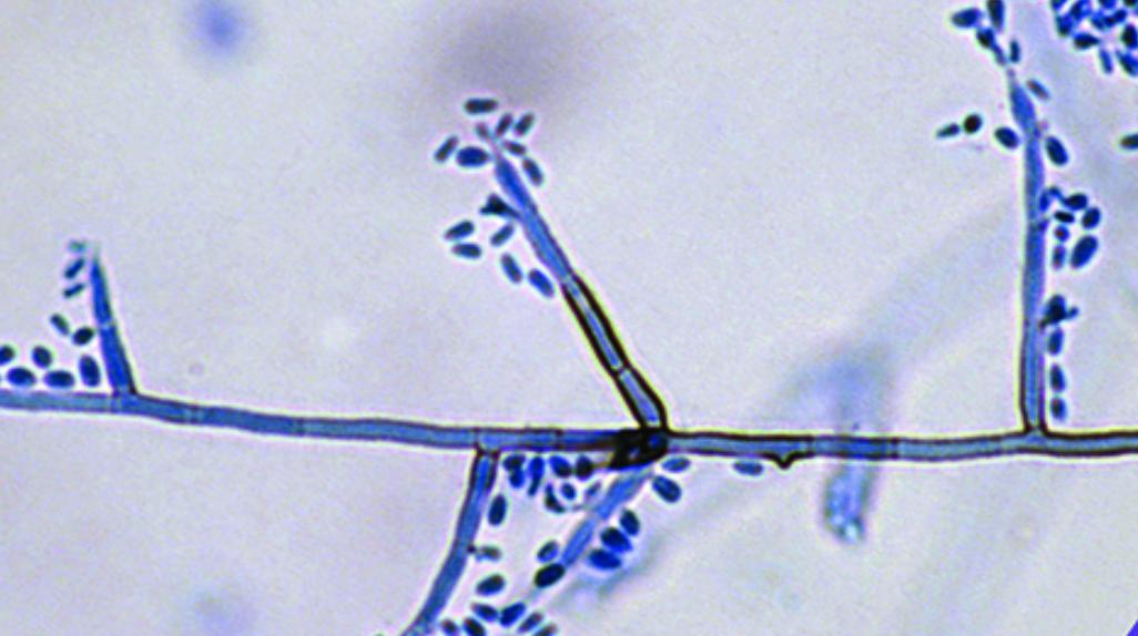
The Karius Test first detected Exophiala spinifera in 2017. This is an anamorphic found in soil and decaying wood. It is associated with and phaeohyphomycosis, and can infect both and immunocompetent individuals.

The Karius Test first detected Exophiala spinifera in 2017. This is an anamorphic #fungus found in soil and decaying wood. It is associated with #mycetoma and phaeohyphomycosis, and can infect both #immunocompromised and immunocompetent individuals.  #PathogenProfile #mcfDNA
account_circle
Michael P. Lee MD(@MPLEEMD) 's Twitter Profile Photo

Phaeohyphomycosis. This is an infection caused by dermatiaceous - darkly melanin pigmented - fungi. These organisms are naturally found in soil and wood. Local trauma is usually the method of infection. Jerad Gardner, MD Sara Shalin Nicole D. Riddle, MD, MSHI, FCAP (she/her)
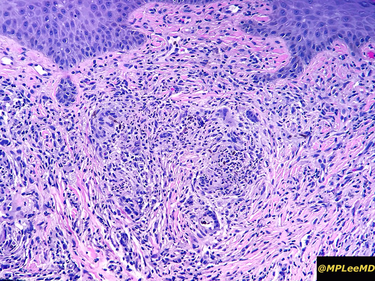
Phaeohyphomycosis. This is an infection caused by dermatiaceous - darkly melanin pigmented - fungi. These organisms are naturally found in soil and wood. Local trauma is usually the method of infection. #dermatology #dermpath #fungi @JMGardnerMD @MightyDermPath  @NRiddleMD
account_circle
Journal of Neurosurgery(@TheJNS) 's Twitter Profile Photo

: An illustrative case demonstrating the utility of liquid biopsy in diagnosing isolated cerebral phaeohyphomycosis.
thejns.org/caselessons/vi…
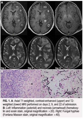
#JNSCaseLessons: An illustrative case demonstrating the utility of liquid biopsy in diagnosing isolated cerebral phaeohyphomycosis.
thejns.org/caselessons/vi…
account_circle