

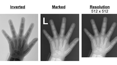
Evaluating the Robustness of a #DeepLearning Bone Age Algorithm to Clinical Image Variation Using Computational Stress Testing doi.org/10.1148/ryai.2… Samantha Santomartino PaulYiMD Vishwa Parekh #PedsRad #AI #MachineLearning


Computational 'stress tests' evaluated robustness of bone age #DL model to simulated real-world image variations doi.org/10.1148/ryai.2… Samantha Santomartino Elham Beheshtian Vishwa Parekh #PedsRad #MSKRad #ML

Follow along with Teddy’s story and learn about the people and places you’ll see while getting an x-ray, CT, and ultrasound! Dr. Sherry Wang presents all three videos in English and Mandarin at bit.ly/3aEbZBp 🐻 #RadInfo4Kids #PedsRad #RadiologyInfo


Evaluating the Robustness of a #DeepLearning Bone Age Algorithm to Clinical Image Variation Using Computational Stress Testing doi.org/10.1148/ryai.2… Samantha Santomartino Vishwa Parekh UM2ii Center #PedsRad #AI #MachineLearning
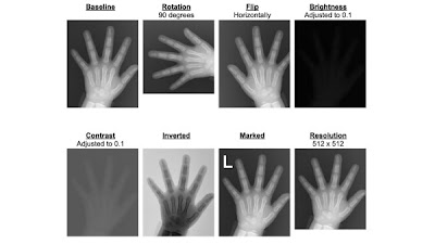


Shahriar Faghani MD @slowvak highlight the need for meticulous stress testing of #DeepLearning models for medical imaging doi.org/10.1148/ryai.2… MayoAILab #PedsRad #robustness #AI


Don’t miss this webinar from The Society for Pediatric Radiology on career development with #NemoursRad Dr Kathleen Schenker, MD as a panelist! #RadRes #RadFellow #pedsrad

Computational 'stress tests' evaluated robustness of bone age #DL model to simulated real-world image variations doi.org/10.1148/ryai.2… Elham Beheshtian PaulYiMD Vishwa Parekh #BoneAge #PedsRad #MachineLearning


Stress testing on an award-winning #DeepLearning model for bone age doi.org/10.1148/ryai.2… Shahriar Faghani MD MayoAILab Brad Erickson #PedsRad #BoneAge #MachineLearning


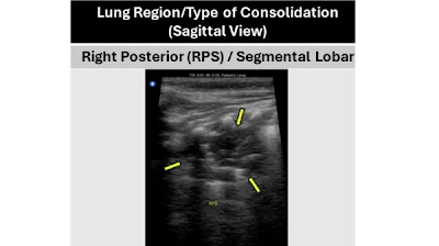
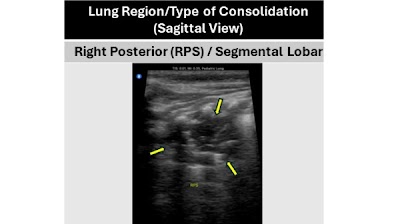
Dataset of lung ultrasound acquired with a #POCUS device in Zambian children with clinical symptoms of pneumonia doi.org/10.1148/ryai.2… Boston University School of Public Health Ultrasound Laboratory Trento MCG #PedsRad #DeepLearning #ML


Dataset of lung ultrasound acquired with a #POCUS device in Zambian children with clinical symptoms of pneumonia doi.org/10.1148/ryai.2… Boston University School of Public Health Boston Medical Center MCG #PedsRad #AI #MachineLearning
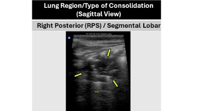


Curated Dataset of Lung US Images of Zambian Children with Clinical Pneumonia Containing Annotations for Consolidation Patterns doi.org/10.1148/ryai.2… Boston University School of Public Health Boston Medical Center MCG #PedsRad #POCUS #ML
