SVI Huygens (@svi_huygens) 's Twitter Profile
SVI Huygens

@svi_huygens

Pushing the limits of image resolution and quality with #deconvolution, restoration, visualization and analysis.
Test Huygens from: svi.nl/download

ID: 39725174

linkhttp://www.svi.nl calendar_today13-05-2009 10:25:25

771 Tweet

1,1K Followers

1,1K Following

SVI Huygens (@svi_huygens) 's Twitter Profile Photo

Matrix STED image reveals detail of Nuclear pore complex protein fluorescence using @abberior STAR-RED after Huygens unique SuperXY #deconvolution. Thanks to Marie Olšinová & Aleš Benda, Imaging Methods Core Facility – BIOCEV Charles University for providing the raw data

SVI Huygens (@svi_huygens) 's Twitter Profile Photo

Our latest research on how image quality can affect colocalization analysis in Microscopy Today: academic.oup.com/mt/article/32/… Thank you to our authors: R Pelle Faculty of Science, Utrecht University, M Ram, P Hemmerich, AL Bottomly, Louise Cole, R Berlinguer-Palmini, HTM, NTM vd Voort, V Schoonderwoert

SVI Huygens (@svi_huygens) 's Twitter Profile Photo

A cryostat muscle section, immunostained for various myosin isoforms, was imaged as a 4-color z-stack tile series. Congrats to Dr. Daniela Malide, Dr. Yingfan Zhang, NHLBI Light microscopy core facility, NIH Bethesda for their prize-winning image. #ScienceArt

A cryostat muscle section, immunostained for various myosin isoforms, was imaged as a 4-color z-stack tile series.

Congrats to Dr. Daniela Malide, Dr. Yingfan Zhang, NHLBI Light microscopy core facility, <a href="/NIH/">NIH</a> Bethesda for their prize-winning image.

#ScienceArt
SVI Huygens (@svi_huygens) 's Twitter Profile Photo

Congratulations to Dr Kseniia Bondarenko, Institute of Immunology and Infection Research, Edinburgh Uni for this inredible image! It shows an acute-stage, overgrown vacuoles filled with Toxoplasma parasites. See how she described it herself here: linkedin.com/posts/ksena-bo…

Congratulations to Dr Kseniia Bondarenko, Institute of Immunology and Infection Research, Edinburgh Uni for this inredible image!

It shows an acute-stage, overgrown vacuoles filled with Toxoplasma parasites.

See how she described it herself here:

linkedin.com/posts/ksena-bo…
SVI Huygens (@svi_huygens) 's Twitter Profile Photo

Large biological samples often require stitching to create high-resolution images of the entire sample. 🔬 Join our free webinar February 18th to learn more about Tile stitching and deconvolution. Sign up here: svi.nl/Webinarinvitat…. #microscopy #webinar #Science

SVI Huygens (@svi_huygens) 's Twitter Profile Photo

A team from Queensland Brain Institute, UQ, UQ Research Computing Centre, and SVI Huygens has harnessed the power of advanced HPC-GPUs  to enhance microscopy image quality. This breakthrough accelerates scientific discoveries in brain development and disorders at QBI, paving the way for new insights. NVIDIA

A team from <a href="/QldBrainInst/">Queensland Brain Institute, UQ</a>, <a href="/RCCUQ/">UQ Research Computing Centre</a>, and <a href="/svi_huygens/">SVI Huygens</a> has harnessed the power of advanced HPC-GPUs  to enhance microscopy image quality. This breakthrough accelerates scientific discoveries in brain development and disorders at QBI, paving the way for new insights.
<a href="/nvidia/">NVIDIA</a>
SVI Huygens (@svi_huygens) 's Twitter Profile Photo

Who left these microscopic fingerprints? Depth-encoded coloring of Drosophila melanogaster nephrocytes showing the typical "fingerprint" pattern on their surface. Sample preparation by Johanna Odenthal (Nephrolab Cologne) and Dr. Christian Jüngst, Christian Jüngst CECAD Cologne

Who left these microscopic fingerprints?
Depth-encoded coloring of Drosophila melanogaster nephrocytes showing the typical "fingerprint" pattern on their surface. Sample preparation by Johanna Odenthal (Nephrolab Cologne) and Dr. Christian Jüngst, <a href="/ChrisJ90/">Christian Jüngst</a> <a href="/CECAD_/">CECAD Cologne</a>
SVI Huygens (@svi_huygens) 's Twitter Profile Photo

Live Webinar, March 18th: Matrix STED deconvolution with Huygens. Nuclear pore complex proteins images with Abberior FL + Matrix Detector and 100% 3D #STED, Huygens deconvolved and visualized. Courtesy: Chi Leung So, PTC International Ltd. Hongkong. Join svi.nl/webinarinvitat…

SVI Huygens (@svi_huygens) 's Twitter Profile Photo

Join our 3-hour hands-on workshop for beginners on May 15, 2025! 🎉 - Two sessions to accommodate all time zones! - Free manual, Huygens Everywhere trial, and demo images included! - Perfect for beginners eager to learn image processing and analysis. svi.nl/Huygens-Worksh…

Join our 3-hour hands-on workshop for beginners on May 15, 2025! 🎉

- Two sessions to accommodate all time zones!

- Free manual, Huygens Everywhere trial, and demo images included!

- Perfect for beginners eager to learn image processing and analysis.

svi.nl/Huygens-Worksh…
SVI Huygens (@svi_huygens) 's Twitter Profile Photo

Automatically process all your microscopy data (confocal, widefield, STED, etc.) with optimal deconvolution! Join our webinar "Image Restoration and Analysis Pipelines with the Huygens Workflow Processor" on April 22nd at 8:00 or 18:00 CEST: svi.nl/WebinarSchedule.

SVI Huygens (@svi_huygens) 's Twitter Profile Photo

Interested to increase signal 7x and resolution by > 2x? Raw and deconvolved MIP rendering of a two channel tubulin labeled COS7 cell image, acq. w\ Olympus IXplore SpinSR10. Thanks to Nicolas Schilling & Joana Delgado Martins, Center for Microscopy and Image Analysis Universität Zürich

SVI Huygens (@svi_huygens) 's Twitter Profile Photo

(Aspiring) microscopists, learn more about image restoration, deconvolution and analysis during our hands-on 3h free workshop May 15th!

SVI Huygens (@svi_huygens) 's Twitter Profile Photo

NEW: AI-powered segmentation with Meta’s Segment Anything Model 2 (SAM2) is now in Huygens Object Analyzer! No coding needed—toggle between classic & AI for best results. Built-in 3D visualization, object stats & top-tier image restoration. Try it: 👉 svi.nl/lLicenserequest

SVI Huygens (@svi_huygens) 's Twitter Profile Photo

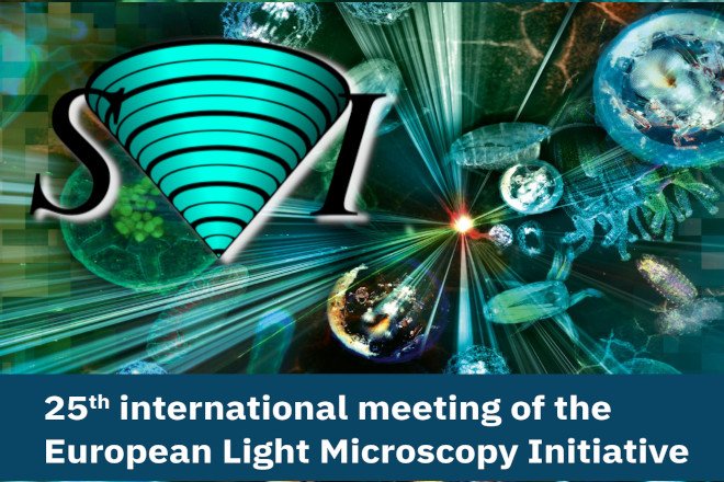
Attending #ELMI2025? Join us at booth F17, and have a chat with Wouter & Melvin about the new AI-segmentation option in Huygens Object Analyzer, and how to import, restore, quality check, and analyze all your microscopy image data with the Huygens Software. EMBL EMBL Events

Attending #ELMI2025? Join us at booth F17, and have a chat with Wouter &amp; Melvin about the new AI-segmentation option in Huygens Object Analyzer, and how to import, restore, quality check, and analyze all your microscopy image data with the Huygens Software.

 <a href="/embl/">EMBL</a> <a href="/EMBLEvents/">EMBL Events</a>
SVI Huygens (@svi_huygens) 's Twitter Profile Photo

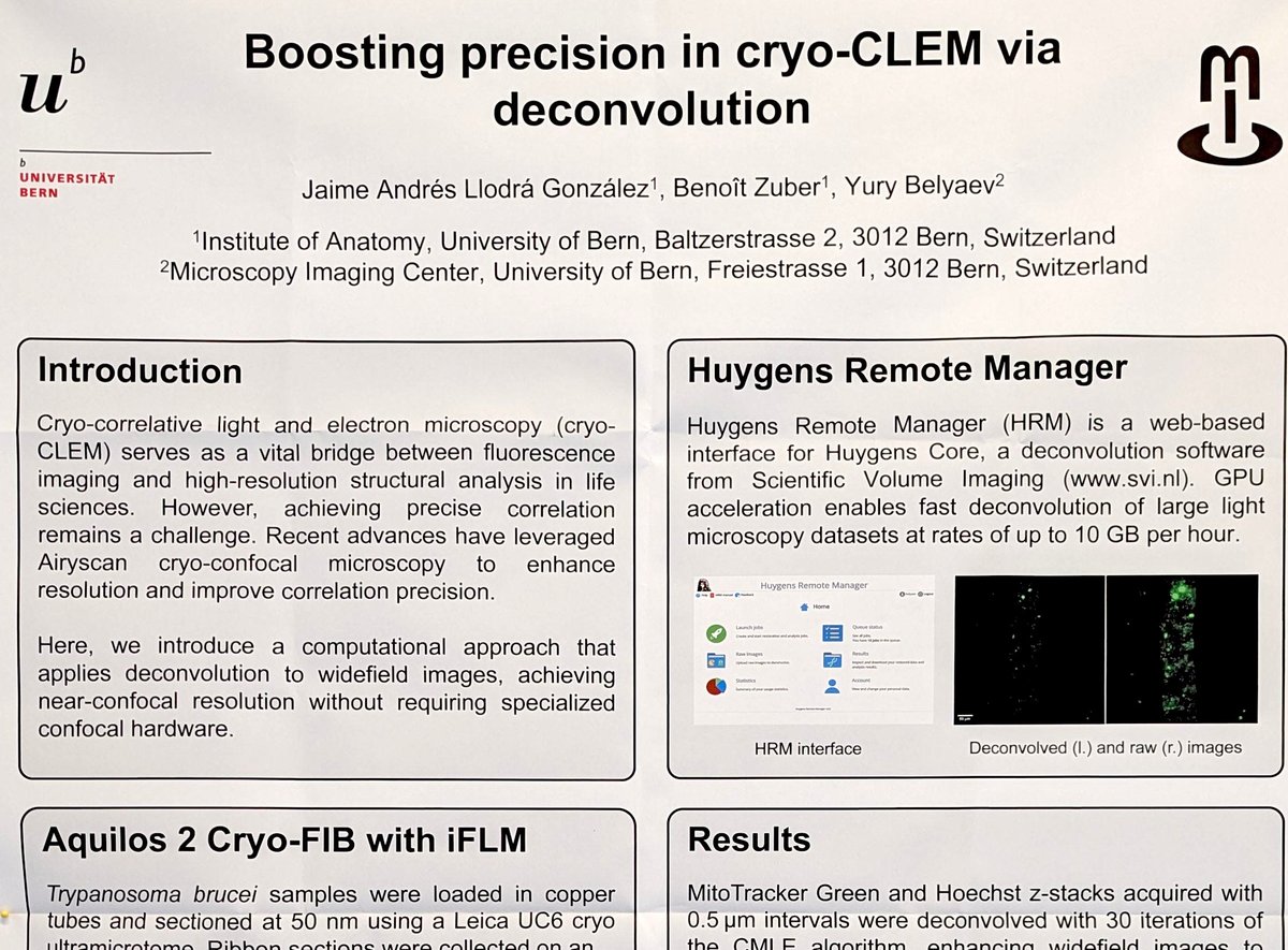
Increasing accuracy of CLEM by reaching near confocal quality from widefield images using Huygens Remote Manager (huygens-rm.org/wp/) as a web-based for deconvolution. An interesting poster #ELMI2025 from the Dept of Anatomy and MIC @MedFacultyUniBEn.

Increasing accuracy of CLEM by reaching near confocal quality from widefield images using Huygens Remote Manager (huygens-rm.org/wp/) as a web-based for  deconvolution. An interesting poster #ELMI2025 from the Dept of Anatomy  and MIC @MedFacultyUniBEn.
SVI Huygens (@svi_huygens) 's Twitter Profile Photo

#Superresolution and Huygens deconvolution discriminates “between ‘true’ and ‘false’ nuclear colocalization. Want to see more? Follow #AccurateColocalizationAnalysis, read doi.org/10.1093/mictod… and Register for our live webinar at svi.nl/webinarinvitat… #fuoresencefriday

#Superresolution and Huygens deconvolution discriminates “between ‘true’ and ‘false’ nuclear colocalization.

Want to see more? Follow #AccurateColocalizationAnalysis, read doi.org/10.1093/mictod… and Register for our live webinar at svi.nl/webinarinvitat…

#fuoresencefriday