Sebastian Isaza
@suizacolombiana
MSK Radiologist
ID:219487714
25-11-2010 00:36:34
113 Tweets
242 Followers
165 Following
 on Twitter photo 2024-05-12 23:11:24 Double Trouble. Lateral and medial meniscus Bucket Handle Tear, in the setting of ACL rupture. Double PCL sign: MM bucket handle Double anterior horn sign: LM bucket handle")
 on Twitter photo 2024-05-11 21:53:07 The A.L.L always gives me Joy. Acute ACL rupture Grade I ALL lesion Lateral femoral condyle contusion (pivot shift mechanism)")
 on Twitter photo 2024-05-10 20:36:21 Wrisber Rip - longitudinal vertical tear at the junction of ligament of Wrisberg and posterior horn of the LM")
 on Twitter photo 2024-05-10 20:30:26 FBI - Fat Blood Interface Lipohemartrosis")
 on Twitter photo 2024-05-07 01:10:56 31 y/o Male. Motorcycle accident. Acute meniscotibial ligament rupture")
 on Twitter photo 2024-05-02 21:39:26 13 y/o. Rule out 5th MTT base mass. Iselin disease - apophysitis of the base of the 5th metatarsal. Edema in STIR and FSPD. Low signal in T1.")
 on Twitter photo 2024-05-02 15:12:14 Recreational Cyclist. Lateral knee pain. Iliotibial band syndrome: High signal in STIR due to Inflammation of the fat adjacent to the iliotibial band.")
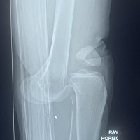
 on Twitter photo 2024-04-13 13:46:59 Deep Sulcus sign of the Knee. ACL rupture sign in XRays. Caused by Pivot Shift trauma mechanism")
 on Twitter photo 2024-04-12 14:56:41 Deep venous thrombosis (DVT) of medial astrocnemius vein. Axial PDFS. Distended gastrocnemius vein (arrow) with increased signal in the medial head of the gastrocnemius muscle")
 on Twitter photo 2024-04-11 20:32:56 So called Proximal ITB syndrome. More of a enthesopathy, with thickening and microtearring of the origin of the ITB. Prominent edema and inflamatory response around it. Looks painful.")
 on Twitter photo 2024-04-10 14:59:17 Medial Gastrocnemius origin avulsion fracture. MCL and MPFL Avulsion fracture Medial capsular defect")
 on Twitter photo 2024-03-18 14:29:15 Segond fx. ALL avulsion fx. When present, anterolateral ligament is clearly depicted as the hipointense lineal structure attaching to the bone fragment. Of course, ACL is torn. #msk #knee")
 on Twitter photo 2024-03-17 18:29:52 PLC Injury Tear of LCL and Popliteal tendon. Ill defined arcuate lig. Partial Thickness tear ACL.")
 on Twitter photo 2024-03-12 01:45:08 Something is missing ... Greater tuberosity avulsion fracture. Concave well defined defect superolateral margin of humeral head. T1 imaging reveals bone fragment attached to Supra and infra tendons, with displacement and retraction. #shoulder #msk #mskrad")
 on Twitter photo 2024-03-08 00:20:10 Walch B2 glenoid dysplasia and glenohumerar OA Graphic from DOI10.1007/s00590-012-1119-4")
 on Twitter photo 2024-03-07 16:31:52 Rings, arcs and pop corns. Characteristic of chondroid lesions. Chondrosarcoma of the left Shoulder Girdle")
 on Twitter photo 2024-03-06 22:39:43 Peripheral nerve sheath tumor High signal fusiform mass with tapered ends, with posterior tibialis nerve leading into and out of the mass")
@suizacolombiana
MSK Radiologist
ID:219487714
25-11-2010 00:36:34
113 Tweets
242 Followers
165 Following
 on Twitter photo 2024-03-06 21:37:40 Lisfranc Injury - Weightbearing AP XR: asymmetric widening of intermetatarsal space - CT: Fracture of lateral aspect of C1 caused by avulsion of Lisfranc ligament (C1-M1) - MR: Lisfranc ligament appears preserved and cortical C1 avulsion is inferred.")