Radiology: Cardiothoracic Imaging
@radiologycti
Insights from Radiology: Cardiothoracic Imaging’s associate social media editors, in collaboration with Suhny Abbara, MD; Editor. Follow @radiology_rsna
ID: 1357830447670116364
05-02-2021 23:16:48
200 Tweet
2,2K Takipçi
246 Takip Edilen

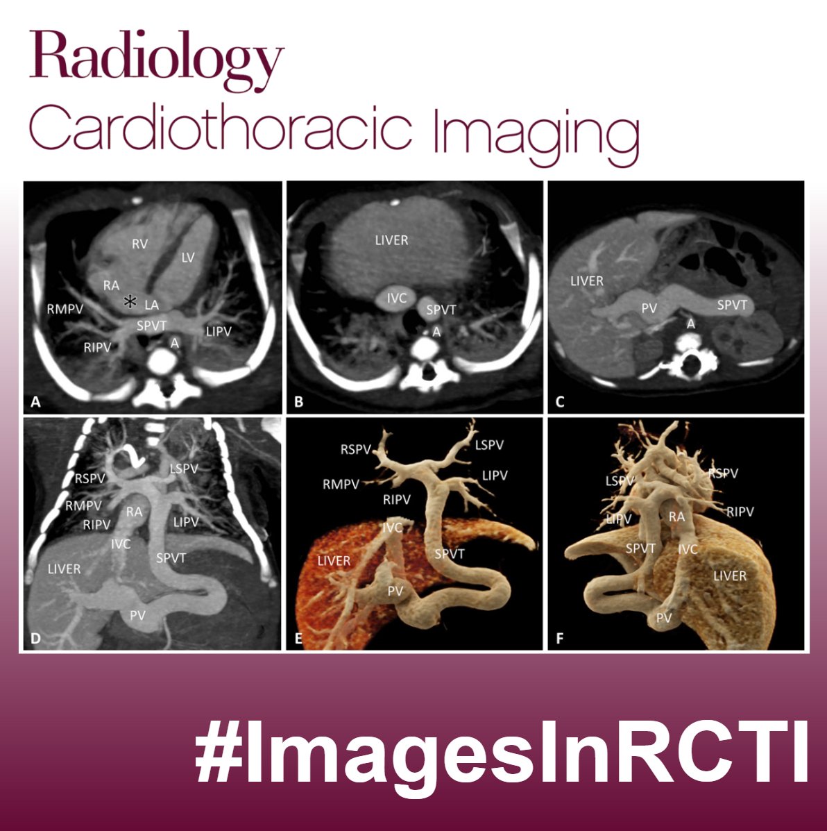
Excited to launch #ImagesInRCTI and #InTrainingRCTI posts, highlighting Images in Publications from Radiology: Cardiothoracic Imaging ! 🫀🫁🚀 1/2) An 11-day-old male neonate presented with cyanosis and dyspnea What is the diagnosis? What are the findings?
Honored to be selected as a board member of the Radiology: Cardiothoracic Imaging editorial board! Excited to develop my editorial skills and deepen my knowledge in cardiac imaging. Looking forward to contributing to the field and learning from esteemed mentors. Radiology: Cardiothoracic Imaging


Can MR elastography be used as an early biomarker of heart failure in women?🚨 📢 check the latest paper by Arani et al. pubs.rsna.org/doi/epdf/10.11… #InTrainingRCTI #TweetorialRCTI #CardiacImaging #Cvimaging #MedTwitter Mayo Clinic Radiology Radiology: Cardiothoracic Imaging

Happy to share our review highlighting adverse cardiovascular health effects of climate change and opportunities to improve sustainability in cardiac imaging 🌎🩻❤️🔥 Led by outstanding Radiology: Cardiothoracic Imaging TEB deputy editor Suvai Gunasekaran 😊 pubs.rsna.org/doi/10.1148/ry… @HeartDocSubha MIЯVΛƬ #IC


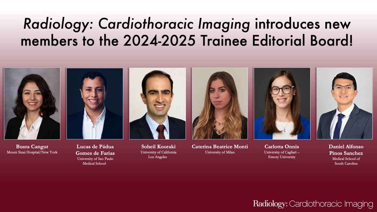
Congratulations to the incoming cohort of Radiology: Cardiothoracic Imaging trainee editorial board members! Welcome to the #InTrainingRCTI team ☺️ Busra Cangut, MD, MS Lucas de Pádua, MD PhD Soheil Kooraki, MD Carlotta Onnis Daniel P. Daniel Vargas, MD Ritu R Gill MD, MPH FACR @bdallen6 Dianna Bardo Suhny Abbara MD Aprateem Mukherjee, MBBS, MD, DM, FRCR RSNA

We're back with another #InTrainingRCTI #ImagesInRCTI challenge highlighting Images in Publications from Radiology: Cardiothoracic Imaging !! 🫀📷💪
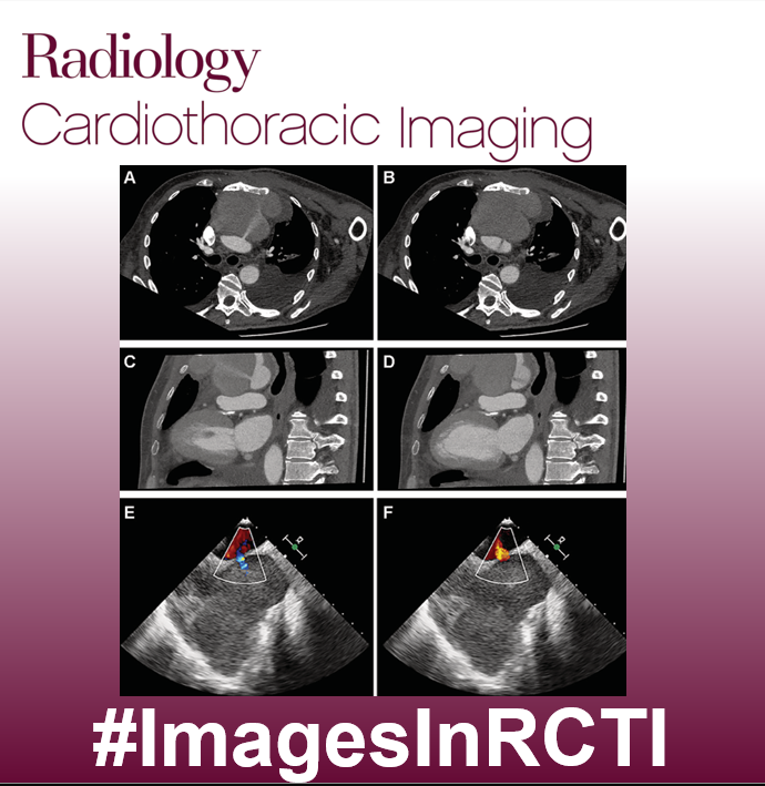

Feel like tackling a mid-week image challenge? A 20-year-old male individual was admitted due to dyspnea on exertion and easy fatigability. What is the diagnosis? What are the findings? #InTrainingRCTI #ImagesInRCTI Radiology: Cardiothoracic Imaging


🚨 RCTI Image Challenge !🚨 1/2) A 28-year-old male patient with antisynthetase syndrome with dermatopolymyositis, polyarthritis, and dyspnea. What is the diagnosis? What are the findings? #InTrainingRCTI #ImagesInRCTI Radiology: Cardiothoracic Imaging


📢 Radiology: Cardiothoracic Imaging is preparing a Special Collection on Quantitative Imaging. Submit your research on: ☑️quantitative CT plaque analysis, ☑️intracardiac 4D flow, ☑️lung disease/fibrosis measurement, ☑️quantitative myocardial perfusion, ☑️myocardial strain analysis, and more! 👇🏻

NEW PODCAST EP 🔊Cardiovascular Imaging and Environmental Sustainability 🔷Intersection of CV imaging and sustainability with expert guests Suvai Gunasekaran and Kate Hanneman ⛓️rsnaradiologycti.libsyn.com/episode-27-car…

🫀 New In a Minute Video 🎥 highlighting “Improved Detection of Small and Low-Density Plaques in Virtual Noncontrast Imaging–based Calcium Scoring at Photon-Counting Detector CT” by Fink et al. Radiology: Cardiothoracic Imaging #InTrainingRCTI #InAMinuteRCTI

It's #InternationalPodcastDay! Check out these podcasts from Radiology, RadioGraphics, Radiology: Artificial Intelligence, Radiology: Cardiothoracic Imaging and RadIC_Editor. bit.ly/RYpodcasts bit.ly/RGpodcasts bit.ly/RadAiPodcasts bit.ly/RYCardioPodcas… bit.ly/RadICpod


🚨 Ready to test your skills again? 🚨 57-years-old male with progressive worsening scrotal edema. Findings? Diagnosis? #InTrainingRCTI #ImagesInRCTI Radiology: Cardiothoracic Imaging
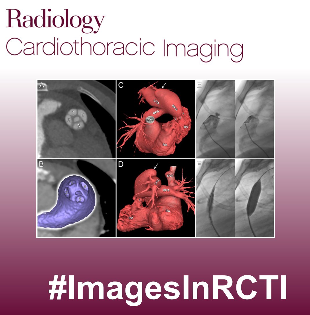


This week’s Radiology: Cardiothoracic Imaging tweetorial brings you a special look at MV geometry after cardiac resynchronization (CS). We take a deeper look at changes in MV structure after CS in detail never seen before with #YesCCT. (1/10) pubs.rsna.org/doi/10.1148/ry… #InTrainingRCTI #TweetorialRCTI



🚨 New episode of the Radiology: Cardiothoracic Imaging podcast is now available! 🚨 Dr. Ranganath talks with Drs.England, Richard Friesen, Brown, Barker from CU Pediatric Radiology about using 4D Flow MRI at 3T to advance fetal cardiac imaging and improve prenatal care. bit.ly/4mjP7HM


