Radiology (@radiology_rsna) 's Twitter Profile
Radiology

@radiology_rsna

A monthly journal devoted to clinical radiology and allied sciences, owned and published by RSNA.

Follow @RSNA for organizational updates from RSNA.

ID: 31073292

linkhttp://radiology.rsna.org/ calendar_today14-04-2009 06:26:09

4,4K Tweet

58,58K Followers

359 Following

Radiology (@radiology_rsna) 's Twitter Profile Photo

Check out this editorial by Dr. Midot, an abdominal and vascular interventional radiologist from Lyon, France, on the study by Williams and Omer et al that assessed restaging MRI in patients of the OPRA trial. bit.ly/4ebcJJQ

Radiology (@radiology_rsna) 's Twitter Profile Photo

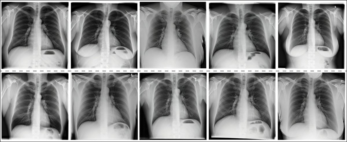
In this study of patients in the Boston Lung Cancer Study, Hata et al built an automated model and assessed its performance in estimating the probability of interstitial lung abnormality. bit.ly/3Tfnse6

In this study of patients in the Boston Lung Cancer Study, Hata et al built an automated model and assessed its performance in estimating the probability of interstitial lung abnormality. bit.ly/3Tfnse6
Radiology (@radiology_rsna) 's Twitter Profile Photo

In this targeted editorial, Dr. Zagurovskaya from IU Radiology and Imaging Sciences comments on the use of machine learning on prediction of interstitial lung abnormality and summarizes the study by Hata et al. bit.ly/4cUGwFw

Radiology (@radiology_rsna) 's Twitter Profile Photo

Check out this original research study by Ramtohul et al from Paris, France, which uses radiomics scores to assess for predictive features of treatment response in triple-negative breast cancer, based on pretreatment perfusion MRI. bit.ly/3Xulfhf

Check out this original research study by Ramtohul et al from Paris, France, which uses radiomics scores to assess for predictive features of treatment response in triple-negative breast cancer, based on pretreatment perfusion MRI. bit.ly/3Xulfhf
Radiology (@radiology_rsna) 's Twitter Profile Photo

Dr. Rauch from MD Anderson Cancer Center comments on the biomarkers in the study by Ramtohul et al and future directions of research in personalized neoadjuvant chemotherapy for triple-negative breast cancer. bit.ly/3zdL2Ry

Radiology (@radiology_rsna) 's Twitter Profile Photo

The parallel #HOPE4LIVER trials demonstrated the safety and technical success of the HistoSonics System for the destruction of liver tumors using histotripsy. UW Radiology Michigan Radiology Mishal Mendiratta-Lala Tim bit.ly/3XenaFv

The parallel #HOPE4LIVER trials demonstrated the safety and technical success of the HistoSonics System for the destruction of liver tumors using histotripsy. <a href="/UWiscRadiology/">UW Radiology</a> <a href="/UMichRadiology/">Michigan Radiology</a> <a href="/MishalLala/">Mishal Mendiratta-Lala</a> <a href="/Tim_Ziemlewicz/">Tim</a>  bit.ly/3XenaFv
Radiology (@radiology_rsna) 's Twitter Profile Photo

This study defines expected US elastography measurement variability to guide clinical interpretation, enable diagnostic enrichment in clinical trials, and support FDA biomarker validation for NAFLD. UW Radiology Mass General Imaging Mass General Research Institute bit.ly/4d1UqG3

This study defines expected US elastography measurement variability to guide clinical interpretation, enable diagnostic enrichment in clinical trials, and support FDA biomarker validation for NAFLD. <a href="/UWRadiology/">UW Radiology</a> <a href="/MGHImaging/">Mass General Imaging</a> <a href="/MGH_RI/">Mass General Research Institute</a> bit.ly/4d1UqG3
Radiology (@radiology_rsna) 's Twitter Profile Photo

Does photon-counting CT have an application in lung transplant? Ultralow-dose photon-counting CT yielded appropriate image quality to show anatomic structures and detect interstitial lung abnormalities after transplant. bit.ly/4ejcjRG

Does photon-counting CT have an application in lung transplant? Ultralow-dose photon-counting CT yielded appropriate image quality to show anatomic structures and detect interstitial lung abnormalities after transplant. bit.ly/4ejcjRG
Radiology (@radiology_rsna) 's Twitter Profile Photo

🚨 A new episode of the Radiology podcast is now available! 🚨 Linda Chu, MD discusses the evolving role of neuroimaging in Alzheimer's care with Drs. Esposito, Torres-Yaghi, and Wood. Sponsored by GE HealthCare bit.ly/4gsdIHB

Radiology (@radiology_rsna) 's Twitter Profile Photo

In this targeted editorial, Dr. Ciet provides background info on lung transplant and discusses how photon-counting CT can revolutionize follow-up for this intervention. bit.ly/47FAGXL

Radiology (@radiology_rsna) 's Twitter Profile Photo

A VI-RADS developed based on features on contrast-enhanced US images acquired via transabdominal and intracavity approaches showed good performance and interreader agreement for the assessment of muscle invasion in patients with bladder cancer. bit.ly/3XDuHiw

A VI-RADS developed based on features on contrast-enhanced US images acquired via transabdominal and intracavity approaches showed good performance and interreader agreement for the assessment of muscle invasion in patients with bladder cancer. bit.ly/3XDuHiw
Radiology (@radiology_rsna) 's Twitter Profile Photo

In this targeted editorial, Dr. Morrell of @uofuradiology discusses the benefits and shortcomings of reporting systems and how the contrast-enhanced US VI-RADS developed by Han and Lin et al may help with assessment of muscle invasion in bladder cancer. pubs.rsna.org/doi/10.1148/ra…

Radiology (@radiology_rsna) 's Twitter Profile Photo

Drs. Santiago and Joshua Shur discuss in this editorial the retrospective substudy by Shen and Gong et al in 224 participants with locally advanced rectal cancer from the expanded phase II trial and the COPEC trial.​ The Royal Marsden Imaging Department bit.ly/3B0iHio

Radiology (@radiology_rsna) 's Twitter Profile Photo

In this targeted editorial, Dr. Francis Deng, MD of Johns Hopkins Radiology comments on the study by Hayden et al and the current status of GPT-4 in its ability to interpret radiologic images as compared with text-based questions. bit.ly/3XnhB7G

Radiology (@radiology_rsna) 's Twitter Profile Photo

When used for uterine artery embolization for symptomatic fibroids, resorbable microspheres showed no evidence of a difference from trisacryl gelatin microspheres in terms of therapeutic effectiveness or postprocedural pain scores. bit.ly/3ZftjUF

When used for uterine artery embolization for symptomatic fibroids, resorbable microspheres showed no evidence of a difference from trisacryl gelatin microspheres in terms of therapeutic effectiveness or postprocedural pain scores. bit.ly/3ZftjUF