3D Protein Imaging (@proteinimaging) 's Twitter Profile
3D Protein Imaging

@proteinimaging

Protein Imager - Online platform for molecular illustrations | Scientific Illustration services

ID: 299532068

linkhttps://3dproteinimaging.com calendar_today16-05-2011 07:55:13

3,3K Tweet

4,4K Takipçi

1,1K Takip Edilen

3D Protein Imaging (@proteinimaging) 's Twitter Profile Photo

Here you can see a crystal structure of the mouse VDAC1 channel (PDB code: 9GNG) Rendering by Francisco J. Enguita (Francisco J. Enguita) made with #ProteinImager 3dproteinimaging.com/protein-imager… #SciArt #molecularart #channel #vdac1 #mouse #membrane #xray
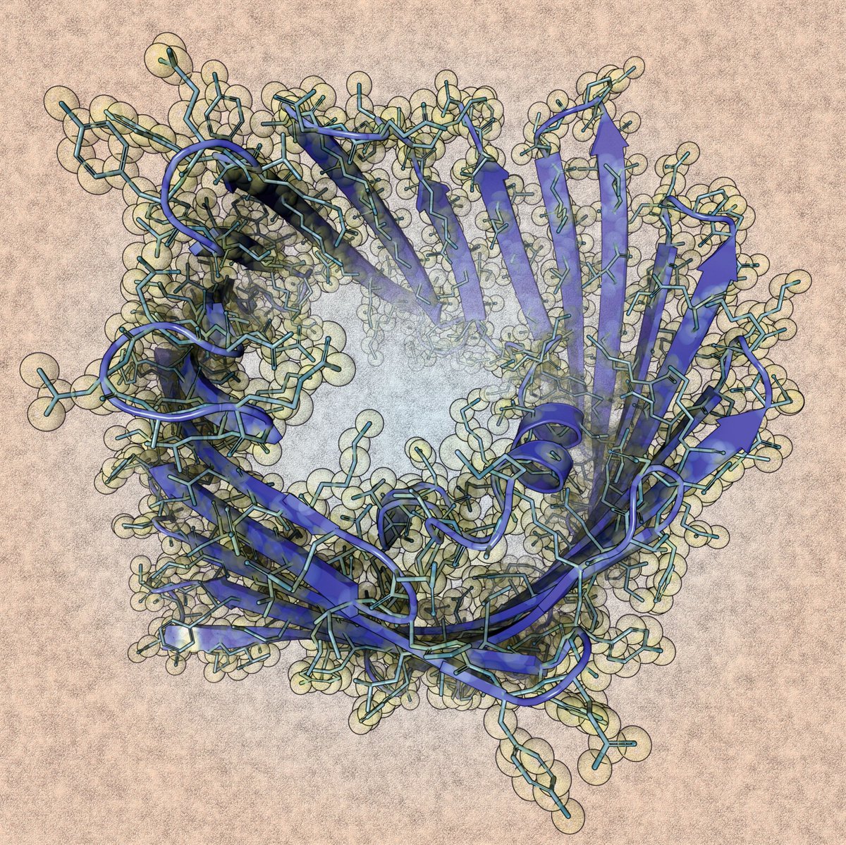
Here you can see a crystal structure of the mouse VDAC1 channel (PDB code: 9GNG)

Rendering by Francisco J. Enguita (<a href="/fenguita/">Francisco J. Enguita</a>) made with #ProteinImager

3dproteinimaging.com/protein-imager…

#SciArt #molecularart #channel #vdac1 #mouse #membrane #xray
3D Protein Imaging (@proteinimaging) 's Twitter Profile Photo

Here you can see a cryoEM structure of Cas9d from Deltaproteobacteria in complex to its gRNA and a target dsDNA (PDB code: 8W2Z) Rendering by Francisco J. Enguita (Francisco J. Enguita) made with #ProteinImager 3dproteinimaging.com/protein-imager… #SciArt #molecularart #crispr #dcas9 #dna #cryoem

Here you can see a cryoEM structure of Cas9d from Deltaproteobacteria in complex to its gRNA and a target dsDNA (PDB code: 8W2Z)

Rendering by Francisco J. Enguita (<a href="/fenguita/">Francisco J. Enguita</a>) made with #ProteinImager

3dproteinimaging.com/protein-imager…

#SciArt #molecularart #crispr #dcas9 #dna #cryoem
3D Protein Imaging (@proteinimaging) 's Twitter Profile Photo

Here you can see an x-ray structure of a rationally-designed protein binder in complex to CYTX component of the Naja pallida venom (PDB code: 9BK6) Rendering by Francisco J. Enguita made with #ProteinImager 3dproteinimaging.com/protein-imager… #SciArt #molecularart #venom #toxin #binder #design

Here you can see an x-ray structure of a rationally-designed protein binder in complex to CYTX component of the Naja pallida venom (PDB code: 9BK6)

Rendering by <a href="/fenguita/">Francisco J. Enguita</a> made with #ProteinImager

3dproteinimaging.com/protein-imager…

#SciArt #molecularart #venom #toxin #binder #design
3D Protein Imaging (@proteinimaging) 's Twitter Profile Photo

Here you can see a crystal structure of the SPG glyoxalase enzyme from Gossypium hirsutum (PDB code: 7VQ6) Rendering by Francisco J. Enguita (Francisco J. Enguita) made with #ProteinImager 3dproteinimaging.com/protein-imager… #SciArt #molecularart #deoxynivalenol #mycotoxin #glyoxalase #xray

Here you can see a crystal structure of the SPG glyoxalase enzyme from Gossypium hirsutum (PDB code: 7VQ6)

Rendering by Francisco J. Enguita (<a href="/fenguita/">Francisco J. Enguita</a>) made with #ProteinImager

3dproteinimaging.com/protein-imager…

#SciArt #molecularart #deoxynivalenol #mycotoxin #glyoxalase #xray
3D Protein Imaging (@proteinimaging) 's Twitter Profile Photo

Here you can see a recent cryoEM structure of the light-driven sodium-pumping rhodopsin KR2 (PDB code: 8RQ5) Rendering by Francisco J. Enguita (Francisco J. Enguita) made with #ProteinImager 3dproteinimaging.com/protein-imager… #SciArt #molecularart #pump #ion #sodium #light #rhodopsin #cryoem
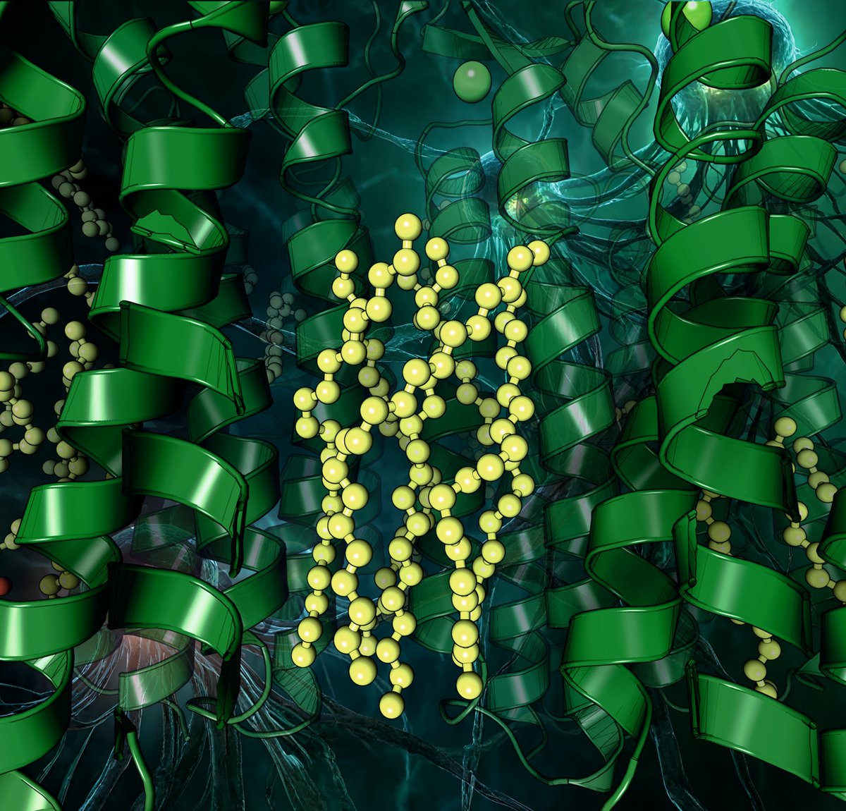
Here you can see a recent cryoEM structure of the light-driven sodium-pumping rhodopsin KR2 (PDB code: 8RQ5)

Rendering by Francisco J. Enguita (<a href="/fenguita/">Francisco J. Enguita</a>) made with #ProteinImager

3dproteinimaging.com/protein-imager…

#SciArt #molecularart #pump #ion #sodium #light #rhodopsin #cryoem
3D Protein Imaging (@proteinimaging) 's Twitter Profile Photo

Here you can see a cryoEM structure of human FAN1 protein in complex with 5' flap DNA substrate (PDB code: 9EO1) Rendering by Francisco J. Enguita (Francisco J. Enguita) made with #ProteinImager 3dproteinimaging.com/protein-imager… #SciArt #molecularart #fanconi #dna #huntington #cryoem
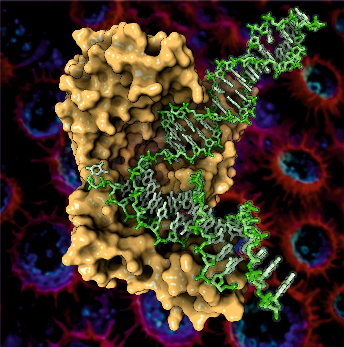
Here you can see a cryoEM structure of human FAN1 protein in complex with 5' flap DNA substrate (PDB code: 9EO1)

Rendering by Francisco J. Enguita (<a href="/fenguita/">Francisco J. Enguita</a>) made with #ProteinImager

3dproteinimaging.com/protein-imager…

#SciArt #molecularart #fanconi #dna #huntington #cryoem
3D Protein Imaging (@proteinimaging) 's Twitter Profile Photo

Rendering by Francisco J. Enguita (Francisco J. Enguita) made with #ProteinImager 3dproteinimaging.com/protein-imager… #SciArt #molecularart #virus #cancer #replication #transformation #cryoem

Rendering by Francisco J. Enguita (<a href="/fenguita/">Francisco J. Enguita</a>) made with #ProteinImager

3dproteinimaging.com/protein-imager…

#SciArt #molecularart #virus #cancer #replication #transformation #cryoem
3D Protein Imaging (@proteinimaging) 's Twitter Profile Photo

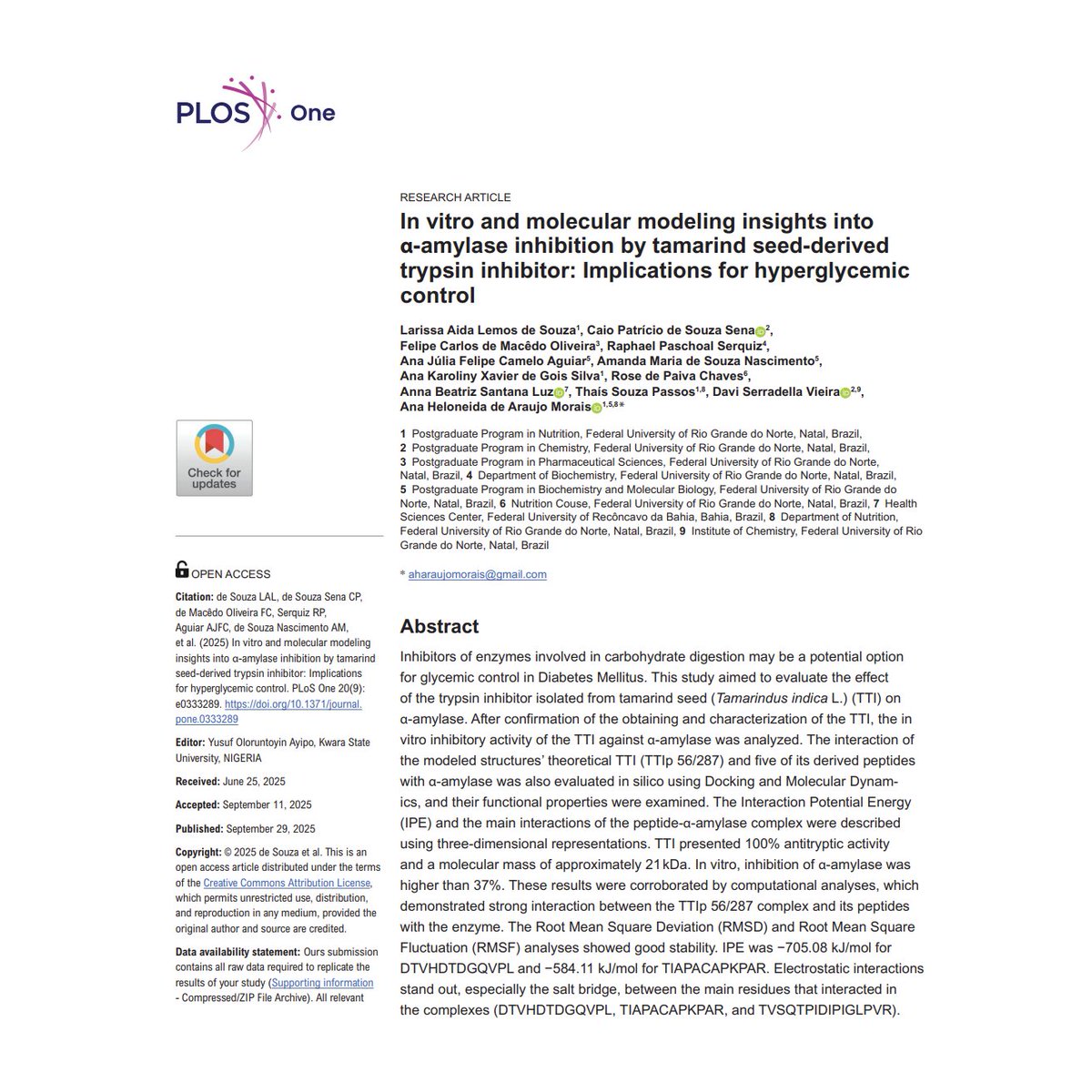
New study by Caio Sena & coauthors reveals tamarind seed trypsin inhibitor can also inhibit α-amylase, a key enzyme in carb digestion Illustrations made using #ProtienImager 3dproteinimaging.com/protein-imager/ doi.org/10.1371/journa… #DiabetesResearch #Tamarind #GlycemicControl

New study by Caio Sena &amp; coauthors reveals tamarind seed trypsin inhibitor can also inhibit α-amylase, a key enzyme in carb digestion

Illustrations made using #ProtienImager 3dproteinimaging.com/protein-imager/

doi.org/10.1371/journa…

#DiabetesResearch #Tamarind #GlycemicControl
3D Protein Imaging (@proteinimaging) 's Twitter Profile Photo

Here you can see a crystal structure of a ppApp synthetase from Streptomyces albidoflavus (PDB code: 8VX3) Rendering by Francisco J. Enguita (Francisco J. Enguita) made with #ProteinImager 3dproteinimaging.com/protein-imager… #SciArt #molecularart #ppApp #streptomyces #second #messenger #xray

Here you can see a crystal structure of a ppApp synthetase from Streptomyces albidoflavus (PDB code: 8VX3)

Rendering by Francisco J. Enguita (<a href="/fenguita/">Francisco J. Enguita</a>) made with #ProteinImager

3dproteinimaging.com/protein-imager…

#SciArt #molecularart #ppApp #streptomyces #second #messenger #xray
3D Protein Imaging (@proteinimaging) 's Twitter Profile Photo

Here you can see a cryoEM structure of the human TLR3 receptor in complex to a de novo designed binder (PDB code: 8YHU) Rendering by Francisco J. Enguita (Francisco J. Enguita) made with #ProteinImager 3dproteinimaging.com/protein-imager… #SciArt #molecularart #virus #binder #design #cryoem
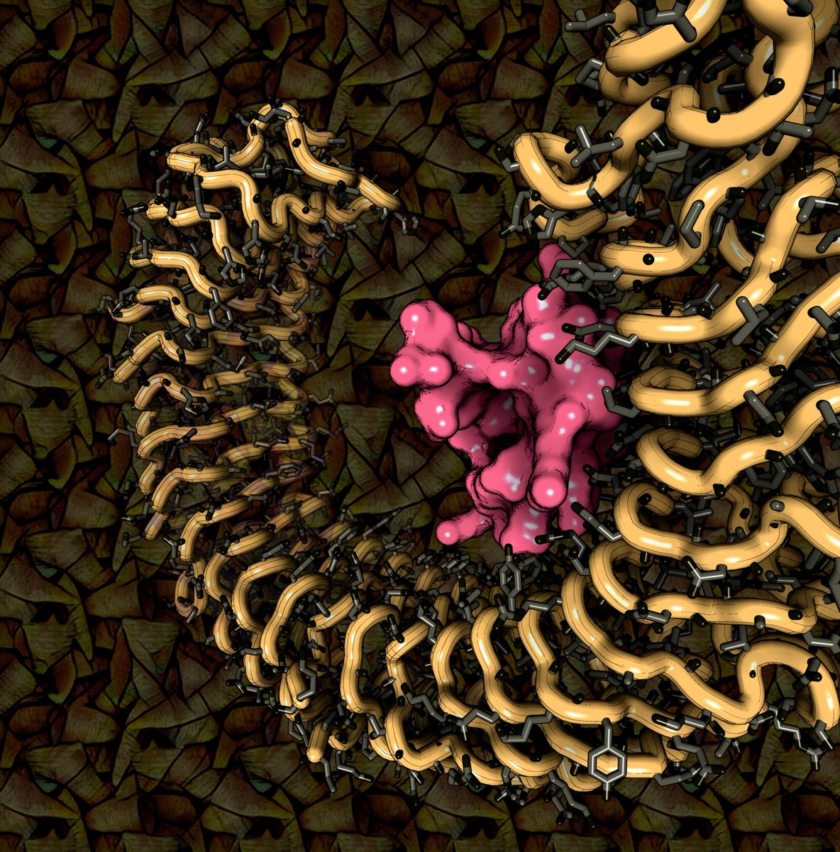
Here you can see a cryoEM structure of the human TLR3 receptor in complex to a de novo designed binder (PDB code: 8YHU)

Rendering by Francisco J. Enguita (<a href="/fenguita/">Francisco J. Enguita</a>) made with #ProteinImager

3dproteinimaging.com/protein-imager…

#SciArt #molecularart #virus #binder #design #cryoem
Uninsubria (@uni_insubria) 's Twitter Profile Photo

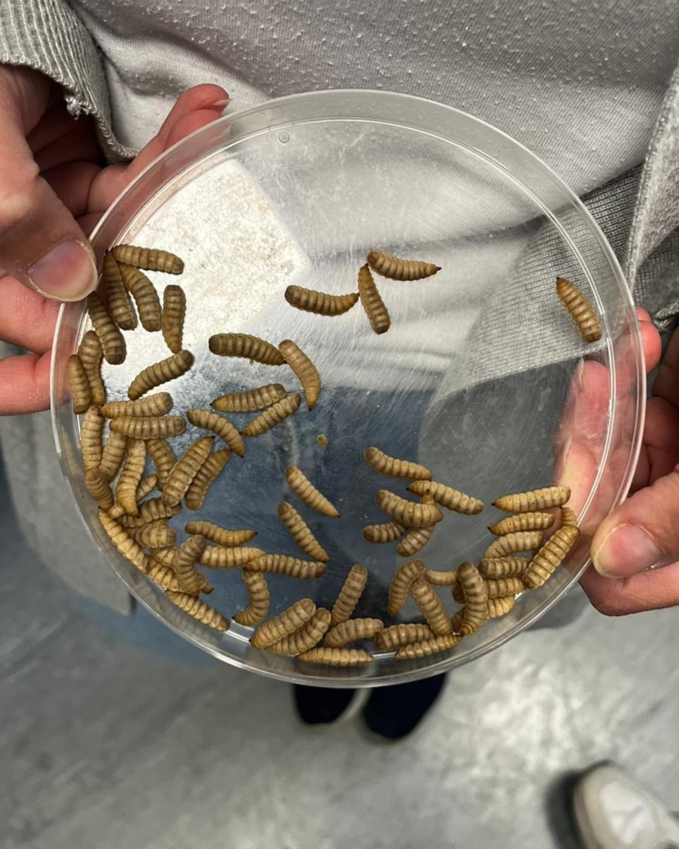
🪰♻️ La ricercatrice Ilaria Armenia ottiene il finanziamento #MarieSkłodowskaCurie per #Plastifly: un metodo sostenibile per degradare la plastica con la mosca soldato nera 👉 uninsubria.it/plastifly #uninsubria #sceglinsubria

🪰♻️ La ricercatrice Ilaria Armenia ottiene il finanziamento #MarieSkłodowskaCurie per #Plastifly: un metodo sostenibile per degradare la plastica con la mosca soldato nera
👉 uninsubria.it/plastifly

#uninsubria #sceglinsubria
3D Protein Imaging (@proteinimaging) 's Twitter Profile Photo

Here you can see a cryoEM structure of the Hermes transposase from the house fly Musca domestica (PDB code: 8EDG) Rendering by Francisco J. Enguita (Francisco J. Enguita) made with #ProteinImager 3dproteinimaging.com/protein-imager… #SciArt #molecularart #musca #transposase #hermes #dna #cryoem
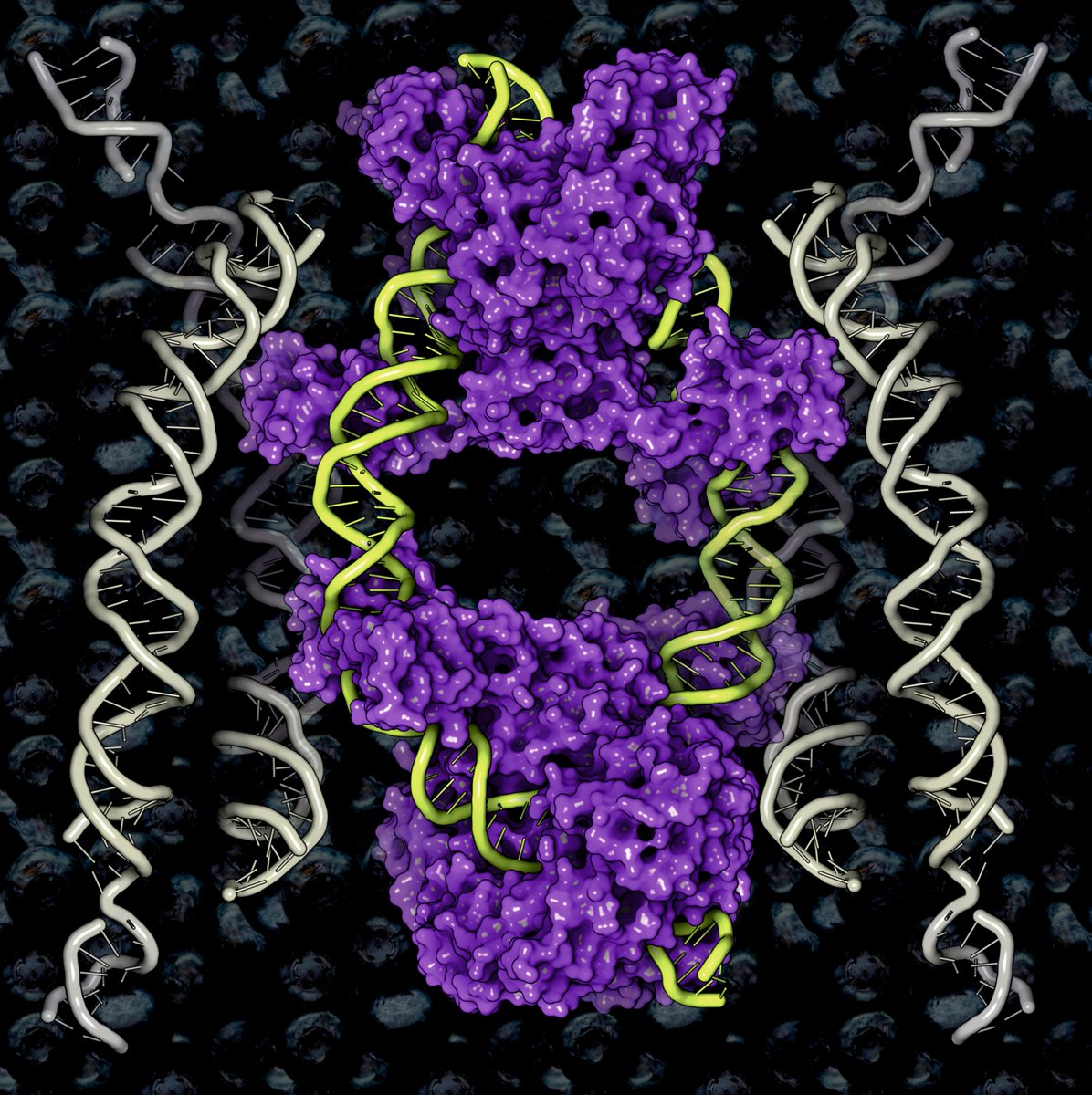
Here you can see a cryoEM structure of the Hermes transposase from the house fly Musca domestica (PDB code: 8EDG)

Rendering by Francisco J. Enguita (<a href="/fenguita/">Francisco J. Enguita</a>) made with #ProteinImager

3dproteinimaging.com/protein-imager…

#SciArt #molecularart #musca #transposase #hermes #dna #cryoem
3D Protein Imaging (@proteinimaging) 's Twitter Profile Photo

Here you can see a cryoEM structure of Comamonas testosteroni KF-1 circularly permuted group II intron Pre-1S state (PDB code: 8XTS) Rendering by Francisco J. Enguita (Francisco J. Enguita) made with #ProteinImager 3dproteinimaging.com/protein-imager… #SciArt #intron #precursor #RNA #cryoem
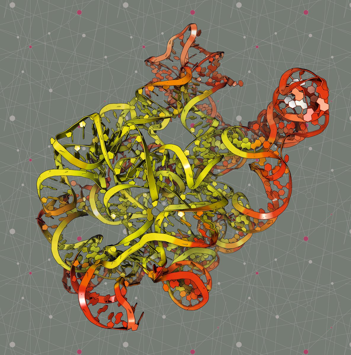
Here you can see a cryoEM structure of Comamonas testosteroni KF-1 circularly permuted group II intron Pre-1S state (PDB code: 8XTS)

Rendering by Francisco J. Enguita (<a href="/fenguita/">Francisco J. Enguita</a>) made with #ProteinImager

3dproteinimaging.com/protein-imager…

#SciArt #intron #precursor #RNA #cryoem
3D Protein Imaging (@proteinimaging) 's Twitter Profile Photo

New study by Francisco J. Enguita (Francisco J. Enguita) et al. reveals cross-kingdom enzymatic strategies for deoxynivalenol (DON) detoxification. 🌾🔬 Illustrations made using #ProtienImager 3dproteinimaging.com/protein-imager article doi.org/10.3390/microo… #Mycotoxins #FoodSafety #Microbiology
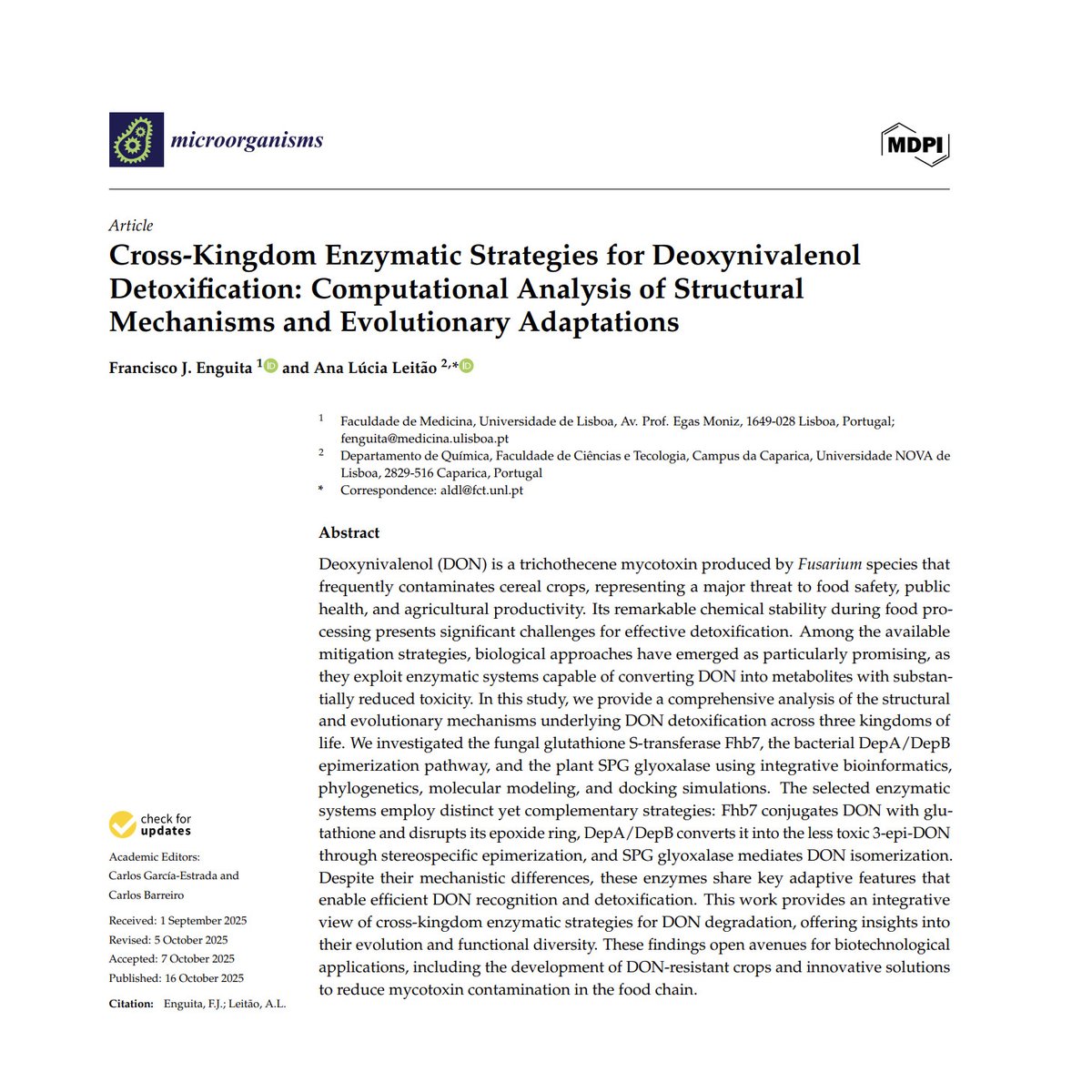
New study by Francisco J. Enguita (<a href="/fenguita/">Francisco J. Enguita</a>) et al. reveals cross-kingdom enzymatic strategies for deoxynivalenol (DON) detoxification. 🌾🔬

Illustrations made using #ProtienImager 3dproteinimaging.com/protein-imager

article doi.org/10.3390/microo…

#Mycotoxins #FoodSafety #Microbiology
3D Protein Imaging (@proteinimaging) 's Twitter Profile Photo

Here you can see a cryoEM structure of the Rep68-AAVS1 heptameric complex of the AAV-2 adenovirus (PDB code: 9BC5) Rendering by Francisco J. Enguita (Francisco J. Enguita) made with #ProteinImager 3dproteinimaging.com/protein-imager… #SciArt #molecularart #adenovirus #DNA #integrase #cryoem
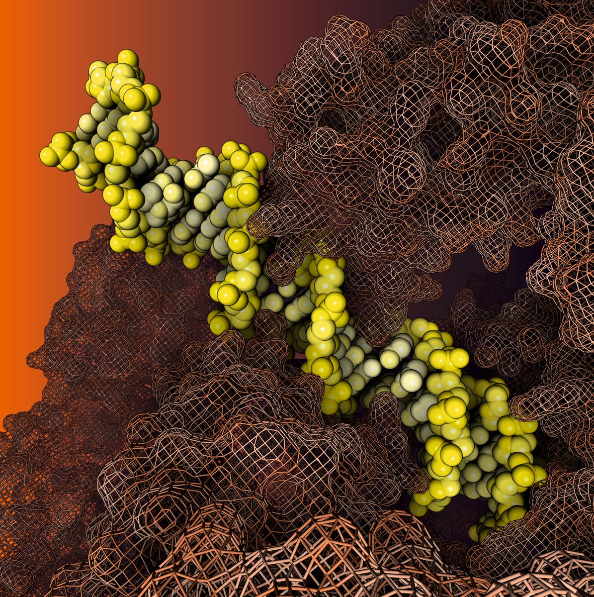
Here you can see a cryoEM structure of the Rep68-AAVS1 heptameric complex of the AAV-2 adenovirus (PDB code: 9BC5)

Rendering by Francisco J. Enguita (<a href="/fenguita/">Francisco J. Enguita</a>) made with #ProteinImager

3dproteinimaging.com/protein-imager…

#SciArt #molecularart #adenovirus #DNA #integrase #cryoem
3D Protein Imaging (@proteinimaging) 's Twitter Profile Photo

Here you can see a cryoEM structure of the Kalium channelrhodopsin 1 from Hyphochytrium catenoides (PDB code: 9CDD) Rendering by Francisco J. Enguita (Francisco J. Enguita) made with #ProteinImager 3dproteinimaging.com/protein-imager… #SciArt #molecularart #channel #potasion #hypochytrium #cryoem
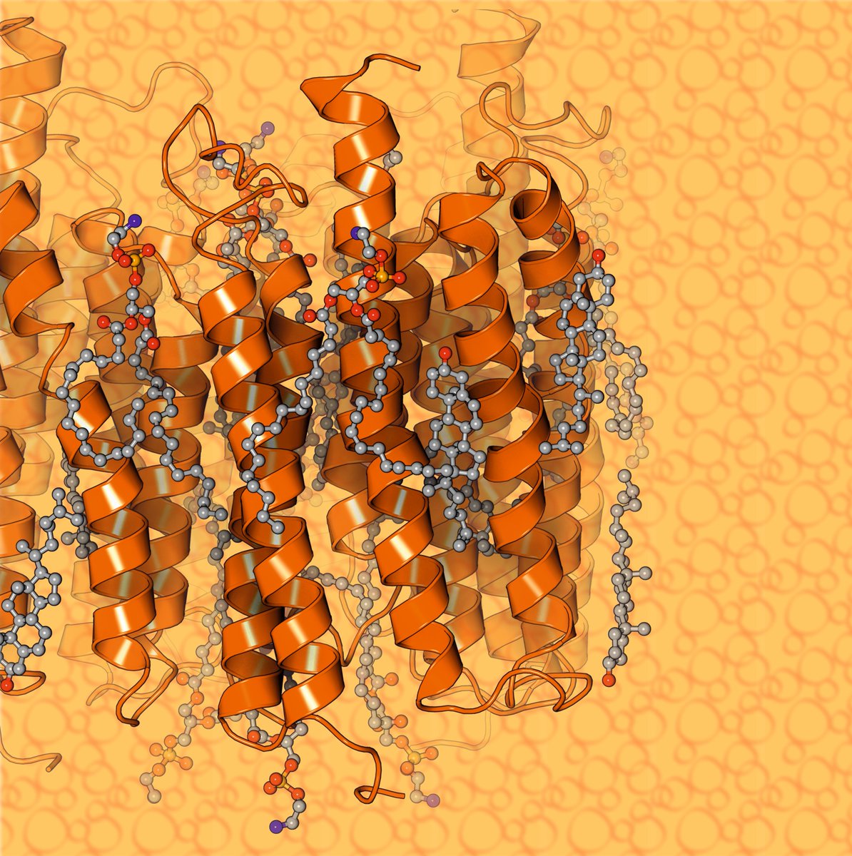
Here you can see a cryoEM structure of the Kalium channelrhodopsin 1 from Hyphochytrium catenoides (PDB code: 9CDD)
Rendering by Francisco J. Enguita (<a href="/fenguita/">Francisco J. Enguita</a>) made with #ProteinImager

3dproteinimaging.com/protein-imager…

#SciArt #molecularart #channel #potasion #hypochytrium #cryoem
3D Protein Imaging (@proteinimaging) 's Twitter Profile Photo

Here you have a cryoEM structure of the photosynthetic LH2-LH1 complex from the purple bacterium Halorhodospira halophila (PDB code: 8Z81) Rendering by Francisco J. Enguita (Francisco J. Enguita) made with #ProteinImager 3dproteinimaging.com/protein-imager… #SciArt #molecularart #photosystem #lh2
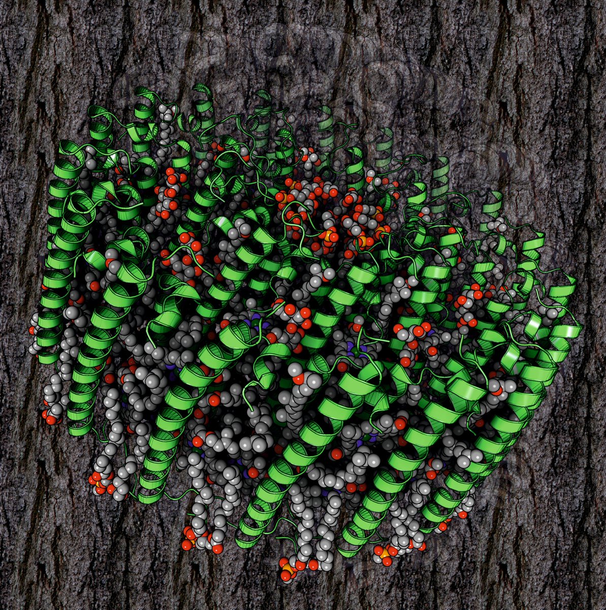
Here you have a cryoEM structure of the photosynthetic LH2-LH1 complex from the purple bacterium Halorhodospira halophila (PDB code: 8Z81)

Rendering by Francisco J. Enguita (<a href="/fenguita/">Francisco J. Enguita</a>) made with #ProteinImager

3dproteinimaging.com/protein-imager…

#SciArt #molecularart #photosystem #lh2
3D Protein Imaging (@proteinimaging) 's Twitter Profile Photo

Here you can see a cryoEM structure of the human BAF-Lamin A/C IgF-H1-nucleosome complex (PDB code: 9J8O) Rendering by Francisco J. Enguita (Francisco J. Enguita) made with #ProteinImager 3dproteinimaging.com/protein-imager… #SciArt #chromatin #nucleosome #BAF #lamin #transcription #cryoem
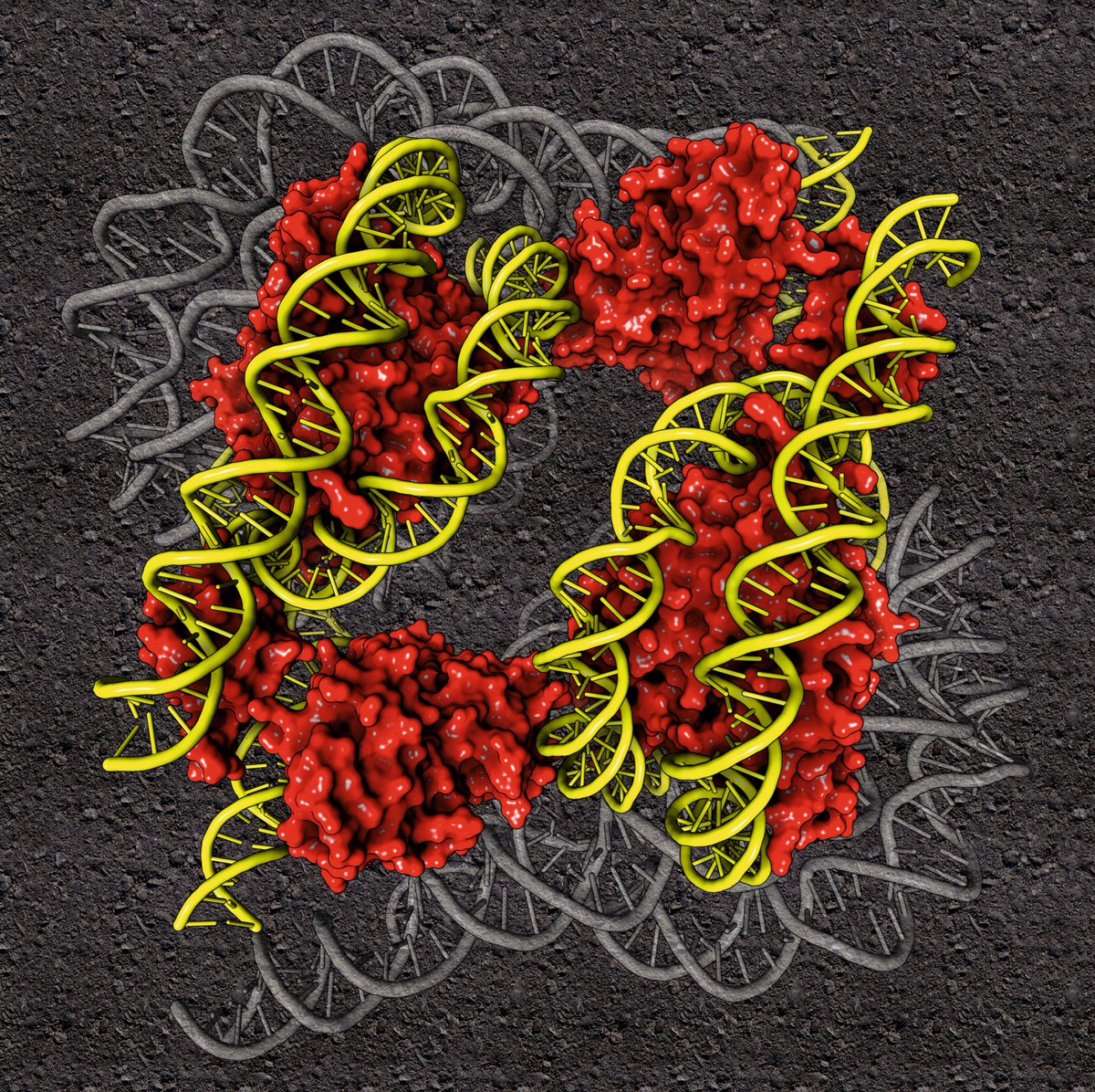
Here you can see a cryoEM structure of the human BAF-Lamin A/C IgF-H1-nucleosome complex (PDB code: 9J8O)

Rendering by Francisco J. Enguita (<a href="/fenguita/">Francisco J. Enguita</a>) made with #ProteinImager

3dproteinimaging.com/protein-imager…

#SciArt #chromatin #nucleosome #BAF #lamin #transcription #cryoem
3D Protein Imaging (@proteinimaging) 's Twitter Profile Photo

Here you can see a cryoEM structure of the SARS-CoV-2 S 6P trimer complexed with a human neutralizing antibody Fab fragment (PDB code: 9BJ3) Rendering by Francisco J. Enguita made with #ProteinImager 3dproteinimaging.com/protein-imager… #SciArt #molecularart #sarscov2 #antibody #neutralizing #cryoem
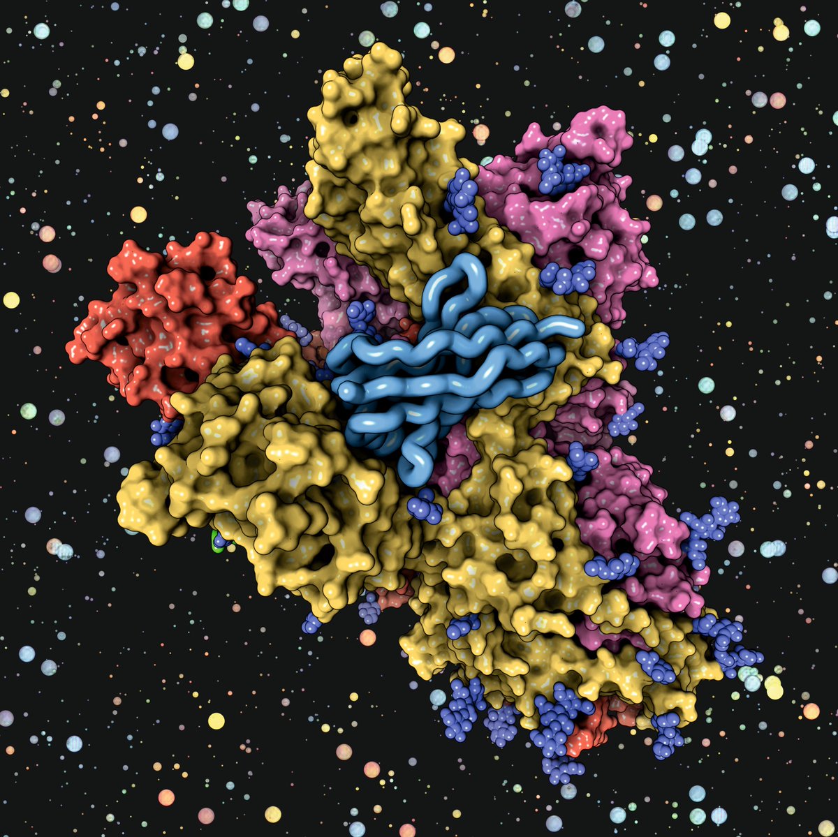
Here you can see a cryoEM structure of the SARS-CoV-2 S 6P trimer complexed with a human neutralizing antibody Fab fragment (PDB code: 9BJ3)

Rendering by <a href="/fenguita/">Francisco J. Enguita</a> made with #ProteinImager

3dproteinimaging.com/protein-imager…

#SciArt #molecularart #sarscov2 #antibody #neutralizing #cryoem