Jonathan Zuckerman MD PhD (@jzrenalpath) 's Twitter Profile
Jonathan Zuckerman MD PhD

@jzrenalpath

Service Chief, Renal Pathology and Residency Program Director UCLA Department of Pathology and Laboratory Medicine. Views are my own.

ID: 1021092105609883648

linkhttp://pathology.ucla.edu/renal-pathology-outreach calendar_today22-07-2018 17:58:23

4,4K Tweet

5,5K Takipçi

344 Takip Edilen

Grace Choung, MD 박영이 (@grace_choung) 's Twitter Profile Photo

A membranous pattern is occasionally present in Fibrillary GN Notice randomly arranged fibrils in the subepithelial regions. DNAJB9-positivity is seen in a similar distribution by IHC. Can be mistaken for membranous nephropathy: doi.org/10.1016/j.ekir…. #renalpath #PathTwitter

A membranous pattern is occasionally present in Fibrillary GN
Notice randomly arranged fibrils in the subepithelial regions. DNAJB9-positivity is seen in a similar distribution by IHC. Can be mistaken for membranous nephropathy: doi.org/10.1016/j.ekir….
#renalpath #PathTwitter
Renal Path Labs (@renalpathlabs) 's Twitter Profile Photo

Images say it all. The disease,the process,possible culprit, the misery of that organ system & about the nicely done stain..Residents,want to describe this pic?Native Bx #PathoResidents #NephrologyResidents #PathTwitter #Nephtwitter #AskRenal #RenalPathLabsGurugram Renal Pathology Society
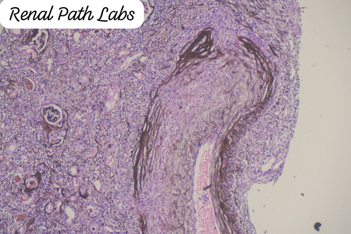
Images say it all. The disease,the process,possible culprit, the misery of that organ system & about the nicely done stain..Residents,want to describe this pic?Native Bx
#PathoResidents #NephrologyResidents #PathTwitter #Nephtwitter #AskRenal #RenalPathLabsGurugram <a href="/Renalpathsoc/">Renal Pathology Society</a>
Sanjeev Sethi (@sethirenalpath) 's Twitter Profile Photo

Membranous nephropathy (MN) antigens from a single center in Portugal. Starting point=negative serum anti-PLA2R titer cases. We detected MN antigens: 3 PLA2R, 2 NELL1, & 1 each EXT1/EXT2, HTRA1, THSD7A. Antigens correlated with underlying disease. 1/2 kireports.org/article/S2468-…

Grace Choung, MD 박영이 (@grace_choung) 's Twitter Profile Photo

Anti-tubular basement membrane (TBM) disease 🔸Rare type of tubulointerstitial nephritis 🔸Strong linear polyclonal IgG restricted to TBMs 🔸No deposits by EM 🔸Autoantibody to TIN-ag in proximal TBM 🔸AKI/CKD, polyuria, polydipsia #renalpath #PathTwitter (1/2)

Anti-tubular basement membrane (TBM) disease
🔸Rare type of tubulointerstitial nephritis
🔸Strong linear polyclonal IgG restricted to TBMs
🔸No deposits by EM
🔸Autoantibody to TIN-ag in proximal TBM
🔸AKI/CKD, polyuria, polydipsia 
#renalpath #PathTwitter 
(1/2)
OHSU Pathology & Laboratory Medicine (@ohsupathology) 's Twitter Profile Photo

New publication alert!🎉 In collaboration with PDXkidney and OHSU Infectious Diseases Division we are excited to share "Bartonella Endocarditis-Associated Glomerulonephritis: A Mimicker of Autoimmunity and Vasculitis". sciencedirect.com/science/articl…

A Tchakarov (@atchakarov1) 's Twitter Profile Photo

Middle age F with cardiac tamponade, new onset heart failure, edema, Raynaud phenomenon, anemia and positive ANA and Anti-RNA polymerase III, presenting with AKI. #PathX #PathTwitter #RenalPath #Nephropath #Nephrology

Middle age F with cardiac tamponade, new onset heart failure, edema, Raynaud phenomenon, anemia and positive ANA and Anti-RNA polymerase III, presenting with AKI. #PathX #PathTwitter #RenalPath #Nephropath #Nephrology
Jonathan Zuckerman MD PhD (@jzrenalpath) 's Twitter Profile Photo

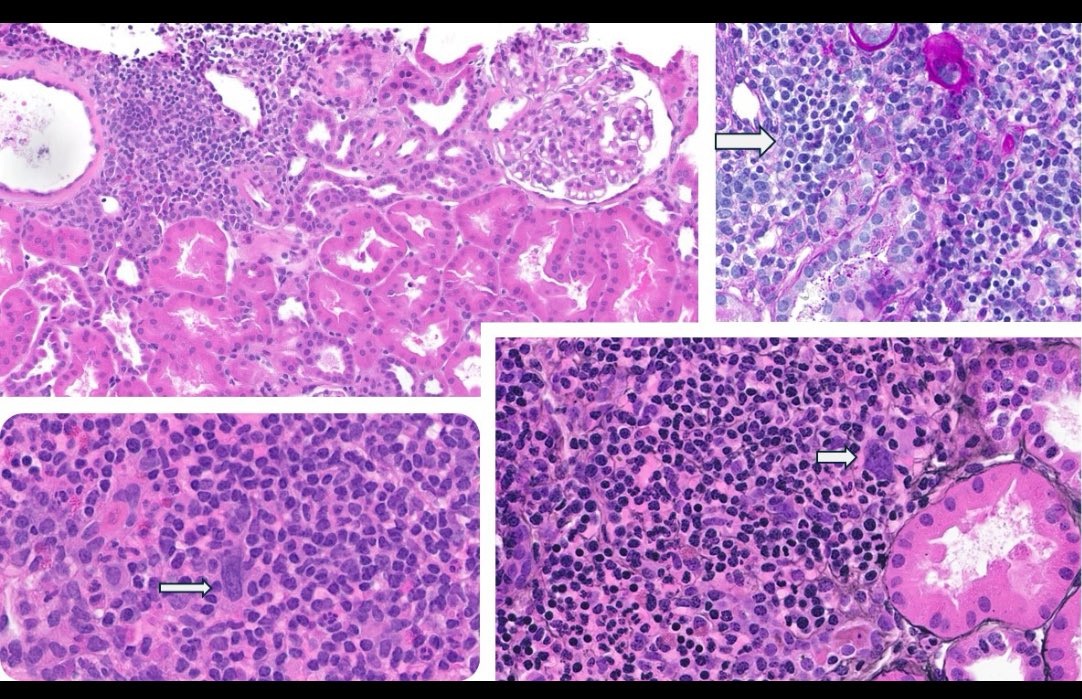
Some nice examples of severe tubulitis in this case of ACR 1B. Interestingly bx for new de novo DSA with with stable allograft function (~1 year post txp). No significant MVI. #renalpath #nephrology #PathTwitter

Some nice examples of severe tubulitis in this case of ACR 1B. Interestingly bx for new de novo DSA with with stable allograft function (~1 year post txp). No significant MVI.  #renalpath #nephrology  #PathTwitter
Anthony Chang, MD (張賀文) (@changucanspare) 's Twitter Profile Photo

Complement touches everything. Might just need complement inhibitors in the water supply Rationale for Targeting Complement to Mitigate Renal... : Journal of the American Society of Nephrology journals.lww.com/jasn/abstract/…

Renal Path Labs (@renalpathlabs) 's Twitter Profile Photo

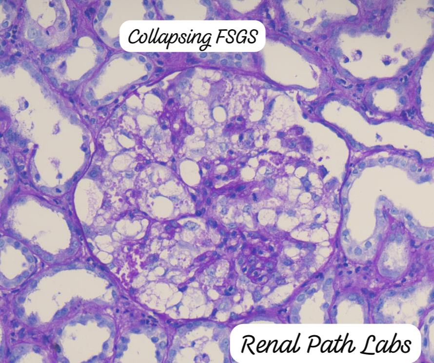
This FSGS was scarier than any crescent…fierce looking, activated podocytes as if ready to engulf the tuft.. And look at the tuft-scared to the core.. 🥹😱 #CollapsingFsgs #RenalPath #PathTwitter #Nephtwitter #RenalPathSociety #AskRenal #RenalPathLabsGurugram #KidneyBiopsy

This FSGS was scarier than any crescent…fierce looking, activated podocytes as if ready to engulf the tuft..
And look at the tuft-scared to the core..
🥹😱
#CollapsingFsgs #RenalPath #PathTwitter #Nephtwitter #RenalPathSociety #AskRenal #RenalPathLabsGurugram #KidneyBiopsy
Ziad El-Zaatari (@ziad_zaatari) 's Twitter Profile Photo

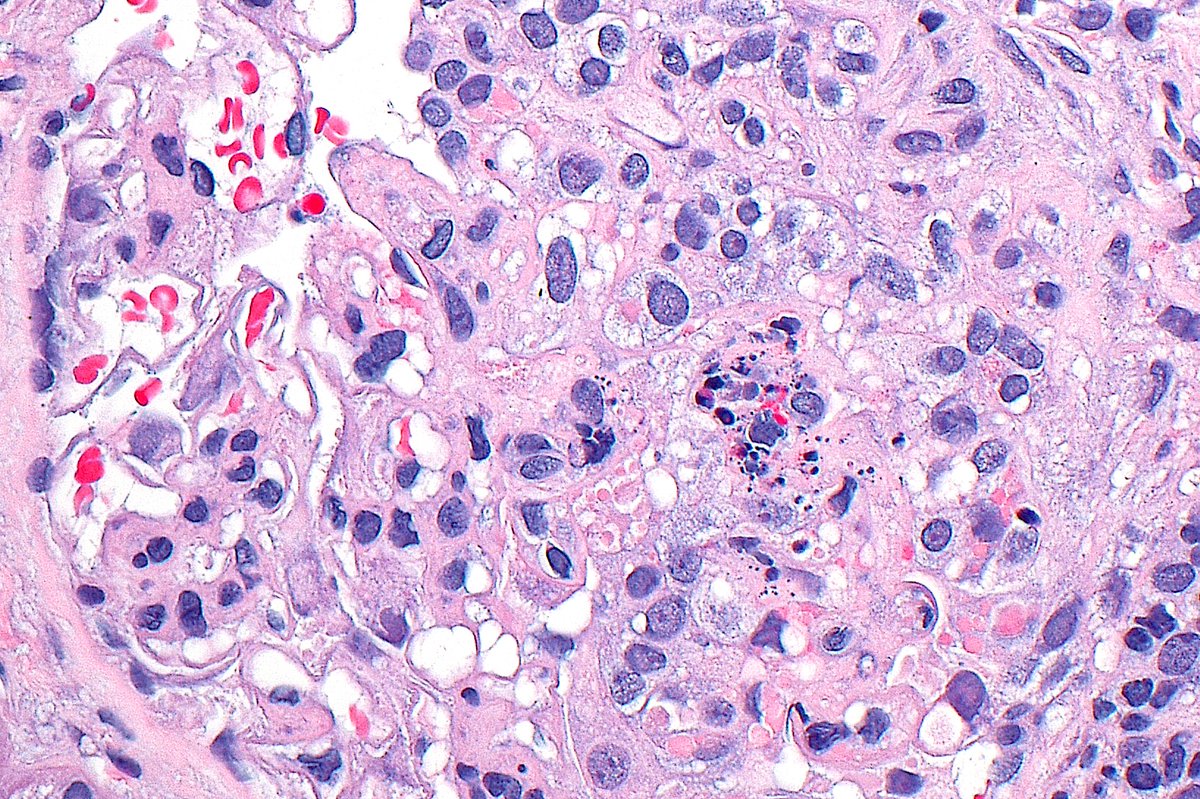
🔬📷 Glomerulus with karyorrhexis, hyaline thrombi, and a cellular crescent in patient with class III lupus nephritis ~ #RenalPath #Pathology #Kidney #Nephrology

🔬📷 Glomerulus with karyorrhexis, hyaline thrombi, and a cellular crescent in patient with class III lupus nephritis ~ #RenalPath #Pathology #Kidney #Nephrology
Priya Alexander, MD (@mpalexandermd) 's Twitter Profile Photo

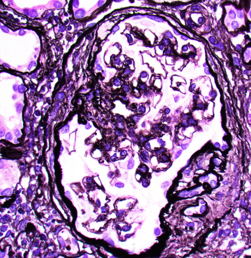
Kidney bx for RPGN. MPO ANCA +++ Temporal heterogeneity of crescents seen. Fibrinogen highlights a crescent (grey arrow). IgG shows trace to 1+ deposits in glomerulus. Remember its PAUCI immune crescentic GN - we can have “some” immune deposits!! What do you think?

Kidney bx for RPGN. MPO ANCA +++
Temporal heterogeneity of crescents seen. Fibrinogen highlights a crescent (grey arrow). IgG shows trace to 1+ deposits in glomerulus.
Remember its PAUCI immune crescentic GN - we can have “some” immune deposits!!  What do you think?
Jonathan Zuckerman MD PhD (@jzrenalpath) 's Twitter Profile Photo

Classic example of Alport spectrum disease in a pt with X-linked Alport syndrome. Lesions of segmental glomerulosclerosis, numerous interstitial foam cells, and atypical GBMs with lamina densa splitting, subepithelial scalloping, and thinning. #renalpath #pathtwitter #nephrology

Classic example of Alport spectrum disease in a pt with X-linked Alport syndrome. Lesions of segmental glomerulosclerosis, numerous interstitial foam cells, and atypical GBMs with lamina densa splitting, subepithelial scalloping, and thinning. #renalpath #pathtwitter #nephrology
Jonathan Zuckerman MD PhD (@jzrenalpath) 's Twitter Profile Photo

Some well formed "eye lashes" on these capillary loops in a case of AL-amyloidosis. Podocytes getting skewered by the spicules in the EM. #renalpath #pathtwitter #nephrology.

Some well formed "eye lashes" on these capillary loops in a case of AL-amyloidosis.  Podocytes getting skewered by the spicules in the EM. #renalpath #pathtwitter #nephrology.
Ladan Zand (@ladanzand) 's Twitter Profile Photo

I am excited to share our latest paper on the potential association between monoclonal gammopathy and ANCA negative vasculitis. Monoclonal testing should be considered in ANCA negative patients. maria jose vargas fernando fervenza journals.lww.com/kidney360/abst…