Juan Edo Rodriguez
@juanedorg
Postdoc @ University Hospital Bonn | Passionate about Light sheet & Expansion microscopy | Neuroscien enthusiast | Exploring organoids, mouse brain, and beyond
ID: 4927679411
18-02-2016 12:57:03
71 Tweet
84 Takipçi
271 Takip Edilen

🔴 Alerta de Workshop! Todo sobre clarificación de tejidos mediante iDISCO+ y🔬Light-sheet en el hub de microscopía del Center for Integrative Biology U. Mayor con el apoyo de CZI Science 5-8 de Nov. ¡No te lo puedes perder! Márcalo en tu calendario e inscríbete antes del 26/07 👇 docs.google.com/forms/d/e/1FAI…
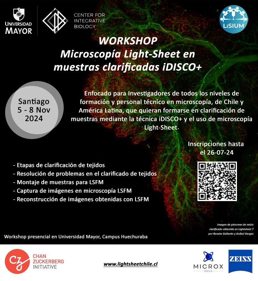

Alerta de Workshop! Todo sobre clarificación de tejidos mediante iDISCO+ y📷Light-sheet en el hub de microscopía del Center for Integrative Biology U. Mayor con el apoyo de CZI Science 5-8 de Nov. ¡No te lo puedes perder! Márcalo en tu calendario e inscríbete antes del 26/07 📷docs.google.com/forms/d/e/1FAI…


Save the date 7-8/10/2024 : GDRImabio will co-organize with France-BioImaging Research Infrastructure a workshop on "High Resolution 3D Microscopy in Biology: Development & Applications", happy to welcome Jungmann Lab Erdinc Sezgin Sophie Brasselet Gaëlle Recher and Virgile Viasnoff, call for abstract soon !

Just out in Development. With our Go-Nuclear models, it is now possible to reliably 3D segment nuclei from 3D digital images. bit.ly/4dcYBz0 bit.ly/3zLS3ZO Athul Vijayan, Tejasvinee Mody, Qin Yu, Soeren Strauss, Adrian Wolny, Lorenzo Cerrone, Richard Smith Lab 🇺🇦
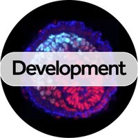
A deep learning-based toolkit for 3D nuclei segmentation and quantitative analysis in cellular and tissue context Read this Techniques and Resources Article by Athul Vijayan, Tejasvinee Mody, Qin Yu, Kay Schneitz Kay Schneitz and colleagues: journals.biologists.com/dev/article/15…




Huge thanks to @mariana_deniz for translating this into Spanish! Find it at bioimagebook.github.io/es/ BioImaging North America Center for Open Bioimage Analysis Edinburgh Univ OERs Executable Books #bioimageanalysis #imagej


#FluorescenceFriday #cellbiology 3D dynamics of six organelles in a COS-7 cell as revealed by lattice #lightsheet microscopy with multichannel unmixing. In collaboration with Valm Lab and Sarah Cohen, then in Lippincott-Schwartz Lab. doi.org/10.1038/nature…

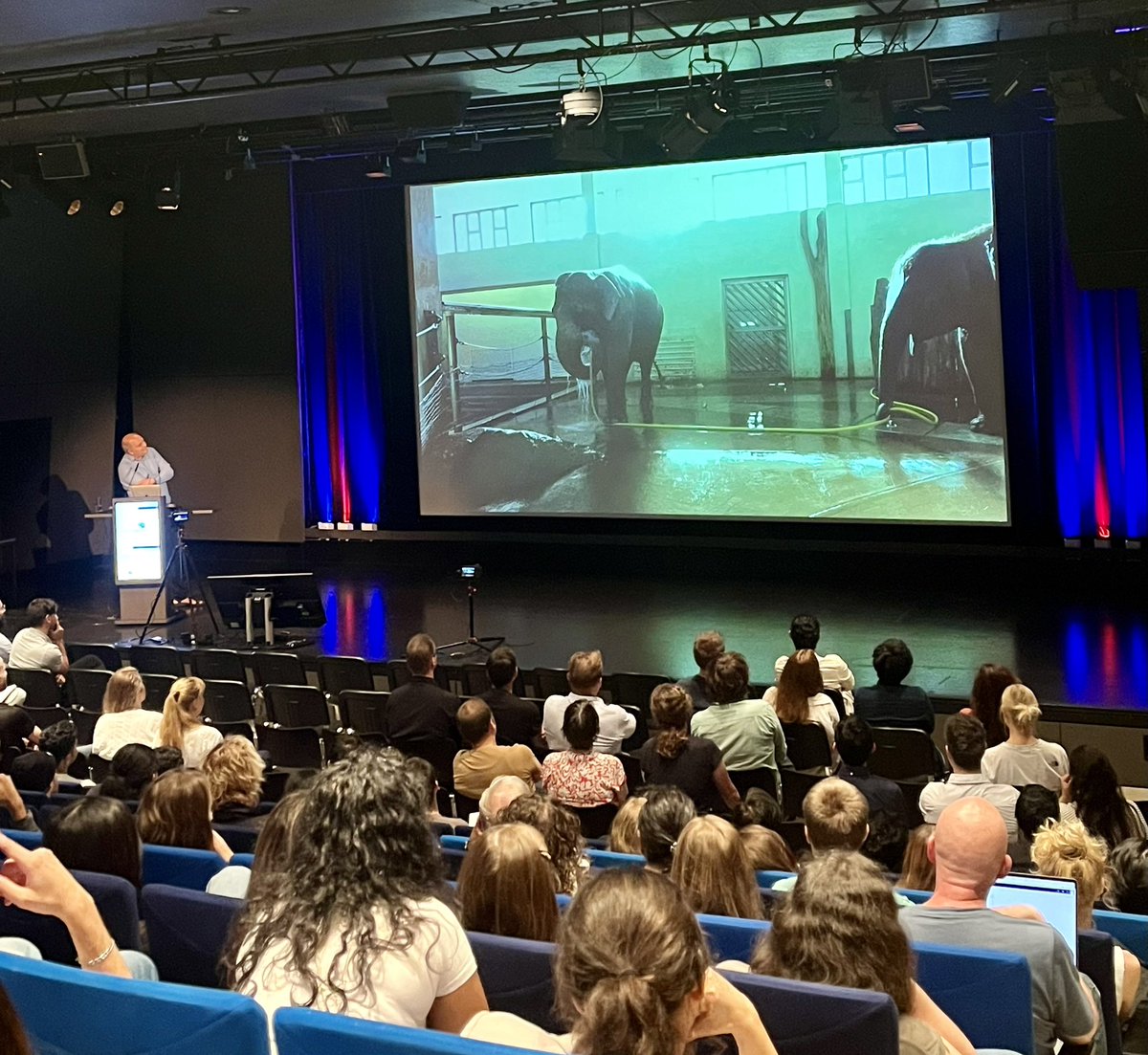
Large brain #neuroscience in 🐘. Enjoying a fantastic keynote lecture by Brecht Lab at #BonnBrain! iBehave Network Uniklinik Bonn @DZNE_en

Proud to present this incredible image of human iPSC-derived forebrain #organoids made by Clara Hayn for drug neurotoxicity testing #LSFM #ExM reveal condensed chromosomes coated with Ki-67🟢in mitotic neural #stemcells (Sox2⚪️Zo1🔴) within a neural rosette #FluoresenceFriday


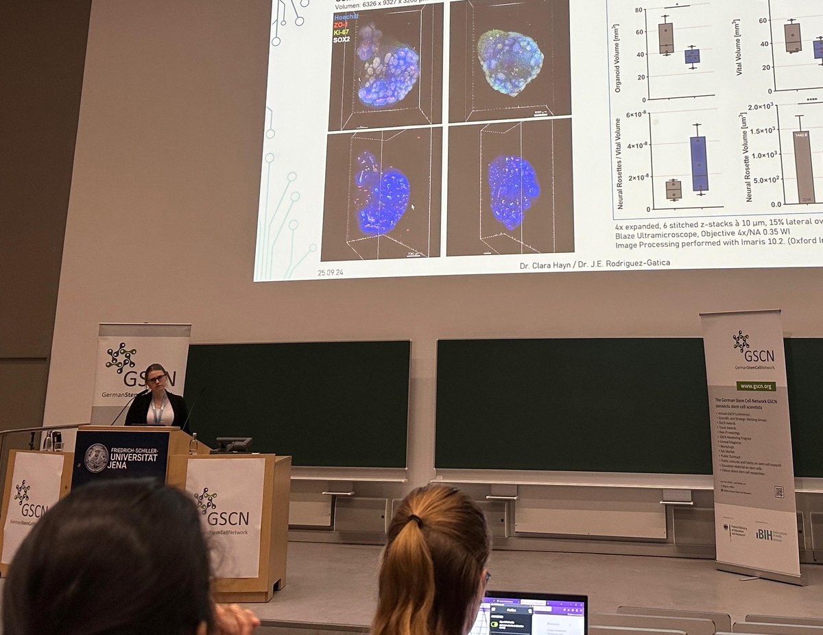
Proud to have been selected to present my data on testing drug neurotoxicity using human iPSC derived forebrain #organoids at #GSCN2024. Many thanks to Juan Edo Rodriguez for the amazing #LSFM + #ExM based characterisation of their 3D morphology that we achieved together 🥳🍀🔬🎉

Descubre un nuevo horizonte en la🔬#microscopíaconfocal En este doc de ZEISS Microscopy encontrarás la explicación de los principios de la microscopía de barrido láser confocal #LSM, #imágenesconfocales MP, Multiphoton, MP, Multiphoton, Airyscan, LSM Plus. 👉shorturl.at/ewTmh

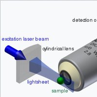
Please RT!We are proud to announce LSFM 2025!Held this year June 1st-7th,2025,at the Mount Desert Island Biological Laboratory (MDIBL)MDI Biological Lab in Bar Harbor, ME. Application portal open now! Application deadline (rolling) May 12th, 2025. Limited aid avlble mdibl.org/course/lsfm-20…

Excited to share how light-sheet and expansion microscopy help unravel long-range neuronal circuits at the German Neuroscience Society - NWG in Göttingen! 🧠🔬 Come visit poster T27-3A today—happy to chat about it! #NWG2025


