X Paul Ph.D ๐Ÿ‡ซ๐Ÿ‡ฎ๐Ÿ‡บ๐Ÿ‡ฆ (@rollingmeow) 's Twitter Profile
X Paul Ph.D ๐Ÿ‡ซ๐Ÿ‡ฎ๐Ÿ‡บ๐Ÿ‡ฆ

@rollingmeow

Feline fanatic. Lipid membrane lover. Science believer. Amateur photographer. Postdoctoral researcher in @IkonenLab.

ID: 3123545005

calendar_today01-04-2015 08:28:21

386 Tweet

216 Takipรงi

923 Takip Edilen

joe (@jtyler91) 's Twitter Profile Photo

PI3K dependent rearrangement of the actin cytoskeleton following the addition of Class I PAK kinase-inhibitor. Please read about it in our pre-print!

Ricardo Henriques (@henriqueslab) 's Twitter Profile Photo

If robots could dream of microtubules, how would they look like? An amazing story by Alon Saguy, Nano Bio Optics Lab (Yoav Shechtman) and colleagues. Proud we could contribute to it onlinelibrary.wiley.com/doi/10.1002/smโ€ฆ

If robots could dream of microtubules, how would they look like? An amazing story by Alon Saguy, <a href="/ShechtmanLab/">Nano Bio Optics Lab (Yoav Shechtman)</a> and colleagues. Proud we could contribute to it
onlinelibrary.wiley.com/doi/10.1002/smโ€ฆ
Reika Tei (@reikatei) 's Twitter Profile Photo

Excited to share this (my first single-authored) #lipidtime minireview!๐ŸŽ‰ Iโ€™ve synthesized all the insights from >5 years of experiments (including some unpublished work) to explore the dynamic regulatory network of phosphatidic acid (PA) metabolism โœ…๐Ÿ”—๐Ÿ‘‡ doi.org/10.1042/BST202โ€ฆ

Excited to share this (my first single-authored) #lipidtime minireview!๐ŸŽ‰ Iโ€™ve synthesized all the insights from &gt;5 years of experiments (including some unpublished work) to explore the dynamic regulatory network of phosphatidic acid (PA) metabolism
โœ…๐Ÿ”—๐Ÿ‘‡
doi.org/10.1042/BST202โ€ฆ
Lorena Benedetti (@lorenabenedet15) 's Twitter Profile Photo

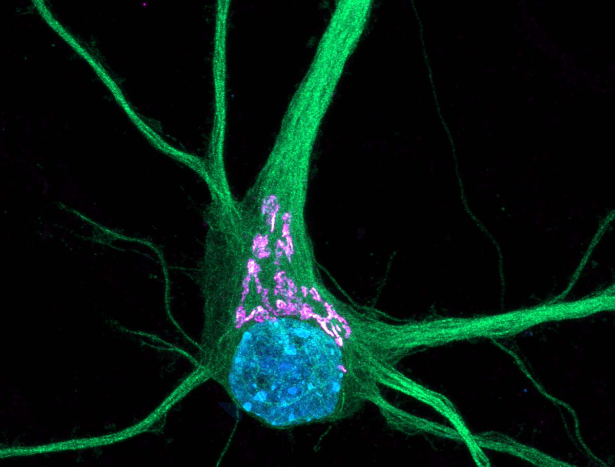
โœจ Excited to kick off #CellSpeak with American Society for Cell Biology! Letโ€™s dive into the breathtaking world of cellsโ€”tiny universes shaping life, health, and discovery. ๐Ÿ”ฌ I'll start with my favorite cell: a neuron (microtubules in green, Golgi complex in magenta, and nucleus in blue). ๐Ÿง ๐Ÿ’™

โœจ Excited to kick off #CellSpeak with <a href="/ASCBiology/">American Society for Cell Biology</a>! Letโ€™s dive into the breathtaking world of cellsโ€”tiny universes shaping life, health, and discovery. ๐Ÿ”ฌ

I'll start with my favorite cell: a neuron (microtubules in green, Golgi complex in magenta, and nucleus in blue). ๐Ÿง ๐Ÿ’™
Danielle Beckman (@danibeckman) 's Twitter Profile Photo

It's been a while since I posted a #MicroscopyMonday ๐Ÿค“๐Ÿ”ฌ. I took this photo on our confocal microscope on Friday. Check these beauties in the repair shop. Microglia (๐ŸŸกiba1 marker) are busy doing synaptic pruning in these neurons (๐Ÿ”ตmap2 marker).

It's been a while since I posted a #MicroscopyMonday ๐Ÿค“๐Ÿ”ฌ. I took this photo on our confocal microscope on Friday. Check these beauties in the repair shop. Microglia (๐ŸŸกiba1 marker) are busy doing synaptic pruning in these neurons (๐Ÿ”ตmap2 marker).
Francesco Padovani (@frank_pado) 's Twitter Profile Photo

SpotMAX, our software tool for analysing multi-dimensional microscopy data, is finally out! ๐ŸŽ‰ And conveniently, I just presented it at the #I2K conference ๐Ÿ˜€ A multi-year-long effort of many great collaborators and users. But what can SpotMAX do? A small thread

Mambo Italiano (@mamboitaliano__) 's Twitter Profile Photo

The Devilโ€™s Bridge in Tuscany, Italy ๐Ÿ‡ฎ๐Ÿ‡น Nestled in the province of Lucca, showcasing its full medieval and slightly eerie beauty

The Devilโ€™s Bridge in Tuscany, Italy ๐Ÿ‡ฎ๐Ÿ‡น

Nestled in the province of Lucca, showcasing its full medieval and slightly eerie beauty
The Culturist (@the_culturist_) 's Twitter Profile Photo

Which of these paintings of Mary is better? Pre-Renaissance art might look primitive to our eyes, but it in fact contains a hidden beauty. Because this isn't a painting of Mary and Jesus โ€” but of the entire universe... (thread) ๐Ÿงต

Which of these paintings of Mary is better?

Pre-Renaissance art might look primitive to our eyes, but it in fact contains a hidden beauty.

Because this isn't a painting of Mary and Jesus โ€” but of the entire universe... (thread) ๐Ÿงต
epithelial mechanics fan club (@epimechfc) 's Twitter Profile Photo

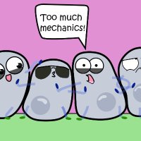
How do tissues maintain their integrity? Let's discuss epithelial fractures! High tissue tension can cause cells to break their contacts, leading to rupture!

Razvan Borza (@rborza_) 's Twitter Profile Photo

AlphaFold can predict protein, DNA, or RNA complexes.. BUT is it easy to determine the reliable interactions?๐Ÿค” โžก๏ธ AlphaBridge will identify the most confident interactions, making your analysis easier! ๐ŸŒTry it here: alpha-bridge.eu ๐Ÿ‘€Read more: biorxiv.org/content/10.110โ€ฆ

AlphaFold can predict protein, DNA, or RNA complexes.. BUT is it easy to determine the reliable interactions?๐Ÿค”

โžก๏ธ AlphaBridge will identify the most confident interactions, making your analysis easier!

๐ŸŒTry it here: alpha-bridge.eu
๐Ÿ‘€Read more: biorxiv.org/content/10.110โ€ฆ
X Paul Ph.D ๐Ÿ‡ซ๐Ÿ‡ฎ๐Ÿ‡บ๐Ÿ‡ฆ (@rollingmeow) 's Twitter Profile Photo

๐ŸŽ‰Thrilled to share our most recent work on how the lipid environment finely regulates the Golgi V-ATPase. A big THANK YOU to everyone in the Ikonen lab IkonenLab @MiesvdStoel X Paul Ph.D ๐Ÿ‡ซ๐Ÿ‡ฎ๐Ÿ‡บ๐Ÿ‡ฆ, and our collaborators. ๐Ÿ”ฌRead more: biorxiv.org/content/10.110โ€ฆ

Journal of Cell Biology (@jcellbiol) 's Twitter Profile Photo

Feng, Cai, Xu et al. Zhejiang University report that saturated fatty acids cause mitochondrial damage and ROS elevation in human endothelial cells. Activation of #lysosome biogenesis using TRPML1 agonists is sufficient to mitigate SFA-induced #mitochondria damage. hubs.la/Q02X6t1l0

Feng, Cai, Xu et al. <a href="/ZJU_China/">Zhejiang University</a> report that saturated fatty acids cause mitochondrial damage and ROS elevation in human endothelial cells. Activation of #lysosome biogenesis using TRPML1 agonists is sufficient to mitigate SFA-induced #mitochondria damage. hubs.la/Q02X6t1l0
Leonel Malacrida (@malacridalab) 's Twitter Profile Photo

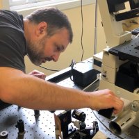
Extremely happy to share this review #FLIM at Nature Reviews Methods Primers in collaboration between #LFD Michelle Digman Belรฉn Torrado and Advanced Bioimaging Unit #BrunoPannunzio. Special thanks to editor #NatalieBarnes, who help us to shape this review. Take a look ๐Ÿ‘‰ rdcu.be/dZqtr

Extremely happy to share this review #FLIM at <a href="/MethodsPrimers/">Nature Reviews Methods Primers</a> in collaboration between #LFD <a href="/michelledigman/">Michelle Digman</a> <a href="/torrado_belen/">Belรฉn Torrado</a> and <a href="/advanced_unit/">Advanced Bioimaging Unit</a> #BrunoPannunzio. Special thanks to editor #NatalieBarnes, who help us to shape this review.
Take a look ๐Ÿ‘‰ rdcu.be/dZqtr
Journal of Cell Biology (@jcellbiol) 's Twitter Profile Photo

Casler, @LLLackner et al. Lackner Lab describe how tripartite membrane contacts btwn #mitochondria, ER & plasma membrane regulate mitochondrial division & PI(4)P #metabolism. hubs.la/Q02_58RP0 Part of the Year in Cell Biology: hubs.la/Q02_5gf90 #CellBio2024
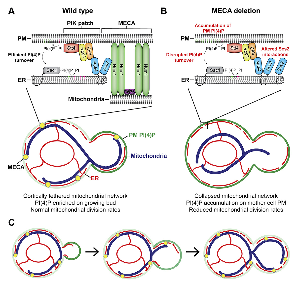
Casler, @LLLackner et al. <a href="/LacknerLabNU/">Lackner Lab</a> describe how tripartite membrane contacts btwn #mitochondria, ER &amp; plasma membrane regulate mitochondrial division &amp; PI(4)P #metabolism. hubs.la/Q02_58RP0

Part of the Year in Cell Biology: hubs.la/Q02_5gf90
#CellBio2024
Journal of Cell Biology (@jcellbiol) 's Twitter Profile Photo

An example 12ร— 3D-ExM 3D movie of a PtK2 #metaphase cell with #aneuploidy (gain). Roshan Norman Chen, Mark Burkard, Aussie Suzuki et al. UWโ€“Madison demonstrate that 3D-ExM provides cost-effective and user-friendly super-resolution #microscopy. hubs.la/Q02-9f1H0

Journal of Cell Biology (@jcellbiol) 's Twitter Profile Photo

Valenti, David, Schuldiner et al. Weizmann Institute created a proteome-wide yeast degron library for rapid in vivo protein depletion. This tool enables dynamic protein function studies with minimal cellular rewiring, advancing beyond prior collections. hubs.la/Q0302VkW0
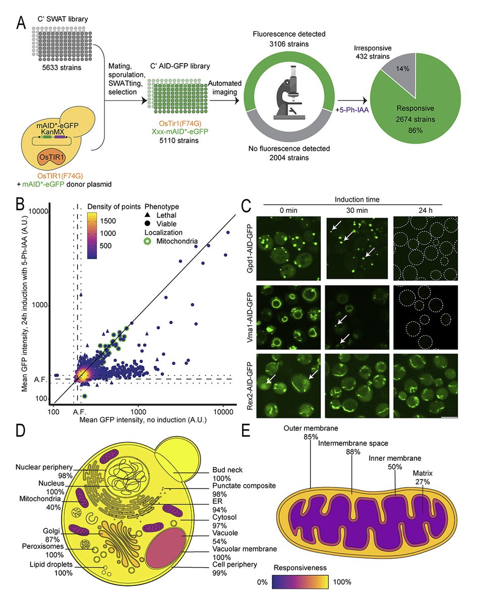
Valenti, David, Schuldiner et al. <a href="/WeizmannScience/">Weizmann Institute</a> created a proteome-wide yeast degron library for rapid in vivo protein depletion. This tool enables dynamic protein function studies with minimal cellular rewiring, advancing beyond prior collections. hubs.la/Q0302VkW0