Debora A. Ledesma, MD (@ledesmadeboraa) 's Twitter Profile
Debora A. Ledesma, MD

@ledesmadeboraa

PGY-1 @UTMB_PATHOLOGY🔬. Former Research Scientist @MDANDERSONNEWS.Dermatologist 🇦🇷. Marathoner🏃‍♀️

ID: 1366243419568373763

calendar_today01-03-2021 04:27:06

82 Tweet

154 Takipçi

405 Takip Edilen

Kurt Schaberg (@kurtschaberg) 's Twitter Profile Photo

I just updated my Thyroid Cytology notes on my teaching website, "Kurt's Notes" (kurtsnotes.net) to incorporate the newest Bethesda system. 🔬🔬🔬🔬🔬🔬🔬🔬🔬🔬🔬🔬🔬🔬🔬🔬🔬🔬🔬🔬🔬🔬🔬🔬🔬🔬🔬 Full notes available here: schaberg.faculty.ucdavis.edu/wp-content/upl…

I just updated my Thyroid Cytology notes on my teaching website, "Kurt's Notes" (kurtsnotes.net) to incorporate the newest Bethesda system.

🔬🔬🔬🔬🔬🔬🔬🔬🔬🔬🔬🔬🔬🔬🔬🔬🔬🔬🔬🔬🔬🔬🔬🔬🔬🔬🔬

Full notes available here: schaberg.faculty.ucdavis.edu/wp-content/upl…
Andres Matoso (@andres_matoso) 's Twitter Profile Photo

Renal medullary carcinoma usually presents with metastasis. Located in the medullary region shows tubulopapillary, cords or solid sheets of cells w/ high nuclear grade and eosinophilic cytoplasm. Neutrophilic infiltration is characteristic. #gupath

Renal medullary carcinoma usually presents with metastasis. Located in the medullary region shows tubulopapillary, cords or solid sheets of cells w/ high nuclear grade and eosinophilic cytoplasm. Neutrophilic infiltration is characteristic. #gupath
Jerad Gardner, MD (@jmgardnermd) 's Twitter Profile Photo

What are these cells & how do you know? Answer: kikoxp.com/posts/3910/ #Pathology #dermpath #dermatology #dermatologia #dermtwitter #pathologists #pathTwitter #histology

What are these cells & how do you know? Answer: kikoxp.com/posts/3910/ #Pathology #dermpath #dermatology #dermatologia #dermtwitter #pathologists #pathTwitter #histology
Elizabeth Montgomery, MD (@lizmontgomerymd) 's Twitter Profile Photo

This pancreatic solitary fibrous tumor is completely sclerotic. Perhaps the pancreatic enzymes destroyed it as it intercalated amongst the acini. #UMiamiPath

This pancreatic solitary fibrous tumor is completely sclerotic.  Perhaps the pancreatic enzymes destroyed it as it intercalated amongst the acini. #UMiamiPath
Andres Matoso (@andres_matoso) 's Twitter Profile Photo

Metastatic breast carcinoma to the bladder can mimic urothelial carcinoma. As with all secondary tumors, it is critical to have a high level of clinical suspicion and do the markers. Morphologic clue: tumor under normal urothelium, no CIS or papillary structures. #gupath

Metastatic breast carcinoma to the bladder can mimic urothelial carcinoma. As with all secondary tumors, it is critical to have a high level of clinical suspicion and do the markers. Morphologic clue: tumor under normal urothelium, no CIS or papillary structures. #gupath
Debora A. Ledesma, MD (@ledesmadeboraa) 's Twitter Profile Photo

Beyond excited to join the amazing UTMB Pathology family!!! UTMB Pathology ❤️🔬🎉🎉🎉 Feeling grateful and blessed🥹🥰🙌 #Match2024 #PathTwitter #pathmatch24

American Journal of Dermatopathology (@ajdp_journal) 's Twitter Profile Photo

#WhatsNewInDermpath Debora A. Ledesma, MD and team Dr Phyu P Aung Carlos Torres-Cabala, MD MD Anderson Cancer Center assessed protein expression of BCL2, MCL1, BIM, and BRAF V600E by IHC in 32 melanoma samples, including Acral Lentiginous Melanoma (ALM) and non-ALM (NALM). journals.lww.com/amjdermatopath…

#WhatsNewInDermpath
<a href="/LedesmaDeboraA/">Debora A. Ledesma, MD</a> and team <a href="/DrPhyuPAung2/">Dr Phyu P Aung</a> <a href="/TorresCabalaMD/">Carlos Torres-Cabala, MD</a> <a href="/MDAndersonNews/">MD Anderson Cancer Center</a> assessed protein expression of BCL2, MCL1, BIM, and BRAF V600E by IHC in 32 melanoma samples, including Acral Lentiginous Melanoma (ALM) and non-ALM (NALM).

journals.lww.com/amjdermatopath…
Dr Phyu P Aung (@drphyupaung2) 's Twitter Profile Photo

We are recruiting our future surgical pathology fellows (2026-2027) to join our wonderful MD Anderson family. Don’t miss the opportunity to learn from our world renowned pathologists.

UTMB Pathology (@utmb_pathology) 's Twitter Profile Photo

Please join us in congratulating our newly selected Chiefs for the 2025-2026 academic year Dr. Peter Powell and Dr. Gilberto Vento!

Please join us in congratulating our newly selected Chiefs for the 2025-2026 academic year Dr. Peter Powell and Dr. Gilberto Vento!
Dr Phyu P Aung (@drphyupaung2) 's Twitter Profile Photo

We are now accepting applications for the 2027–2028 Surgical Pathology Fellowship at MD Anderson! Join our dynamic and supportive Pathology family—only a few positions remain! If you're interested, we encourage you to apply soon before the remaining spots are filled.

Celeste Wagner, MD (@wagceleste) 's Twitter Profile Photo

🎉Excited to share our latest publication on familial pancreatic neoplasms, co-authored with my mentor, Jing He, MD. UTMB Pathology #PancreaticCancer #Pathology pathologyoutlines.com/topic/pancreas…

🎉Excited to share our latest publication on familial pancreatic neoplasms, co-authored with my mentor, <a href="/JingHeMD/">Jing He, MD</a>.
<a href="/UTMB_Pathology/">UTMB Pathology</a> 

#PancreaticCancer #Pathology

pathologyoutlines.com/topic/pancreas…
Zahra Sarrafan Chaharsoughi, MD (@zsarrafan) 's Twitter Profile Photo

Extremely grateful for the opportunity to present three posters at #CAP2025 in Orlando, connect with brilliant minds in the field, and be part of such a wonderful community CAPathologists! Huge thanks to my attendings and co-authors UTMB Pathology for their support.
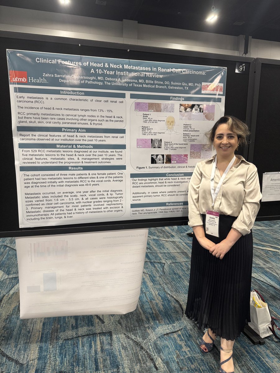
Extremely grateful for the opportunity to present three posters at #CAP2025 in Orlando, connect with brilliant minds in the field, and be part of such a wonderful community <a href="/Pathologists/">CAPathologists</a>! 

Huge thanks to my attendings and co-authors <a href="/UTMB_Pathology/">UTMB Pathology</a> for their support.