CSHL Mouse Course
@cshlmousecourse
Intensive lecture and lab course @CSHL designed for scientists interested in using mouse models to study mammalian development, stem cells and cancer.
ID: 875764660296970240
https://meetings.cshl.edu/courses.aspx?course=C-MOUS&year=22 16-06-2017 17:19:00
413 Tweet
605 Takipçi
591 Takip Edilen


Congratulations to Dr. Loydie Jerome-Majewska Research Institute of the MUHC (The Institute) McGill University receiving the Anne McLaren Award for Outstanding Women in Developmental Biology. International Society of Differentiation is sponsoring this named lecture Society for Developmental Biology Annual Meeting this summer in Chicago. bit.ly/3Sl6Boy


Wow! This so amazing! Thanks thank you for this acknowledgment and thank you International Society of Differentiation and Society for Developmental Biology for this recognition. It’s been an amazing year for me and I appreciate it more than I can say.


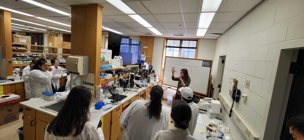
The 2023 CSHL Mouse Course has officially kicked off! Eszter Pos@EPosfai from Princeton University gave an exciting presentation last night and today we are learning IVF from Yojiro Yamanaka and Mitra Cowan from McGill.


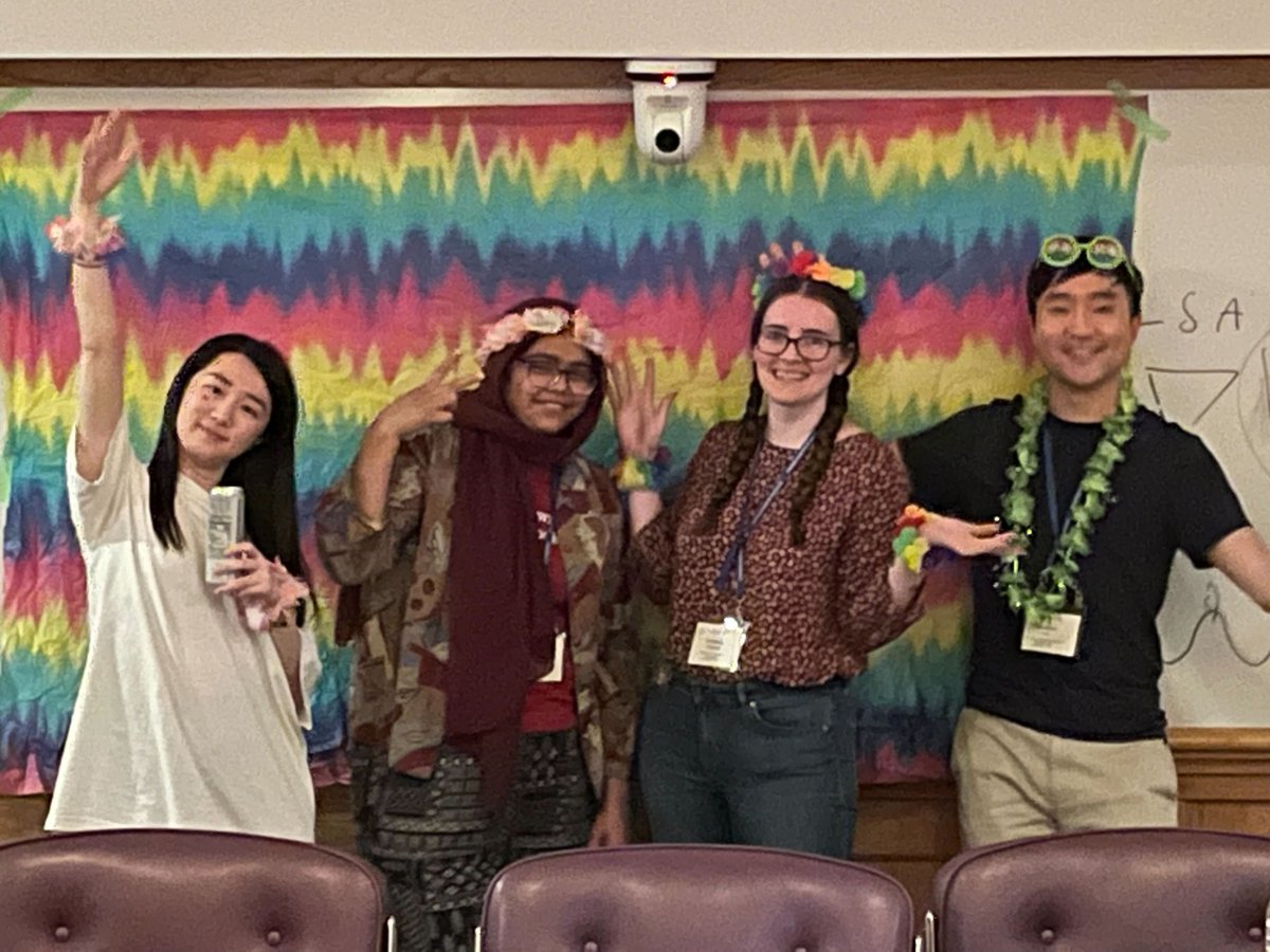
We are enjoying our first party after a fantastic evening lecture from Heiko Lickert Heiko Lickert


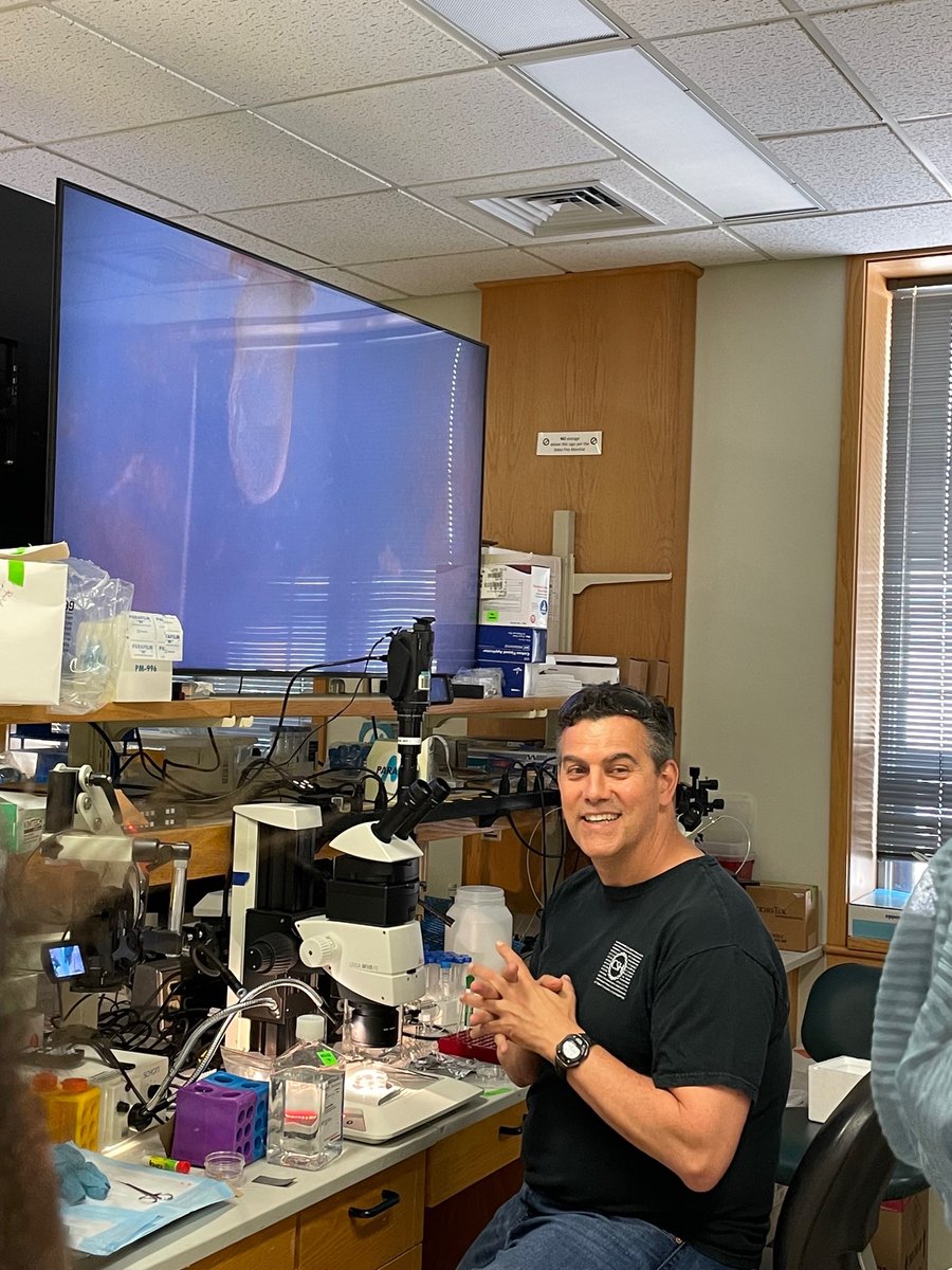
Paul Trainor Paul Trainor Stowers Institute DevelopmentalDynamic arrived last night after 1am, lectured for 3 hours this morning and is currently teaching E7.5 roller culture. #IronMan


This image taken by Dr. Madhulika Pathak Carnegie Embryology shows Sox9 and Twist embryonic gene expression. It was imaged on a Nikon AXR confocal and modeled with Imaris software. Thanks to Molecular Instruments, Nikon Microscope Solutions, Imaris 3D/4D Imaging and Dr. Matt Anderson for their generous support.

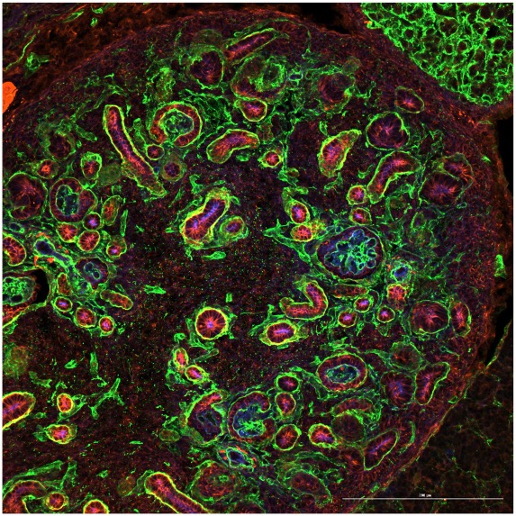
Hyunwook Lee Hyunwook Lee @DrWillZacharias generated this embryonic mouse kidney immunofluorescence image. He sectioned it on an Epredia Cryostar and imaged it with a BioTek Cytation C10 Confocal Imaging Reader. Thank you Agilent Technologies and Epredia for your generous support.


Dr. Günes Taylor Güneş Taylor from The Francis Crick Institute took this lovely picture. It shows Sox9 (red) and Pecam (magenta) detected with HCR reagents provided by Molecular Instruments. It was imaged on a Nikon AXR Confocal microscope loaned by Nikon Microscope Solutions


