Victor Farah, MD (@victorfarah75) 's Twitter Profile
Victor Farah, MD

@victorfarah75

Advanced Cardiac Imaging, ECHO/CMR/CT, Structural and Valvular Heart Disease. FACC, FASE, FSCMR

ID: 351865225

calendar_today09-08-2011 20:50:31

848 Tweet

274 Followers

170 Following

American College of Cardiology (@accintouch) 's Twitter Profile Photo

🔑 Normal-flow low-gradient severe AS is defined as aortic valve area ≤1.0 cm2 w/ low gradient + normal flow and occurs in approx 20-30% of patients w/ severe AS. Read more key points from a review comparing the US & European guidelines for NFLG AS: bit.ly/3wsueF3

🔑 Normal-flow low-gradient severe AS is defined as aortic valve area ≤1.0 cm2 w/ low gradient + normal flow and occurs in approx 20-30% of patients w/ severe AS.

Read more key points from a review comparing the US & European guidelines for NFLG AS: bit.ly/3wsueF3
Tom C. Nguyen, MD (@tomcnguyen) 's Twitter Profile Photo

Ever wonder what an in-situ Amplatzer plug and hole in a bioprosthetic valve looks like? The minimal amount of endothelialization on the plug is interesting.

🇳🇴🇩🇰Asbjørn Støylen 🇬🇱🇵🇸🇺🇦 (@strain_rate) 's Twitter Profile Photo

🧵on ventricular ejection. Does blood always flow downwards a pressure gradient? Certainly not. A pressure gradient accelerates stagnant blood to flow down the gradient, but blood in motion may flow against the pressure gradient (by inertia), being decelerated.

🧵on ventricular ejection. Does blood always flow downwards a pressure gradient? Certainly not. A pressure gradient accelerates stagnant blood to flow down the gradient, but blood in motion may flow against the pressure gradient (by inertia), being decelerated.
Jae K. Oh (@jaekoh2) 's Twitter Profile Photo

1/5 My Dx for the e' velocity of 4 cm/s is reduced myocardial relaxation, which is sine qua non of diastolic dysfunction. It is present in all forms of myocardial disease and also with aging. Let me explain its progression & how to use the information for our pts Mayo Clinic CV

1/5 My Dx for the e' velocity of 4  cm/s  is reduced myocardial relaxation, which is sine qua non of diastolic dysfunction. It is present in all forms of myocardial disease and also with aging. Let me explain its progression &amp; how to use the information for our pts <a href="/MayoClinicCV/">Mayo Clinic CV</a>
The Heart (@zheart11768530) 's Twitter Profile Photo

🔴Where Are We With Treatment and Prevention of Heart Failure in Patients Post–Myocardial Infarction? #openaccess #2024Review JACC Journals jacc.org/doi/10.1016/j.… #internalmedicine #medicalpractice #medx #usmle #MedTwitter #cardiotwitter #FOAMed
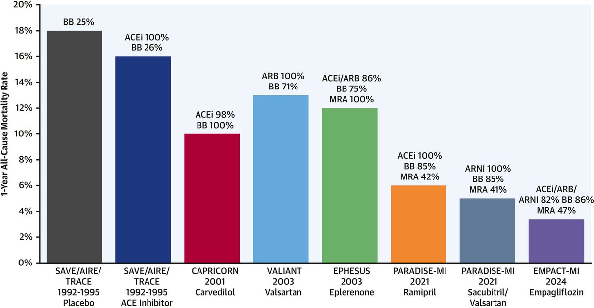
🔴Where Are We With Treatment and Prevention of Heart Failure in Patients Post–Myocardial Infarction? #openaccess #2024Review <a href="/JACCJournals/">JACC Journals</a>

jacc.org/doi/10.1016/j.…
 #internalmedicine #medicalpractice #medx #usmle #MedTwitter #cardiotwitter #FOAMed
The Heart (@zheart11768530) 's Twitter Profile Photo

🔴 Valvular Heart Disease : 🔸Echocardiographic & Hemodynamic findings 🔸The guiding principles for medical and Device Management #internalmedicine #medicalpractice #medx #usmle #MedTwitter #cardiotwitter #FOAMed #echofirst #MedstudentTwitter #paramedic #MedEd #Cardiology

🔴 Valvular Heart  Disease : 

🔸Echocardiographic &amp; Hemodynamic findings 
🔸The guiding principles for medical and  Device Management

#internalmedicine #medicalpractice #medx #usmle #MedTwitter #cardiotwitter #FOAMed #echofirst  #MedstudentTwitter #paramedic  #MedEd #Cardiology
Jae K. Oh (@jaekoh2) 's Twitter Profile Photo

Most of times, we should be able to determine diastolic function and filling pressure based on mitral flow and annulus velocity as in this case of restrictive hemodynamics with rapid rise of LV diastolic pressure. Patient had severe AR due to IE.

Most of times, we should be able to determine diastolic function and filling pressure based on mitral flow and annulus velocity as in this case of restrictive hemodynamics with rapid rise of LV diastolic pressure. Patient had severe AR due to IE.
Jae K. Oh (@jaekoh2) 's Twitter Profile Photo

Majority selected the first option of "75 yo with exertional dyspnea", a typical symptom of HFpEF or diastolic heart failure where diastolic filling pressure is not elevated at rest, usually grade 1 DF, but increases with exercise. Diastolic exercise Echo is helpful.

Majority selected the first option of "75 yo with exertional dyspnea", a typical symptom of HFpEF or diastolic heart failure where diastolic filling pressure is not elevated at rest,  usually grade 1 DF, but  increases with exercise. Diastolic exercise Echo is helpful.
American Society of Echocardiography (@ase360) 's Twitter Profile Photo

Check out the full video, “How to Measure Wall Thickness with Echocardiography,” from our Microlesson Series on Hypertrophic Cardiomyopathy (#HCM)! Watch it here: bit.ly/3LydPTe

AHN_CVI (@ahn_cvi) 's Twitter Profile Photo

Today is HCM Awareness Day! HCM impacts approximately 1 in 500 Americans and we are proud that Allegheny General Hospital is a recognized Hypertrophic Cardiomyopathy Center of Excellence. ❤️👍 AHN To learn more visit: ahn.org/services/cardi…

Today is HCM Awareness Day!  HCM impacts approximately 1 in 500 Americans and we are proud that Allegheny General Hospital is a recognized Hypertrophic Cardiomyopathy Center of Excellence. ❤️👍 <a href="/AHNtoday/">AHN</a>  
To learn more visit: ahn.org/services/cardi…