Spraggins Lab
@spragginslab
Student-run Twitter page for @JeffSpraggins
| @VanderbiltCDB | @VU_BIOMIC
ID: 1421189347668205572
https://medschool.vanderbilt.edu/biomic/ 30-07-2021 19:23:32
202 Tweet
418 Followers
296 Following

The Prentice lab is looking forward to #ASMS2024! Come check out our work on gas-phase ion/ion reactions, imaging, ion mobility, and more! Am Soc for Mass Spec


Always a great time getting together with Spraggins Lab & Van de Plas Lab for our annual SVP dinner! #ASMS2024


Shoutout to Allison Esselman for her informative talk and panel responses yesterday at the workshop on advances in high spatial resolution imaging mass spectrometry!! #ASMS2024


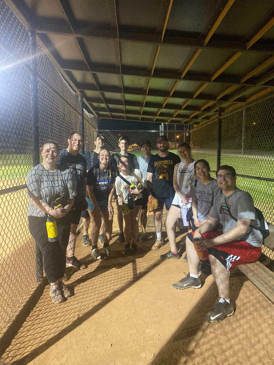
Our collaborations with the Eric Skaar and @CassatLab has expanded from the bench, onto the softball field! Great first game of the season everyone!


Eye hope we can see eye to eye - beauty is in the eye of the beholder & you will see r-eye-t away this CODEX image from #_hubmap researcher Dr. Angela Kruse Spraggins Lab Vanderbilt University is a vision to behold w/ its retinal retinue of proteins! Eye know you’ll like what you see!


Imaging of gangliosides during Staph infections. New paper from Katerina Djambazova and the Spraggins Lab. Happy to be involved in this beautiful work. J. of the American Society for Mass Spectrometry 🔬 🦠 pubs.acs.org/doi/10.1021/ja…

New publication by Katerina Djambazova on MALDI TIMS imaging of gangliosides in kidney s.aureus abscesses pubs.acs.org/doi/10.1021/ja…

Excellent lightning talks from yesterday’s #SBS2024 by Youlim K. from the Amieva lab, Stanford University and Cody and Thai from the Spraggins Lab at Vanderbilt University and many more!


Congrats to Dr. Angela R Kruse and the rest of the team on their work optimizing methods for spatial proteomics using MALDI IMS! Check out our paper below to see how thermal denaturation can improve peptide imaging even in unfixed, frozen tissues. pubs.acs.org/doi/10.1021/ac…

Excited to share our most recent paper by Dr. Christopher Good & our Collaborators in the @CassatLab! This work highlights our ability to map lipid dynamics between bacteria and leukocytes in S.aureus osteomyelitis. @VI4Research Vanderbilt School of Medicine Basic Sciences cell.com/cell-chemical-…

dark blue-nuclei, yellow-smooth muscle cells, red-leukocytes, magenta-Mueller glia, green-bipolar cells, light blue-cone cells, white-fibroblasts, and light pink-retinal pigmented epithelium Spraggins Lab VU Biomolecular Multimodal Imaging Center #WomenInScience #HuBMAP

Harsimran Kaur, Cody Heiser, Simon Vandekar developed Multiplex Image Labelling with Regional Morphology (MILWRM) to identify consensus tissue domains in spatial transcriptomics (ST) and multiplex immunofluorescence data (mIF) @NCIHTAN nature.com/articles/s4200…

If you want to map Cells in situ, then come see #HuBMAP’s OMAPs here: nature.com/articles/s4159… Teal – proximal tubules Dark blue – vasculature Pink – podocytes in glomeruli Yellow – mesangial cells in glomeruli Spraggins Lab VU Biomolecular Multimodal Imaging Center #WomenInScience


