Peter Fiester, MD (@petefiestermd) 's Twitter Profile
Peter Fiester, MD

@petefiestermd

Associate Professor & Director @UFHealth #Neuroradiology @UFHealthjax, images=pubs, #Radiology, #MedEd, #Spine, #Stroke, #Skullbase

ID: 2858720910

linkhttps://med.jax.ufl.edu/directory/bio/2977/peter-fiester/ calendar_today16-10-2014 22:56:29

398 Tweet

2,2K Followers

322 Following

Peter Fiester, MD (@petefiestermd) 's Twitter Profile Photo

🤷‍♂️ Why care about Anterior Atlanto-Occipital Membrane (AAOM) tears? ✅ Routinely visible on #MRI ✅ Tears in 3️⃣ distinct patterns ✅ Presents w/ CCJ prevertebral effusion on #CT & major CCJ lig. injuries on #MRI ➡️ rdcu.be/dawhs #NeuroRad #NeuroSurgery #Spine

🤷‍♂️ Why care about Anterior Atlanto-Occipital Membrane (AAOM) tears? 

✅  Routinely visible on #MRI

✅  Tears in 3️⃣ distinct patterns

✅  Presents w/ CCJ prevertebral effusion on #CT & major CCJ lig. injuries on #MRI 

➡️ rdcu.be/dawhs 

#NeuroRad #NeuroSurgery #Spine
Peter Fiester, MD (@petefiestermd) 's Twitter Profile Photo

📢 Honored to share our latest #Research endeavor on this often overlooked #Ligament in the upper cervical #Spine. Upper prevertebral effusion on #CT = great predictor of AAOMc injury & internal #CCJ derangement in high velocity #Trauma! #Radiology #RadRes #Neurosurgery #Spine

📢 Honored to share our latest #Research endeavor on this often overlooked #Ligament in the upper cervical #Spine.

Upper prevertebral effusion on #CT = great predictor of AAOMc injury & internal #CCJ derangement in high velocity #Trauma!

#Radiology #RadRes #Neurosurgery #Spine
Peter Fiester, MD (@petefiestermd) 's Twitter Profile Photo

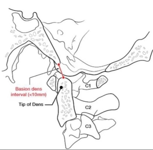
💁‍♂️ Friendly #RadRes reminder ➡️ BDI widening is unreliable in determining #craniocervical dissociation. For some reason, this measurement is the one residents always rememeber. Just a plain film hangover! 😉 #NeuroRad #Radiology #MedEd

💁‍♂️ Friendly #RadRes reminder ➡️ BDI widening is unreliable in determining #craniocervical dissociation. 

For some reason, this measurement is the one residents always rememeber.  Just a plain film hangover! 😉

#NeuroRad #Radiology #MedEd
Peter Fiester, MD (@petefiestermd) 's Twitter Profile Photo

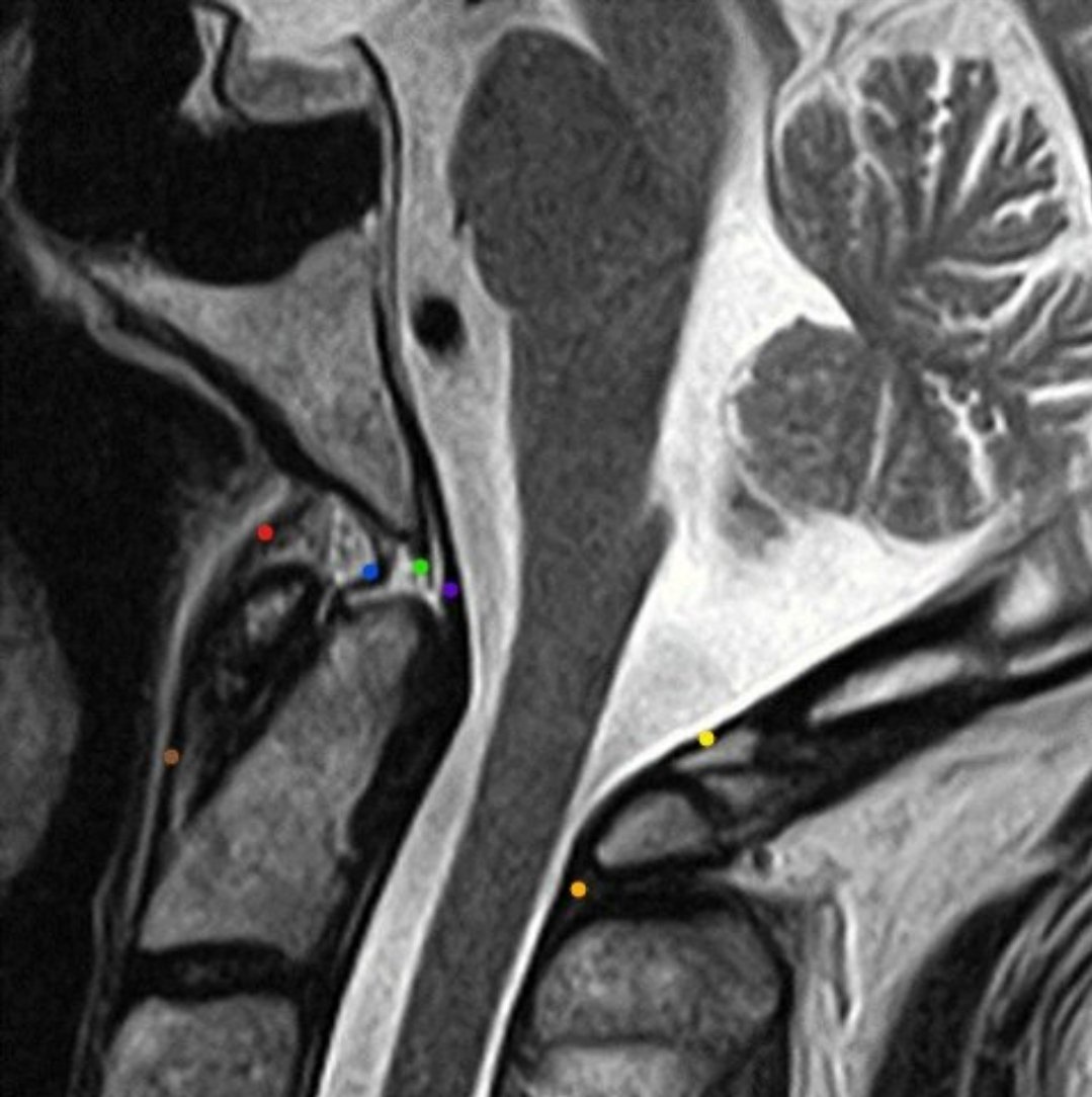
Hx: High velocity MVC #trauma. ➡️ Cervical #MRI: An apical ligament tear & craniocervical injury on the sag. STIR 🤔? ✅ Nope, just a normal venous plexus & a negative MRI. #RadRes #NeuroRad #Spine

Hx: High velocity MVC #trauma.

➡️ Cervical #MRI: An apical ligament tear & craniocervical injury on the sag. STIR 🤔?

✅ Nope, just a normal venous plexus & a negative MRI. 

#RadRes #NeuroRad #Spine
Philip R. Chapman, MD (@philiprchapman1) 's Twitter Profile Photo

Juvenile nasopharyngeal angiofibromas are rare, but are often a board favorite. They are vascular tumors that occur almost exclusively in adolescent males & present with nasal obstruction or epistaxis. Treatment includes endovascular embolization followed by surgical resection.

Juvenile nasopharyngeal angiofibromas are rare, but are often a board favorite. They are vascular tumors that occur almost exclusively in adolescent males & present with nasal obstruction or epistaxis.  Treatment includes endovascular embolization followed by surgical resection.
Peter Fiester, MD (@petefiestermd) 's Twitter Profile Photo

#RadRes: Anterior osteophyte fx. can be tricky in a degenerative spine: ⤵️ Left = acute Right = chronic ✅ Prevertebral effusion & absent cortication along the "lucency" although are super helpful... #NeuroRad #Trauma #Spine

#RadRes: Anterior osteophyte fx. can be tricky in a degenerative spine: ⤵️

Left = acute 
Right = chronic

✅  Prevertebral effusion & absent cortication along the "lucency" although are super helpful...

#NeuroRad #Trauma #Spine
Peter Fiester, MD (@petefiestermd) 's Twitter Profile Photo

🔑 Tectorial membrane interdigitates w/ & forms the dural plane between the retroclival epidural & subdural compartments in peds patients: ✅ Epidural = more common in MVA trauma (sign of CCJ derangement!) ✅ Subdural = more common in Non accidental trauma. #RadRes #NeuroRad

🔑 Tectorial membrane interdigitates w/ & forms the dural plane between the retroclival epidural & subdural compartments in peds patients:

✅ Epidural = more common in MVA trauma (sign of CCJ derangement!)

✅ Subdural = more common in Non accidental trauma.

#RadRes #NeuroRad
Philip R. Chapman, MD (@philiprchapman1) 's Twitter Profile Photo

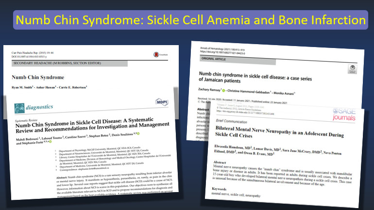
"Numb chin syndrome" , or mental nerve neuropathy, refers to unilateral or bilateral numbness of the lower lip and chin in distribution of the mental or inferior alveolar nerves. It can have a variety of causes. Vaso-occlusion in the maxillofacial region of sickle cell patients

"Numb chin syndrome" , or mental nerve neuropathy, refers to unilateral or bilateral numbness of the lower lip and chin in distribution of the mental or inferior alveolar nerves. It can have a variety of causes.

Vaso-occlusion  in the maxillofacial region of sickle cell patients
Peter Fiester, MD (@petefiestermd) 's Twitter Profile Photo

#RadRes 📚 Central C2 osteochondrosal injury on #MRI in a 5 y/o ⤵️ ✅ Anterior angulation of the dens. ✅ C2 Synchondrosal fluid. ✅ Posterior AO membrane complex injury (low grade) #NeuroRad #Spine #Trauma

#RadRes 📚  Central C2 osteochondrosal injury on #MRI in a 5 y/o ⤵️

✅ Anterior angulation of the dens.
✅ C2 Synchondrosal fluid.
✅ Posterior AO membrane complex injury (low grade)

#NeuroRad #Spine #Trauma
Peter Fiester, MD (@petefiestermd) 's Twitter Profile Photo

Nice depiction of C2 synchondroses in a 2 y/o 😎 (with adult C2 vertebrae for comparison): ⤵️ ✅ Note the "absent" os odontoideum (which hasn't yet ossified) but w/ the apicodental synchondrosis present. #RadRes #NeuroRad #Spine #MedEd

Nice depiction of C2 synchondroses in a 2 y/o 😎 (with adult C2 vertebrae for comparison): ⤵️

✅ Note the "absent" os odontoideum (which hasn't yet ossified) but w/ the apicodental synchondrosis present.

#RadRes #NeuroRad #Spine #MedEd
Peter Fiester, MD (@petefiestermd) 's Twitter Profile Photo

Great work by our #radiology residents UF Jax Radiology Residency in their latest publication on a subtle C2 traumatic injury in the #pediatric population. #RadRes #NeuroRad #Spine #Trauma C2 Synchondrosal Injuries: A Case Report and Anatomic Review cureus.com/articles/17694…

Peter Fiester, MD (@petefiestermd) 's Twitter Profile Photo

Hx: "seizures" Looks up vitals: SBP 220 Dx: HBE (Hypertensive Brainstem Encephalopathy) ➡️ Pontine predominant variant of "PRES" No differential diagnosis needed! 😉

Hx: "seizures"  

Looks up vitals: SBP 220

Dx: HBE (Hypertensive Brainstem Encephalopathy) ➡️  Pontine predominant variant of "PRES" 

No differential diagnosis needed! 😉
Peter Fiester, MD (@petefiestermd) 's Twitter Profile Photo

CT: "Normal" atlanto-occipital joint space. MRI: Unstable craniocervical dissociation injury (same pt.) 🔑: Don't solely rely on the joint space or "lines" to exclude CCJ trauma. Exclude 1. Prevertebral 2. Supradental 3. Retroclival hematomas too!

CT:  "Normal" atlanto-occipital joint space.

MRI:  Unstable craniocervical dissociation injury (same pt.)

🔑:  Don't solely rely on the joint space or "lines" to exclude CCJ trauma.

Exclude 
1.   Prevertebral
2.  Supradental
3.  Retroclival hematomas too!
Peter Fiester, MD (@petefiestermd) 's Twitter Profile Photo

Rule of thumb for detecting M2 MCA vessel occlusion on CTA: ⤵️ ✅ Superior M2 division = territory predominantly ant. to sylvian fissure. ✅ Inferior M2 division = territory predominantly post. to sylvian fissure. Real time saver on those busy call nights 😉!

Rule of thumb for detecting M2 MCA vessel occlusion on CTA:  ⤵️

 ✅  Superior M2 division = territory predominantly ant. to sylvian fissure.

✅  Inferior M2 division = territory predominantly post. to sylvian fissure.

Real time saver on those busy call nights 😉!
Peter Fiester, MD (@petefiestermd) 's Twitter Profile Photo

📢 Not sure who still needs to hear this... But this imaging finding is NEVER (like not ever!) a sign of dural sinus thrombosis (Don't even entertain it! 🤫) Dx: Incidental arachnoid granulation (Image courtesy of Radiopedia)

📢  Not sure who still needs to hear this...

But this imaging finding is NEVER (like not ever!) a sign of dural sinus thrombosis (Don't even entertain it! 🤫)

Dx:  Incidental arachnoid granulation

(Image courtesy of Radiopedia)
Peter Fiester, MD (@petefiestermd) 's Twitter Profile Photo

Cerebellar vascular territories 🧠 ⤵️ Tentorial surface (red) ➡️ Superior cerebellar a. Occipital surface (blue) ➡️ Posterior inferior cerebellar a. Petrosal surface (pink) ➡️ Anterior inferior cerebellar a.

Cerebellar vascular territories 🧠 ⤵️

Tentorial surface (red) ➡️  Superior cerebellar a.

Occipital surface (blue) ➡️  Posterior inferior cerebellar a. 

Petrosal surface (pink) ➡️  Anterior inferior cerebellar a.
Peter Fiester, MD (@petefiestermd) 's Twitter Profile Photo

Actual Hx: "Internuclear Opthalmoplegia (INO)" 😯 🧠 MRI = small right MLF stroke. (Disrupts CN VI nuclei via PPRF ➡️ weakens ipsilateral eye adduction, but preserves contralateral eye abduction.) Shout out to this awesome neurology resident for making my job easy! 😎

Actual Hx:  "Internuclear Opthalmoplegia (INO)" 😯

🧠 MRI = small right MLF stroke.  

(Disrupts CN VI nuclei via PPRF ➡️ weakens ipsilateral eye adduction, but preserves contralateral eye abduction.)

Shout out to this awesome neurology resident for making my job easy! 😎
Peter Fiester, MD (@petefiestermd) 's Twitter Profile Photo

INO can of course be bilateral too! Hx: "Discordant gaze & multiple sclerosis (MS)" 🧠 MRI ➡️ MS plaque in dorsal pons affecting Bilateral MLF (Lack of convergence in horizontal plane causing a characteristic divergence) Aka: "Wall-eyed" 😳

INO can of course be bilateral too!

Hx:  "Discordant gaze & multiple sclerosis (MS)"

🧠 MRI  ➡️  MS plaque in dorsal pons affecting Bilateral MLF (Lack of convergence in horizontal plane causing a characteristic divergence)

Aka: "Wall-eyed" 😳