linda johnston (@oreoimc) 's Twitter Profile
linda johnston

@oreoimc

Cytotech -pathology /science nerd with a healthy spattering of sarcasm ( oh, and a proud Canadian🇨🇦)

ID: 2718467305

calendar_today09-08-2014 02:35:53

4,4K Tweet

2,2K Followers

2,2K Following

Lorand Kis (@kis_lorand) 's Twitter Profile Photo

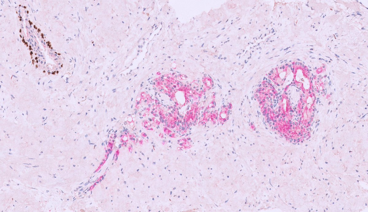
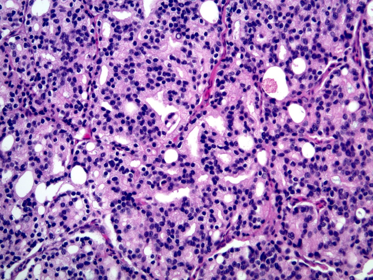
#yourdailyGleasongrading is back with a quizz 😁... Guess how was this tiny cancerfocus measuring 1 mm graded? I hope it is visible that the glands are perineural...The last picture is p63 (brown)-AMACR (red) IHC 👇👇 #GUpath #pathology

#yourdailyGleasongrading is back with a quizz 😁... 
Guess how was this tiny cancerfocus measuring 1 mm graded? I hope it is visible that the glands are perineural...The last picture is p63 (brown)-AMACR (red) IHC 👇👇
#GUpath #pathology
Sam Khader, MD (@samkhader) 's Twitter Profile Photo

Interesting morphology of a squamous cell carcinoma metastatic to the pleural cavity (not common to see SqCC in an effusion #CytoPath specimen). Some of the cells have vacuolated and wispy cytoplasm. Note the combo stain for p40/DSG3. The lung tumor had a papillary architecture.

Interesting morphology of a squamous cell carcinoma metastatic to the pleural cavity (not common to see SqCC in an effusion #CytoPath specimen). Some of the cells have vacuolated and wispy cytoplasm. Note the combo stain for p40/DSG3. The lung tumor had a papillary architecture.
Sanjay Mukhopadhyay (@smlungpathguy) 's Twitter Profile Photo

Pleomorphic carcinoma, lung: Note: this is the thoracic WHO term for sarcomatoid carcinoma. As usual, they threw out a good term that everyone understands for awful, inane terminology.This case also... kikoxp.com/posts/24453

Pleomorphic carcinoma, lung: Note: this is the thoracic WHO term for sarcomatoid carcinoma. As usual, they threw out a good term that everyone understands for awful, inane terminology.This case also... kikoxp.com/posts/24453
Gloria Sura, MD (@gloria_suramd) 's Twitter Profile Photo

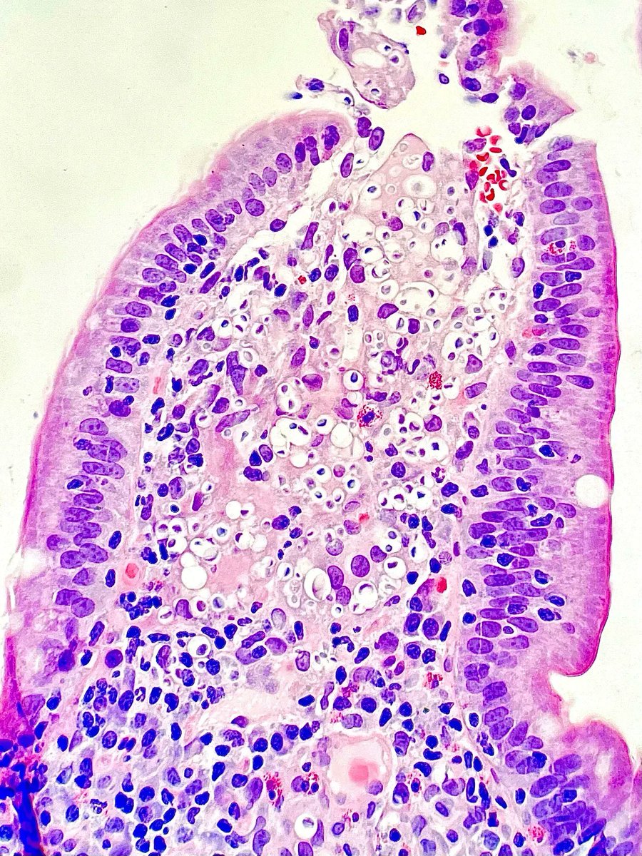
What fungal infection is seen in this biopsy of the small intestine of an immunocompromised patient? Answer & additional stain: buff.ly/3yEAmaZ #PathArt #PathTwitter #IDTwitter #MedTwitter

What fungal infection is seen in this biopsy of the small intestine of an immunocompromised patient? Answer & additional stain: buff.ly/3yEAmaZ
#PathArt #PathTwitter #IDTwitter #MedTwitter
Carlos Miguel Ruiz (@nucleololailo) 's Twitter Profile Photo

✨COLLAGEN BALLS ✨ it is not uncommon to see them in #cytology samples from peritoneal washings, together with large sheets of benign mesothelial cells, perhaps a little angry..

✨COLLAGEN BALLS ✨

it is not uncommon to see them in #cytology samples from peritoneal washings, together with large sheets of benign mesothelial cells, perhaps a little angry..
Sam Khader, MD (@samkhader) 's Twitter Profile Photo

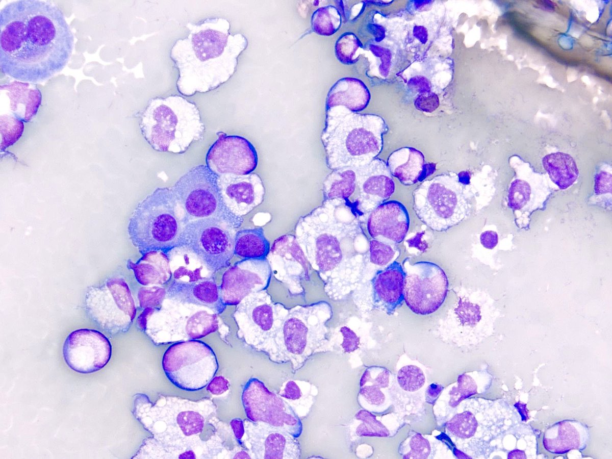
Pleural fluid in a patient with history of of #AML. It’s imperative to review the peripheral blood flow cytometry or smear in cases with abundant red blood cells. Some of these cells are suggestive of blasts; however, peripheral blood contamination cannot be excluded. #CytoPath

Pleural fluid in a patient with history of of #AML. It’s imperative to review the peripheral blood flow cytometry or smear in cases with abundant red blood cells. Some of  these cells are suggestive of blasts; however, peripheral blood contamination cannot be excluded. #CytoPath
Muhammad Ahsan Gill (@ahsanuitis) 's Twitter Profile Photo

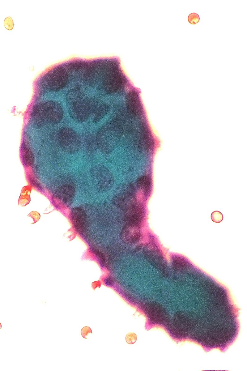
Dr. Zubair Baloch (Zubair Baloch) explaining a helpful cytologic clue to reactive pneumocytes in acute/subacute lung injuries. Their resemblance to Napoleon Bonaparte's iconic hat. Initially described by Natasha Rekhtman MD PhD 🔗 to the article👇: meridian.allenpress.com/aplm/article/1… #IACTutorial
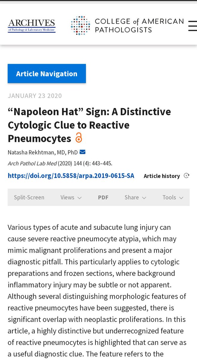
Dr. Zubair Baloch (<a href="/aakasharmand/">Zubair Baloch</a>) explaining a helpful cytologic clue to reactive pneumocytes in acute/subacute lung injuries. Their resemblance to Napoleon Bonaparte's iconic hat. 
 Initially described by <a href="/natasharekhtman/">Natasha Rekhtman MD PhD</a>
🔗 to the article👇: meridian.allenpress.com/aplm/article/1…
#IACTutorial
Sam Khader, MD (@samkhader) 's Twitter Profile Photo

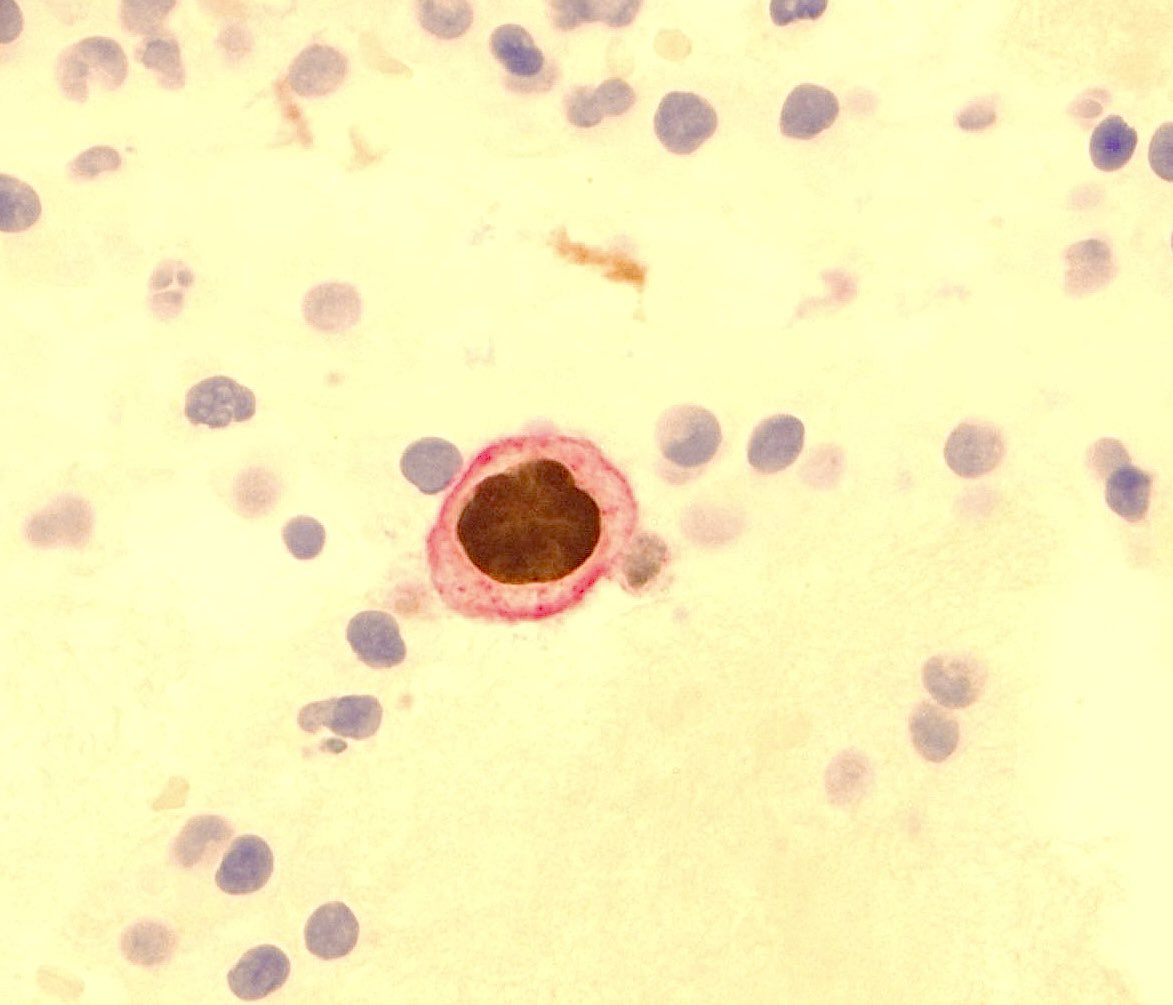
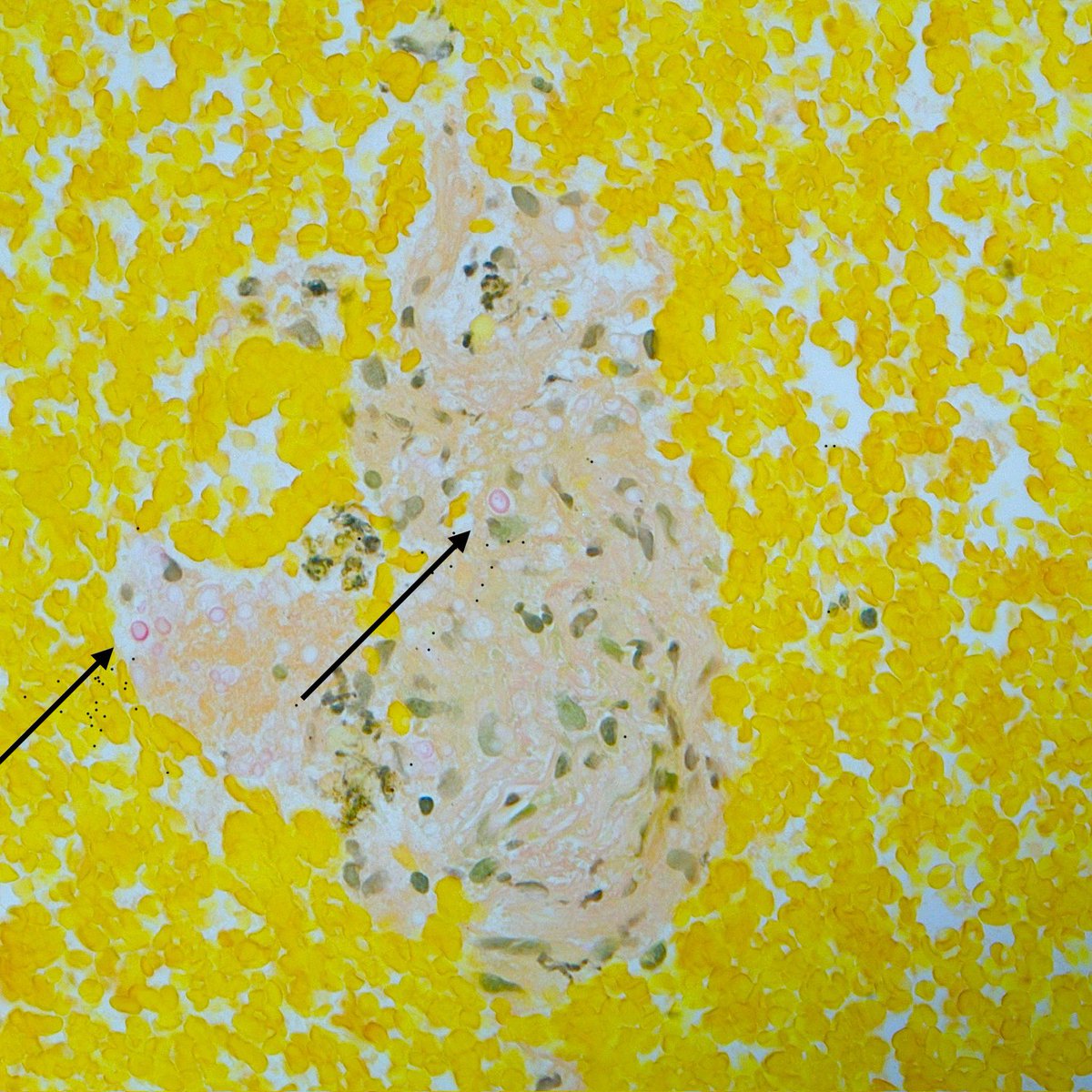
A cell block preparation from a lung nodule FNA in an older man with history of multiple myeloma. Notice the variably sized structures in the H&E stain, which are highlighted by GMS stain. Mucicarmine stain demonstrates the mucin capsule. Diagnosis? #CytoPath

A cell block preparation from a lung nodule FNA in an older man with history of multiple myeloma. Notice the variably sized structures in the H&amp;E stain, which are highlighted by GMS stain. Mucicarmine stain demonstrates the mucin capsule. Diagnosis? #CytoPath
Sam Khader, MD (@samkhader) 's Twitter Profile Photo

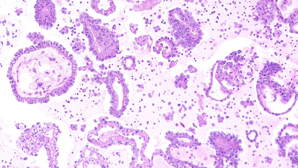
An impressive cell block preparation from a peritoneal fluid #CytoPath specimen in a patient with history of papillary renal cell carcinoma. What #IHC (s) would you order to confirm the diagnosis?

An impressive cell block preparation from a peritoneal fluid #CytoPath specimen in a patient with history of papillary renal cell carcinoma. What #IHC (s) would you order to confirm the diagnosis?