Divya Gunda (@learnneurorad) 's Twitter Profile
Divya Gunda

@learnneurorad

Neuroradiologist @CooperRadRes, here to share #Neurorad cases for #radres #RadEd. Tweets my own, for #MedEd, not medical advice. Alum: @PennRadiology @OUHealth.

ID: 3511086254

linkhttp://instagram.com/learnneurorad calendar_today10-09-2015 03:16:50

566 Tweet

5,5K Followers

434 Following

Divya Gunda (@learnneurorad) 's Twitter Profile Photo

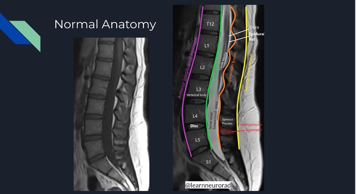
Normal anatomy of the mid-lumbar spine at the L3-4 intervertebral disc level in the axial plane on a T2-weighted MRI. The indicated zones are sites of disc herniations according to Lumbar Disc Nomenclature 2.0. #Neurorad #radres #MedEd #Medtwitter #spine #anatomy #NeuroTwitter

Normal anatomy of the mid-lumbar spine at the L3-4 intervertebral disc level in the axial plane on a T2-weighted MRI. The indicated zones are sites of disc herniations according to Lumbar Disc Nomenclature 2.0. #Neurorad #radres #MedEd #Medtwitter #spine #anatomy #NeuroTwitter
@X (@publishedxprime) 's Twitter Profile Photo

Come join us tomorrow Sep 04 at 10:15am to see these 10 superstars in action at the #ASHNR24 ASHNR T4C - The Cortex Club Crazy cases session! Heralding the future of our specialty! So proud of them!

Come join us tomorrow Sep 04 at 10:15am to see these 10 superstars in action at the #ASHNR24 <a href="/ASHNRSociety/">ASHNR</a> T4C - <a href="/thecortexclub/">The Cortex Club</a> Crazy cases session! 

Heralding the future of our specialty! So proud of them!
Divya Gunda (@learnneurorad) 's Twitter Profile Photo

Sneak peek: Day #2 of the Bootcamp session at #ASHNR24 discussing the Jugular Foramen. The jugular tubercle is a key anatomic structure to distinguish lesions in the jugular foramen (above the tubercle) from lesions in the hypoglossal canal (below the tubercle) #Neurorad #radres

Sneak peek: Day #2 of the Bootcamp session at #ASHNR24 discussing the Jugular Foramen. The jugular tubercle is a key anatomic structure to distinguish lesions in the jugular foramen (above the tubercle) from lesions in the hypoglossal canal (below the tubercle) #Neurorad #radres
Divya Gunda (@learnneurorad) 's Twitter Profile Photo

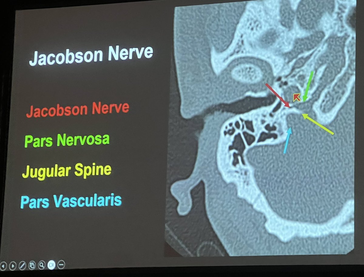
More anatomy of the jugular foramen from #ASHNR24. The jugular foramen is divided into the pars nervosa and the pars vascularis by the jugular spine. #radres #Neurorad

More anatomy of the jugular foramen from #ASHNR24. The jugular foramen is divided into the pars nervosa and the pars vascularis by the jugular spine. #radres #Neurorad
Divya Gunda (@learnneurorad) 's Twitter Profile Photo

MRI brachial plexus shows a distal thoracic duct draining into the IJ & subclavian vein confluence. It can often have a flared or tubular configuration, not to be mistaken for pathology such as lymphadenopathy or brachial plexus injury, which would be more posterior. #radres

MRI brachial plexus shows a distal thoracic duct draining into the IJ &amp; subclavian vein confluence. It can often have a flared or tubular configuration, not to be mistaken for pathology such as lymphadenopathy or brachial plexus injury, which would be more posterior. #radres
Divya Gunda (@learnneurorad) 's Twitter Profile Photo

Unlike other bone lesions, osseous involvement by lymphoma can have a soft tissue mass on either side of the bone, while relatively preserving the adjacent cortex as illustrated 👇#radres #Neurorad

Unlike other bone lesions, osseous involvement by lymphoma can have a soft tissue mass on either side of the bone, while relatively preserving the adjacent cortex as illustrated 👇#radres #Neurorad
Divya Gunda (@learnneurorad) 's Twitter Profile Photo

Fossa navicularis magna is a benign developmental lesion manifesting as an indentation in the anterior inferior clivus. This does not need follow up, biopsy or further imaging. Dr Harun Yıldız illustration of different developmental lesions of the clivus. #Neurorad #radres
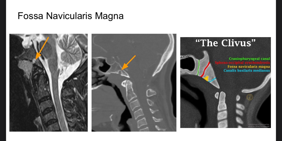
Fossa navicularis magna is a benign developmental lesion manifesting as an indentation in the anterior inferior clivus. This does not need follow up, biopsy or further imaging. <a href="/drharunyildiz/">Dr Harun Yıldız</a> illustration of different developmental lesions of the clivus. #Neurorad #radres
Divya Gunda (@learnneurorad) 's Twitter Profile Photo

This is an easy one for most but still see this overlooked and it helps to see it many times. History of generalized seizures. Findings of left mesial temporal sclerosis with significant atrophy and abnormal signal in the left hippocampus. Image is not pretty but that’s reality!

This is an easy one for most but still see this overlooked and it helps to see it many times. History of generalized seizures. Findings of left mesial temporal sclerosis with significant atrophy and abnormal signal in the left hippocampus. Image is not pretty but that’s reality!