Jack Garnham (@jjgarnham) 's Twitter Profile
Jack Garnham

@jjgarnham

Consultant neuroradiologist @ImperialNHS. Previous pan-London neuroradiology fellow. Winner of the Frank Doyle medal. Interest in medical education.

ID: 1751700029025226752

calendar_today28-01-2024 20:13:32

334 Tweet

691 Followers

277 Following

Jack Garnham (@jjgarnham) 's Twitter Profile Photo

Wilson's disease! Defective ATP7B intracellular copper transporter with accumulation of copper in various organs. Consider in a young patient presenting with neuropsychiatric symptoms. Often affects the basal ganglia and thalami (with swelling acutely that later progresses to

Wilson's disease! Defective ATP7B intracellular copper transporter with accumulation of copper in various organs. Consider in a young patient presenting with neuropsychiatric symptoms. Often affects the basal ganglia and thalami (with swelling acutely that later progresses to
Jack Garnham (@jjgarnham) 's Twitter Profile Photo

Neurosyphilis, a disease in the ascendant in the UK (in 2022 we had the highest number of syphilis diagnoses since 1948!). It has myriad potential manifestations, including cranial neuropathies (here we have bilateral facial and vestibulocochlear nerve enhancement, left

Neurosyphilis, a disease in the ascendant in the UK (in 2022 we had the highest number of syphilis diagnoses since 1948!). It has myriad potential manifestations, including cranial neuropathies (here we have bilateral facial and vestibulocochlear nerve enhancement, left
Jack Garnham (@jjgarnham) 's Twitter Profile Photo

The central tegmental tracts connect the red nuclei and inferior olivary nuclei. Central tegmental tract T2-weighted hyperintensity and diffusion restriction is sometimes seen in young children. We don't know exactly why it occurs or what it implies. It's been described in

The central tegmental tracts connect the red nuclei and inferior olivary nuclei. Central tegmental tract T2-weighted hyperintensity and diffusion restriction is sometimes seen in young children. We don't know exactly why it occurs or what it implies. It's been described in
Jack Garnham (@jjgarnham) 's Twitter Profile Photo

Acute encephalopathy with biphasic seizures and late reduced diffusion (AESD) is a rare parainfectious syndrome. It presents in early childhood with two phases of seizure activity punctuated by a brief period of apparent recovery, firstly febrile seizures, later clusters of focal

Acute encephalopathy with biphasic seizures and late reduced diffusion (AESD) is a rare parainfectious syndrome. It presents in early childhood with two phases of seizure activity punctuated by a brief period of apparent recovery, firstly febrile seizures, later clusters of focal
Jack Garnham (@jjgarnham) 's Twitter Profile Photo

Autoimmune encephalitis (AE) is caused by antibodies directed against intracellular (group I) or extracellular (group II) neuronal antigens. Group I AE is frequently paraneoplastic. Imaging can be normal at first (particularly for anti-NMDAR AE). When abnormal, limbic

Autoimmune encephalitis (AE) is caused by antibodies directed against intracellular (group I) or extracellular (group II) neuronal antigens. Group I AE is frequently paraneoplastic. Imaging can be normal at first (particularly for anti-NMDAR AE). When abnormal, limbic
Jack Garnham (@jjgarnham) 's Twitter Profile Photo

FIG4 is a gene with a role in intracellular transport. Mutations in FIG4 can cause a variety of disorders, including Yunis Varon syndrome, a form of Charcot Marie Tooth disease, and a heterogenous paediatric-onset disease with prominent neurological involvement. This patient with

FIG4 is a gene with a role in intracellular transport. Mutations in FIG4 can cause a variety of disorders, including Yunis Varon syndrome, a form of Charcot Marie Tooth disease, and a heterogenous paediatric-onset disease with prominent neurological involvement. This patient with
Jack Garnham (@jjgarnham) 's Twitter Profile Photo

Superficial siderosis (SS) can be cortical or infratentorial. Cortical SS (cSS) affects the supratentorial sulci while infratentorial SS (iSS) affects the posterior fossa structures and spinal cord, as in this case. Do you see the marked susceptibility lining the surface of the

Superficial siderosis (SS) can be cortical or infratentorial. Cortical SS (cSS) affects the supratentorial sulci while infratentorial SS (iSS) affects the posterior fossa structures and spinal cord, as in this case. Do you see the marked susceptibility lining the surface of the
Jack Garnham (@jjgarnham) 's Twitter Profile Photo

Patient with a post-traumatic headache. The subtle findings were missed on the initial scan. Can you appreciate the enlargement of the right superior ophthalmic vein (SOV)? Look at the tubular structures just inferior to the superior rectus - levator complexes. On the CTA, you

Patient with a post-traumatic headache. The subtle findings were missed on the initial scan. Can you appreciate the enlargement of the right superior ophthalmic vein (SOV)? Look at the tubular structures just inferior to the superior rectus - levator complexes. On the CTA, you
Jack Garnham (@jjgarnham) 's Twitter Profile Photo

One of the worst traumas I've seen. Don't ride motorbikes! Craniocaudal atlanto-axial dissociation. Traumatic blunt cerebrovascular injuries; transections of both V3 segments and dissections of the left V2 and left ICA. Hypoxic brain injury (note the diffuse loss of grey-white

One of the worst traumas I've seen. Don't ride motorbikes! Craniocaudal atlanto-axial dissociation. Traumatic blunt cerebrovascular injuries; transections of both V3 segments and dissections of the left V2 and left ICA. Hypoxic brain injury (note the diffuse loss of grey-white
Jack Garnham (@jjgarnham) 's Twitter Profile Photo

We are all familiar with cavernous malformations (CMs) and their typical "popcorn" appearance and haemorrhagic rim. These benign non-neoplastic vascular lesions are seen in approximately 0.5% of people. CMs are composed of dilated thin-walled capillaries with an endothelial

We are all familiar with cavernous malformations (CMs) and their typical "popcorn" appearance and haemorrhagic rim. These benign non-neoplastic vascular lesions are seen in approximately 0.5% of people. CMs are composed of dilated thin-walled capillaries with an endothelial
Jack Garnham (@jjgarnham) 's Twitter Profile Photo

Early Fahr's disease (primary familial brain calcification or PFBC) with calcification in the lentiform nuclei, globi pallidi, dentate nuclei, and corpus medullare. Typically presents with progressive dystonia, parkinsonism, and neuropsychiatric features in adulthood. Most Fahr's

Early Fahr's disease (primary familial brain calcification or PFBC) with calcification in the lentiform nuclei, globi pallidi, dentate nuclei, and corpus medullare. Typically presents with progressive dystonia, parkinsonism, and neuropsychiatric features in adulthood. Most Fahr's
Jack Garnham (@jjgarnham) 's Twitter Profile Photo

What's your differential for an intraventricular mass in this location? What do you think about this predominantly solid markedly FLAIR hyperintense lesion in the left lateral ventricle? It's non-enhancing and there's no internal haemorrhage or calcification. This turned out to

What's your differential for an intraventricular mass in this location? What do you think about this predominantly solid markedly FLAIR hyperintense lesion in the left lateral ventricle? It's non-enhancing and there's no internal haemorrhage or calcification.

This turned out to
Jack Garnham (@jjgarnham) 's Twitter Profile Photo

You could almost walk past this pre-medullary mass on T2 or FLAIR (is it just flow artefact?) but the DWI hyperintensity is striking and there is positive mass effect. This is an epidermoid cyst, a subtype of ectodermal inclusion cyst. These aren't neoplastic; they arise from

You could almost walk past this pre-medullary mass on T2 or FLAIR (is it just flow artefact?) but the DWI hyperintensity is striking and there is positive mass effect.

This is an epidermoid cyst, a subtype of ectodermal inclusion cyst. These aren't neoplastic; they arise from
Jack Garnham (@jjgarnham) 's Twitter Profile Photo

How do you explain this tiny focus of diffusion restriction in the body of the right hippocampus? The patient was a confused adult with profound anterograde amnesia. This was transient global amnesia! TGA is a rare, benign, self-limited disorder characterised by failure of

How do you explain this tiny focus of diffusion restriction in the body of the right hippocampus? The patient was a confused adult with profound anterograde amnesia.

This was transient global amnesia! TGA is a rare, benign, self-limited disorder characterised by failure of
Jack Garnham (@jjgarnham) 's Twitter Profile Photo

Bilateral medial medullary infarction (BMMI), resembling a heart (or a pair of AirPods, for the technologically inclined). At this level the medial medullary territory is supplied by branches from the anterior spinal artery. Bilateral infarction can occur with occlusion of a

Bilateral medial medullary infarction (BMMI), resembling a heart (or a pair of AirPods, for the technologically inclined). At this level the medial medullary territory is supplied by branches from the anterior spinal artery. Bilateral infarction can occur with occlusion of a
Jack Garnham (@jjgarnham) 's Twitter Profile Photo

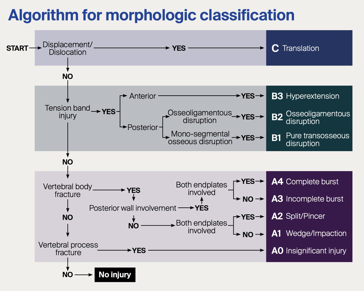
This T7 fracture involves two columns but is it unstable? The Denis three column model would suggest it is, but the reality is more complex. The three column model is now outdated. It doesn’t adequately account for soft tissue injuries, dynamic instability, or neurologic injury,

This T7 fracture involves two columns but is it unstable? The Denis three column model would suggest it is, but the reality is more complex.

The three column model is now outdated. It doesn’t adequately account for soft tissue injuries, dynamic instability, or neurologic injury,
Jack Garnham (@jjgarnham) 's Twitter Profile Photo

A young woman presents with acute, painful, monocular left-sided visual loss, worsening over a few days. On MRI we see enlargement, T2 hyperintensity, and enhancement of the intraorbital segment of the left optic nerve. This was acute optic neuritis. In adults, approximately 30%

A young woman presents with acute, painful, monocular left-sided visual loss, worsening over a few days. On MRI we see enlargement, T2 hyperintensity, and enhancement of the intraorbital segment of the left optic nerve.

This was acute optic neuritis. In adults, approximately 30%
Jack Garnham (@jjgarnham) 's Twitter Profile Photo

On a CT neck you see thickening and medialisation of the left aryepiglottic fold, an enlarged left pyriform sinus, and enlargement of the left laryngeal ventricle. What does this mean and what do you need to do next? The next step is a CT chest. These features suggest a left

On a CT neck you see thickening and medialisation of the left aryepiglottic fold, an enlarged left pyriform sinus, and enlargement of the left laryngeal ventricle. What does this mean and what do you need to do next?

The next step is a CT chest. These features suggest a left
Jack Garnham (@jjgarnham) 's Twitter Profile Photo

Well-defined, ovoid, predominantly intraconal mass in the right orbit, displacing the optic nerve and extraocular muscles. T2 hyperintense with dynamic enhancement, patchy and heterogeneous at first with progressive filling on the delayed images These are the typical features of

Well-defined, ovoid, predominantly intraconal mass in the right orbit, displacing the optic nerve and extraocular muscles. T2 hyperintense with dynamic enhancement, patchy and heterogeneous at first with progressive filling on the delayed images

These are the typical features of