Cardio Med (@cardioeducator) 's Twitter Profile
Cardio Med

@cardioeducator

🐦‍⬛ Tweet and Repost Educational Content of Cardiovascular Medicine 🩻 | 🩺 Follow and Subscribe ⚡

ID: 1870836120600391680

calendar_today22-12-2024 14:17:54

413 Tweet

289 Followers

1,1K Following

CardiovascularCorner (@trackyourheart) 's Twitter Profile Photo

M-mode echocardiography at the mitral valve level provides crucial insights into LV function. - It captures the RV, LV, mitral leaflets, interventricular septum, and posterior LV wall motion. - The E-point represents early diastolic anterior mitral leaflet motion, while the

M-mode echocardiography at the mitral valve level provides crucial insights into LV function.

- It captures the RV, LV, mitral leaflets, interventricular septum, and posterior LV wall motion.

- The E-point represents early diastolic anterior mitral leaflet motion, while the
Dr G Rajesh (Gopalan Nair Rajesh) (@drrajeshg1) 's Twitter Profile Photo

A 48 year old male presented with effort angina, TMT +ve, CT CAG was done, invasive CAG not done yet. I am desperately trying to demonstrate the likely cause for angina in TEE which is already beautifully demonstrated in CTCAG.

Alex Felix (@alexsfelixecho) 's Twitter Profile Photo

✨ASD in the elderly - A thread 🧵 1/6 Please share your thoughts 🧠🌟 🧩70y 👵🏽, previous MI, NYHA II, lipotimia. On a transthoracic #echofirst: ASD ostium secundum + moderate AR / mild-to-moderate MR Qp:Qs = 1.3:1 (reliable?🤔) EF 64% Poll below… Benoy Shah MD

CardiovascularCorner (@trackyourheart) 's Twitter Profile Photo

Understanding Transducer Movements in Transthoracic Echo (TTE) Accurate echocardiographic imaging depends heavily on how the transducer is manipulated on the chest wall. This image illustrates 3 essential movements: (A) Tilting in place changes the angle of the ultrasound beam,

Understanding Transducer Movements in Transthoracic Echo (TTE)

Accurate echocardiographic imaging depends heavily on how the transducer is manipulated on the chest wall. This image illustrates 3 essential movements:

(A) Tilting in place changes the angle of the ultrasound beam,
CardiovascularCorner (@trackyourheart) 's Twitter Profile Photo

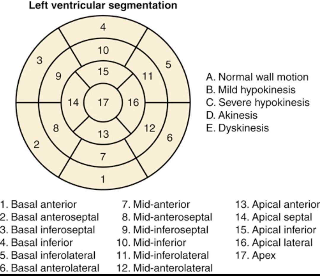
Understanding regional wall motion abnormalities (RWMA) starts with this: the 17-segment LV model. It divides the left ventricle into basal, mid, apical segments for precise localization of dysfunction. Why does it matter? RWMA refers to areas of the LV wall that contract

Understanding regional wall motion abnormalities (RWMA) starts with this: the 17-segment LV model.

It divides the left ventricle into basal, mid, apical segments for precise localization of dysfunction.

Why does it matter?
RWMA refers to areas of the LV wall that contract
CardiovascularCorner (@trackyourheart) 's Twitter Profile Photo

Recognizing PVCs from the Papillary Muscles on ECG – Key to Targeted Therapy Premature Ventricular Complexes (PVCs) can arise from the anterolateral or inferomedial papillary muscles of the left ventricle—both mid-cavity structures. The ECG features help localize the origin and

Recognizing PVCs from the Papillary Muscles on ECG – Key to Targeted Therapy

Premature Ventricular Complexes (PVCs) can arise from the anterolateral or inferomedial papillary muscles of the left ventricle—both mid-cavity structures. The ECG features help localize the origin and
Cardio Med (@cardioeducator) 's Twitter Profile Photo

Myocardial Bridging: A Hidden Cause of Angina 🫀 Myocardial bridging occurs when a segment of a coronary artery, most commonly the left anterior descending (LAD), tunnels through the heart muscle instead of running on the surface.

Myocardial Bridging: A Hidden Cause of Angina 🫀 Myocardial bridging occurs when a segment of a coronary artery, most commonly the left anterior descending (LAD), tunnels through the heart muscle instead of running on the surface.
Mounir Basalus (Ⲃⲁⲥⲓⲗⲓⲟⲥ) (@basalus) 's Twitter Profile Photo

1/⚡️ Ever heard of the Pickelhaube sign? It’s not just history — it’s cardiology! A quick 🧵 on how this spiked helmet shows up in ECGs and echocardiograms.

1/⚡️ Ever heard of the Pickelhaube sign? It’s not just history — it’s cardiology! A quick 🧵 on how this spiked helmet shows up in ECGs and echocardiograms.
EACVI President (@eacvipresident) 's Twitter Profile Photo

New Clinical Consensus on Left Atrium & Left Atrial Appendage Imaging 🚨 The #EACVI with EHRA has released updated guidance on #MMI for the assessment of the Left Atrium (LA) & Left Atrial Appendage (LAA). This evidence-based document is essential for improving diagnosis,

New Clinical Consensus on Left Atrium & Left Atrial Appendage Imaging 🚨

The #EACVI with EHRA has released updated guidance on #MMI for the assessment of the Left Atrium (LA) & Left Atrial Appendage (LAA).

This evidence-based document is essential for improving diagnosis,
Ritika Tuli (@ritikatulimd) 's Twitter Profile Photo

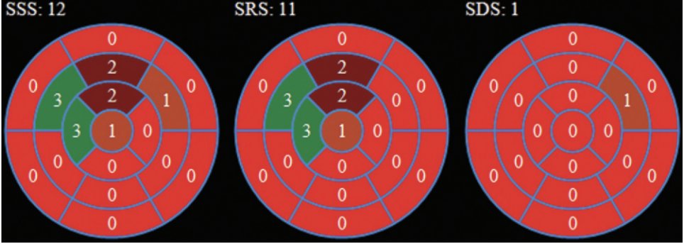
🧠 #CardioNuggets: Summed Stress Score (SSS) in Nuclear Perfusion Imaging 🫀 17 LV segments scored 0–4 (0 = normal, 4 = no uptake) ➕ Total = SSS (max 68) 🔍 Interpretation: 0–3: Normal 4–8: Mild 9–13: Mod ≥14: Severe perfusion defect 📊 SSS = Scar + Ischemia SDS =

🧠 #CardioNuggets: 

Summed Stress Score (SSS) in Nuclear Perfusion Imaging

🫀 17 LV segments scored 0–4 (0 = normal, 4 = no uptake)

➕ Total = SSS (max 68)

🔍 Interpretation:

0–3: Normal
4–8: Mild
9–13: Mod
≥14: Severe perfusion defect

📊 SSS = Scar + Ischemia

SDS =
CardiovascularCorner (@trackyourheart) 's Twitter Profile Photo

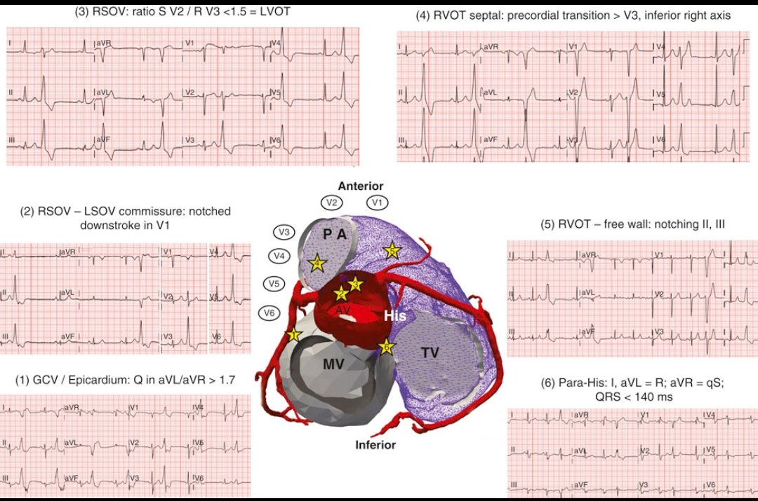
PVC localization on ECG. Premature ventricular complexes (PVCs) are early depolarizations originating from the ventricles, bypassing the His-Purkinje system. This powerful image illustrates PVCs from different anatomical origins➡️outflow tracts (RVOT/LVOT) and the tricuspid

PVC localization on ECG.
Premature ventricular complexes (PVCs) are early depolarizations originating from the ventricles, bypassing the His-Purkinje system.

This powerful image illustrates PVCs from different anatomical origins➡️outflow tracts (RVOT/LVOT) and the tricuspid
CardiovascularCorner (@trackyourheart) 's Twitter Profile Photo

How to semi-quantitatively grade Mitral Regurgitation (MR) on echocardiography? A quick guide using practical Doppler-based parameters: 1️⃣ Color Flow Jet Area (Apical 4-Chamber View): ➡️ Jet area as % of LA area: 🔵 Mild: <20% 🔵 Moderate: 20–39% 🔵 Severe: ≥40% or very

How to semi-quantitatively grade Mitral Regurgitation (MR) on echocardiography?
A quick guide using practical Doppler-based parameters:

1️⃣  Color Flow Jet Area (Apical 4-Chamber View):
➡️ Jet area as % of LA area: 
🔵 Mild: &lt;20%
🔵 Moderate: 20–39%
🔵 Severe: ≥40% or very