Amrit Singh, MD (@amritpsinghmd) 's Twitter Profile
Amrit Singh, MD

@amritpsinghmd

Hi, I'm a pathologist, specializing in #hematopathology and #dermatopathology.

ID: 929740783997083652

linkhttps://scholar.google.com/citations?hl=en&user=BYGt_yIAAAAJ calendar_today12-11-2017 16:00:51

981 Tweet

1,1K Followers

701 Following

Silvija Gottesman MD (@sgottesmanmd) 's Twitter Profile Photo

Elastic fibers pattern in benign nevi vs melanoma #SilviPearls #dermpath: 💠 elastic fibers intermixed bw nests & individual melanocytes in banal nevi 💠 melanoma has lost/nearly lost elastic fibers & pushes the UV altered elastic fibers downwards 💠 elastin stain useful

Elastic fibers pattern in benign nevi vs melanoma #SilviPearls #dermpath:

💠 elastic fibers intermixed bw nests & individual melanocytes in banal nevi 

💠 melanoma has lost/nearly lost elastic fibers & pushes the UV altered elastic fibers downwards 

💠 elastin stain useful
Mark Ong (@drmarkong) 's Twitter Profile Photo

Adult. Biopsy of supraclavicular fossa lymph node. Chromogranin A and synaptophysin both positive. No known primary tumour. How would report this? Poll in next tweet. Would the Ki67 influence what you call it? #pathology

Adult. Biopsy of supraclavicular fossa lymph node.

Chromogranin A and synaptophysin both positive. No known primary tumour.

How would report this? Poll in next tweet.

Would the Ki67 influence what you call it?

#pathology
Mark Ong (@drmarkong) 's Twitter Profile Photo

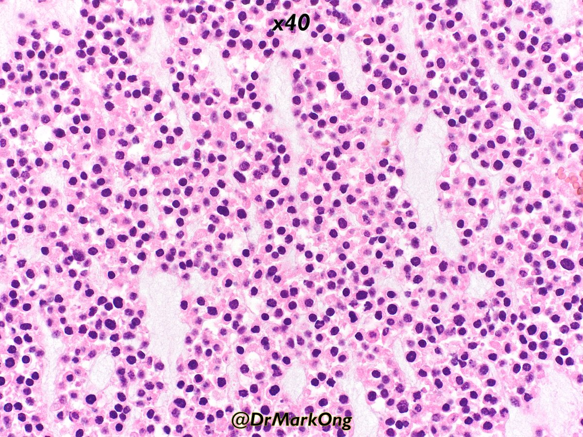
Biopsies of a nasal cavity mass from 40 year old. Unusual case that was eventually diagnosed on the resection specimen. Not my area of expertise, but was sent as possible lymphoma. Differentials and how would you approach this? #pathology #ENTpath

Biopsies of a nasal cavity mass from 40 year old.

Unusual case that was eventually diagnosed on the resection specimen. Not my area of expertise, but was sent as possible lymphoma.

Differentials and how would you approach this?

#pathology #ENTpath
Sanam Loghavi, MD صنم لغوی 🔬🧬 (@sanamloghavi) 's Twitter Profile Photo

A very nice summary of peripheral T cell lymphoma classification by Dr. Barbara Pro at #HOPLive24 Sadly our treatment options remain very limited compared to our understanding of the disease biology. My takeaway: need better biologically-informed clinical trial design!
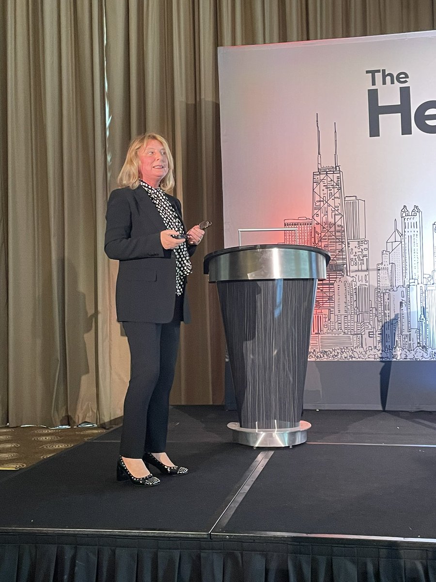
A very nice summary of peripheral T cell lymphoma classification by Dr. <a href="/bpromd/">Barbara Pro</a> at #HOPLive24 
Sadly our treatment options remain very limited compared to our understanding of the disease biology. 
My takeaway: need better biologically-informed clinical trial design!
Christine J. Ko, MD (@christinejkomd) 's Twitter Profile Photo

Cool case! #diagnosis? Yale Dermatology GR had a case of this today! both arms affected, photodistributed; #dermpath "red-orange, regularly spherical, of overall smaller dimension, and negative for Perls iron stain" jaadcasereports.org/article/S2352-… JAAD Journals #dermatology #pathology
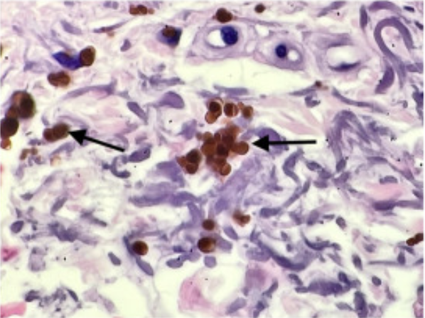
Cool case!  #diagnosis?  <a href="/yalederm/">Yale Dermatology</a> GR had a case of this today! both arms affected, photodistributed; #dermpath "red-orange, regularly spherical, of overall smaller dimension, and negative for Perls iron stain" jaadcasereports.org/article/S2352-… <a href="/JAADjournals/">JAAD Journals</a> #dermatology #pathology
Charles Herndon (@drcyclopath) 's Twitter Profile Photo

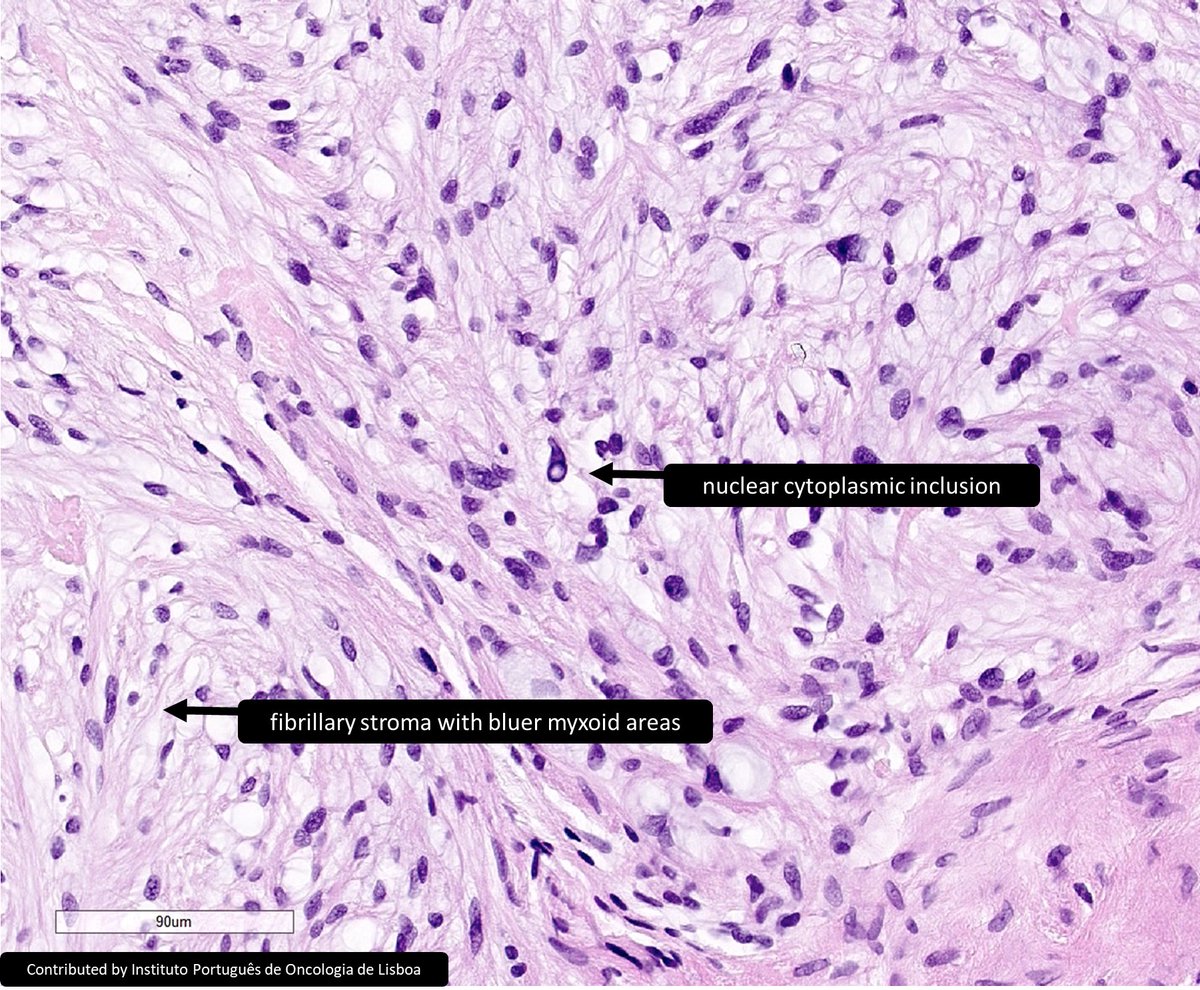
Finger of 20yo M, diagnosis? A) Plexiform neurofibroma B) Cellular neurothekeoma C) Dermal nerve sheath myxoma D) Myxfibrosarcoma #DermPath #BSTPath #Pathology #PathTwitter

Finger of 20yo M, diagnosis?
A) Plexiform neurofibroma
B) Cellular neurothekeoma
C) Dermal nerve sheath myxoma
D) Myxfibrosarcoma
#DermPath #BSTPath #Pathology #PathTwitter
Siba El Hussein, MD (@sibaelhussein) 's Twitter Profile Photo

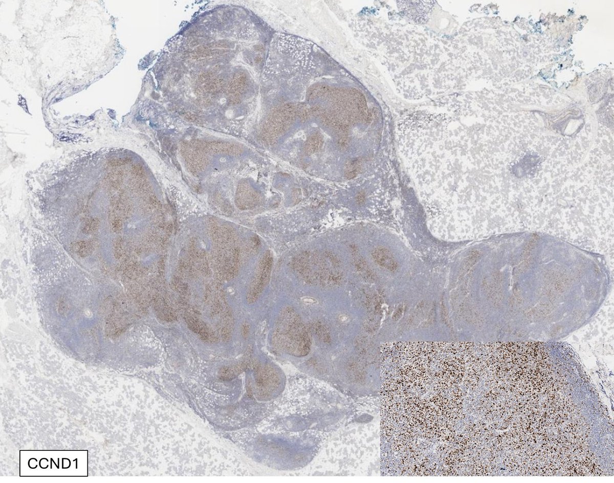
Follicular lymphoma with BCL2 rearrangement by FISH & unusual overexpression of CCND1 by IHC (CD5 & SOX11 negative; CCND1 rearrangement negative by FISH). This rare finding has been reported previously➡️shorturl.at/lzDOU but is worth to be aware of #hemepath #lymsm #PathX

Follicular lymphoma with BCL2 rearrangement by FISH &amp; unusual overexpression of CCND1 by IHC (CD5 &amp; SOX11 negative; CCND1 rearrangement negative by FISH). This rare finding has been reported previously➡️shorturl.at/lzDOU but is worth to be aware of #hemepath #lymsm #PathX
Toby Schmitt, MD (@tobyaschmitt) 's Twitter Profile Photo

p53 in vulvar dysplasia: • A basal sparing pattern supports HSIL (uVIN/HPV-related VIN) • Basal keratinocytes are negative • Remainder of the epithelium shows a wild-type (mosaic) pattern • This pattern is as suggestive of HSIL as a block positive p16 #pathology #pathtwitter

p53 in vulvar dysplasia:
• A basal sparing pattern supports HSIL (uVIN/HPV-related VIN)
• Basal keratinocytes are negative
• Remainder of the epithelium shows a wild-type (mosaic) pattern
• This pattern is as suggestive of HSIL as a block positive p16
#pathology #pathtwitter
Charles Herndon (@drcyclopath) 's Twitter Profile Photo

VAAD/deVIL: basal/scattered wild-type p53 patterns no atypia, anl maturation, hypogranulosis, flat + cytoplasm pallor (VAAD) or exophytic (deVIL) dVIN: mutant p53 (e.g., homogenous basal & midepithelial) basal atypia, anl maturation, hyperchromasia, nucleoli 📽️HPV-independent

VAAD/deVIL: basal/scattered wild-type p53 patterns
no atypia, anl maturation, hypogranulosis, flat + cytoplasm pallor (VAAD) or exophytic (deVIL)
dVIN: mutant p53 (e.g., homogenous basal &amp; midepithelial) 
basal atypia, anl maturation, hyperchromasia, nucleoli
📽️HPV-independent
Jerad Gardner, MD (@jmgardnermd) 's Twitter Profile Photo

20 F arm nodule. Diagnosis? If you could only do ONE STAIN, what would you do? Digital slide🔬 kikoxp.com/posts/17751 Answer ✅ youtu.be/obIibd_Xzlc?si… #pathology #pathologists #pathTwitter #BSTpath #dermpath #dermatology #dermatologia #dermtwitter

20 F arm nodule. Diagnosis? If you could only do ONE STAIN, what would you do?
Digital slide🔬 kikoxp.com/posts/17751
Answer ✅ youtu.be/obIibd_Xzlc?si… 
#pathology #pathologists #pathTwitter #BSTpath #dermpath #dermatology #dermatologia #dermtwitter
Judith Ferry (@judith_ferry) 's Twitter Profile Photo

Pathologists! Check out this fibrin associated large B cell lymphoma in an atrial myxoma. Typical features: incidental finding, large pleomorphic EBV+ B cells with non-GCB phenotype, often CD30+, in a background of debris, typically cured by excision. #lymphoma #hemepath

Pathologists! Check out this fibrin associated large B cell lymphoma in an atrial myxoma. Typical features: incidental finding, large pleomorphic EBV+ B cells with non-GCB phenotype, often CD30+, in a background of debris, typically cured by excision.
#lymphoma #hemepath
Judith Ferry (@judith_ferry) 's Twitter Profile Photo

CD45+, CD43+, so could be ALCL, but CD30 was negative. Note the fine chromatin of the neoplastic cells, suggesting blasts. Additional IHC: CD68+, lysosome+, NPM1+. Dx is myeloid sarcoma, NPM1+, c/w NPM1 mutation. #hemepath #heme #myeloid

CD45+, CD43+, so could be ALCL, but CD30 was negative. Note the fine chromatin of the neoplastic cells, suggesting blasts. Additional IHC: CD68+, lysosome+, NPM1+. Dx is myeloid sarcoma, NPM1+, c/w NPM1 mutation. #hemepath #heme #myeloid
Ifeyinwa Obiorah, MD PhD (@ifeyinwaobiorah) 's Twitter Profile Photo

Happy to conclude the annual #ASCP2024 meeting by presenting our topic on aggressive cutaneous lymphoma with my wonderful colleague UVA Pathology Dr Linglei Ma.
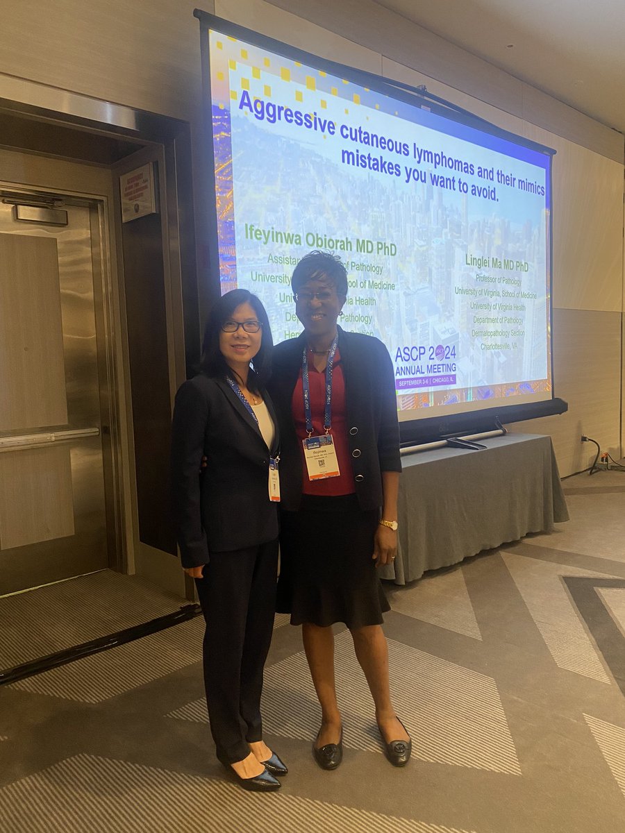
Happy to conclude the annual #ASCP2024 meeting by presenting our topic on aggressive cutaneous lymphoma with my wonderful colleague <a href="/UVA_Pathology/">UVA Pathology</a> Dr Linglei Ma.
Toby Schmitt, MD (@tobyaschmitt) 's Twitter Profile Photo

Aggressive angiomyxoma: - "Aggressively thick-walled" blood vessels - Small, bland spindle cells in a myxoid background - Extravasated red blood cells common WSI and more info: pathlibrary.com/slidebox/?case…

Aggressive angiomyxoma:
- "Aggressively thick-walled" blood vessels
- Small, bland spindle cells in a myxoid background
- Extravasated red blood cells common

WSI and more info: pathlibrary.com/slidebox/?case…
Silvija Gottesman MD (@sgottesmanmd) 's Twitter Profile Photo

Close up of the base of the nevoid melanoma case posted today. Showing the useful parallel theque pattern described by Elston. PMID 26299955

Close up of the base of the nevoid melanoma case posted today. Showing the useful parallel theque pattern described by Elston. PMID 26299955
Sara Zadeh, MD (@missdoctorz) 's Twitter Profile Photo

NUT Carcinoma. 4 cm axillary mass in a 60-year-old man. Small round blue cells with abrupt squamous differentiation. BRD4-NUT fusion detected via FISH.

NUT Carcinoma. 4 cm axillary mass in a 60-year-old man. Small round blue cells with abrupt squamous differentiation. BRD4-NUT fusion detected via FISH.