AmphiSpace
@ebglab
The Benito-Gutierrez Lab uses amphioxus as a model system to understand how vertebrates evolved and conquered water, land and air
ID: 1240700472723492865
https://www.amphispace.com 19-03-2020 18:04:18
222 Tweet
245 Followers
132 Following
 's Twitter Profile Photo")
 on Twitter photo Apical organ development is controlled by an anterior network (aGRN) composed of early repressive interactions between vegetal Wnt and animal Six3/6 and FoxQ2, which drive downstream apical organ genes. Many of the same genes are expressed in the vertebrate brain too!
3/10")
 on Twitter photo Is there a link between apical organs and chordate brains? To answer this question, we investigated the evolution of the aGRN in deuterostomes and traced its conservation in chordates using our favourite #amphioxus, a basally-branching chordate.
4/10")
 on Twitter photo By comparing published scRNAseq datasets we find a biphasic activation of aGRN genes in sea urchin and amphioxus but in not zebrafish, where FoxQ2 is missing and the other markers are expressed later in the hypothalamus. This supports aGRN conservation in deuterostomes.
5/10")
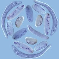
 on Twitter photo Investigation of aGRN genes in amphioxus revealed that early markers are co-expressed on the animal side and restrict anteriorly, while late markers appear in the anterior brain during neurulation, with a similar dynamic to apical organ development.
6/10")
 on Twitter photo These results indicate that the aGRN is conserved in chordates! 🤩 To test whether the amphioxus aGRN is also controlled by Wnt, we overactivated Wnt signalling with Azakenpaullone and saw a downregulation of all aGRN markers in the ectoderm.
7/10")
 on Twitter photo As late Wnt overactivation does not lead to loss of neural cells, we can examine what happens to the brain when the aGRN is removed! We find that the aGRN is necessary to confer anterior identity to the neural plate, as the brain is posteriorized when the aGRN is repressed.
8/10")
 on Twitter photo But what exactly is this anterior identity? Here we describe a hypothalamic-like region in the amphioxus larval brain expressing Otp, FoxD, Bsx, Hmx, miR-7 and calcitonin-type neuropeptides. It forms in the area where aGRN is active and is lost following Wnt overactivation.
9/10")
 on Twitter photo Our work shows that an aGRN patterning the anterior neurectoderm is ancestral to bilaterians. In the chordate lineage the aGRN was incorporated into neurulation to specify and position the forebrain anteriorly, linking the evolution of the AO to that of the chordate brain.
10/10")
I want to thank my wonderful (and patient) labmate Daniel Keitley, all the AmphiSpace, Dept of Zoology, CRUK Cambridge Institute and the Cambridge #EvoDevo community for their help and support! If you have any questions/comments, please let us know! 😄
 's Twitter Profile Photo")
 's Twitter Profile Photo")
☕️NEW: Ton, Keitley et al. provide a rabbit development atlas and propose that combining rabbit and mouse atlases can help dissect early primate development. Mai-Linh Ton Daniel Keitley MarioniLab Bertie Gottgens AmphiSpace rdcu.be/dfalo nature.com/articles/s4155…
 on Twitter photo This feather star has it all: beautifully colored body, 10 arms, great at regeneration. Plus, it’s a #crinoid, the sister group to other living #echinoderms (starfish, sea urchin..), making it an ideal comparative system to understand how the puzzling echinoderm body plan evolved")
 on Twitter photo Next, we focused on our favorite, the nervous system, and looked at the distribution of serotonin, GABA and glutamate 🧠. We characterized the complexity of the larval apical organ (AO), and identified glutamatergic cells in the neural plexus")
We then optimized in situ HCR (Molecular Instruments) to test the molecular patterning of the crinoid AO. 🧬 We found that serotonergic neurons form in a domain co-expressing Six3/6 and FoxQ2 (from early development) and Lhx2/9 as in other echinoderms, supporting AO conservation
 on Twitter photo We then optimized in situ HCR (<a href=\"/HCRimaging/\">Molecular Instruments</a>) to test the molecular patterning of the crinoid AO. 🧬 We found that serotonergic neurons form in a domain co-expressing Six3/6 and FoxQ2 (from early development) and Lhx2/9 as in other echinoderms, supporting AO conservation")
 on Twitter photo At metamorphosis, larvae settle from the anterior pole, neurons degenerate and a new nervous system develops. We showed that cells in the oral/vestibular ectoderm, where the ectoneural system will form, start to express anterior markers in the post-metamorphic cystidean stage!")
This work has been an amazing collaboration between my PhD Dept of Zoology and Master’s Università degli Studi di Milano labs, and I want to thank Silvia, both supervisors AmphiSpace and Roberta Pennati, Maurice Elphick for all his help and support, all the authors, and all members of both labs! 😄
Excited to see last Giacomo's Giacomo Gattoni work in the bioRxiv! Fun collaboration with Silvia Mercurio at Roberta Pennati's lab. Thank you all for your hard work, and giving the entire world another beautiful model system to work with 👏😀