Venkatesh M☯️ (@drvenkimdrd) 's Twitter Profile
Venkatesh M☯️

@drvenkimdrd

@Radiologist ..Never ending learner, educator, #radiology #Erad #traveller , Youtuber for Shades of Radiology, Medical illustrator

ID: 834970302304169984

linkhttp://radnucleus.wordpress.com calendar_today24-02-2017 03:36:47

11,11K Tweet

6,6K Takipçi

1,1K Takip Edilen

Venkatesh M☯️ (@drvenkimdrd) 's Twitter Profile Photo

ECU Tenosynovitis with Tendinosis on MRI The scan revealed ECU tenosynovitis with tendinosis, a condition characterized by inflammation of the tendon sheath and degenerative changes within the tendon itself.

ECU Tenosynovitis with Tendinosis on MRI

The scan revealed ECU tenosynovitis with tendinosis, a condition characterized by inflammation of the tendon sheath and degenerative changes within the tendon itself.
Skeletal Radiology (@skeletaljournal) 's Twitter Profile Photo

🟢 Groin pain in soccer players: anatomy, clinical presentation, biomechanics, pathology and imaging findings ⚽ Read more 👉 rdcu.be/efECx #SkeletalRadiology #Soccer #SportsMedicine

🟢 Groin pain in soccer players: anatomy, clinical presentation, biomechanics, pathology and imaging findings

⚽ Read more 👉  rdcu.be/efECx 

#SkeletalRadiology #Soccer #SportsMedicine
Bhavin Jankharia (@bhavinj) 's Twitter Profile Photo

Case 16: A Scab in a Cavity In patients with post-tuberculosis lung disease (PTLD), if we identify a "scab-like" sign, that may predict hemoptysis and chronic pulmonary aspergillosis in the future. youtu.be/RzPJ0iG0OMU

Case 16: A Scab in a Cavity

In patients with post-tuberculosis lung disease (PTLD), if we identify a "scab-like" sign, that may predict hemoptysis and chronic pulmonary aspergillosis in the future.

youtu.be/RzPJ0iG0OMU
Tapas Kumar Sahu (@doc_tks_rad) 's Twitter Profile Photo

"Snoopy sign" of pericardial agenesis 🔹Cardiac levoposition 🔹Lengthening and flattening of the border of LV 🔹Lucency separating the LV and left hemidiaphragm 🔹Lucency separating the pulmonary artery and aorta (interposed lung) 🔹 Loss of the right heart border PC: REF/CMC

"Snoopy sign" of pericardial agenesis 
🔹Cardiac levoposition 🔹Lengthening and flattening of the border of LV
🔹Lucency separating the LV and left hemidiaphragm 
🔹Lucency separating the pulmonary artery and aorta (interposed lung)
🔹 Loss of the right heart border

PC: REF/CMC
luciana@chamie.com.br (@lucianachamiec1) 's Twitter Profile Photo

I’m pleased to share with you our new publication in RadioGraphics on the imaging features of adenomyosis, including typical and atypical imaging appearances at US and MRI RSNA RadioGraphics Cooky Menias
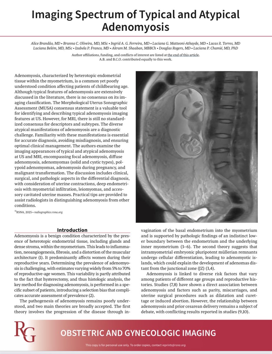
I’m pleased to share with you our new publication in RadioGraphics on the imaging features of adenomyosis, including typical and atypical imaging appearances at US and MRI <a href="/RSNA/">RSNA</a> <a href="/RadioGraphics/">RadioGraphics</a> <a href="/cookyscan1/">Cooky Menias</a>
radRounds Radiology Network (@radrounds) 's Twitter Profile Photo

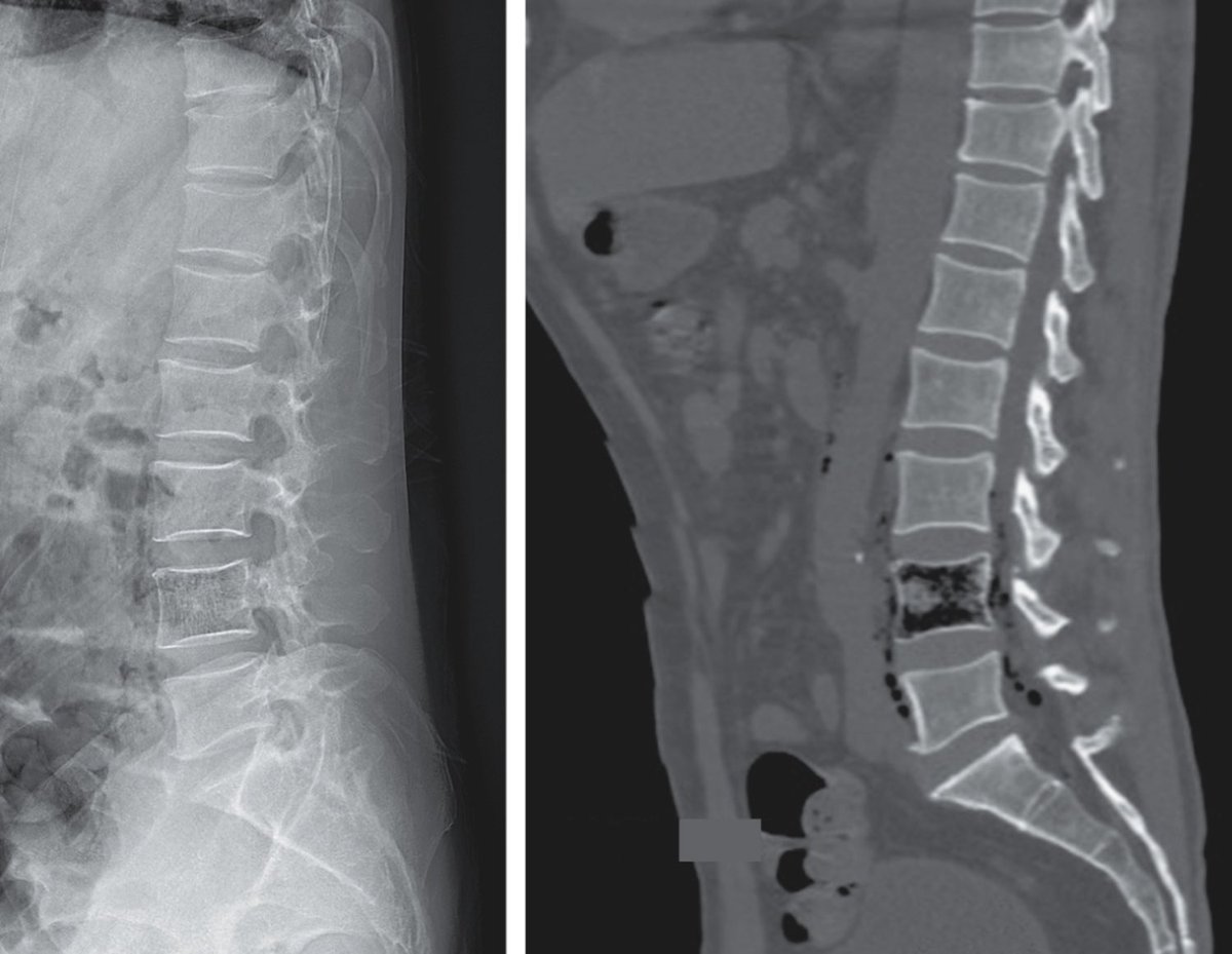
A 62-year-old man presented with a 5-day history of low back pain and a 1-day history of lethargy. Physical examination was notable for tenderness over the lumbar spine and a normal neurologic examination. Radiographs of the lumbar spine showed gas within L4. Computed tomography

A 62-year-old man presented with a 5-day history of low back pain and a 1-day history of lethargy. Physical examination was notable for tenderness over the lumbar spine and a normal neurologic examination. Radiographs of the lumbar spine showed gas within L4. Computed tomography
SPIN | Society of Pediatric Neuroimaging (@spinacademics) 's Twitter Profile Photo

3/ 🧠 Neuroimaging: Hallmarks 🔍 CT ✅ Bilateral hyperdense thalami 🧲 MRI ✅ T1: Hyperintense bulky thalami ✅ T2: Hypointense thalami ✅ T2: Persistent WM hyperintensity ✅ Involves: subcortical U fibers, periventricular WM, internal capsule

3/
🧠 Neuroimaging: Hallmarks

🔍 CT
✅ Bilateral hyperdense thalami

🧲 MRI
✅ T1: Hyperintense bulky thalami
✅ T2: Hypointense thalami
✅ T2: Persistent WM hyperintensity
✅ Involves: subcortical U fibers, periventricular WM, internal capsule