Live Cell Imaging Facility-LCIF (@lcif_uofm) 's Twitter Profile
Live Cell Imaging Facility-LCIF

@lcif_uofm

Advanced microscopy core @umanitoba, @UM_RadyFHS. We provide technical support from experiment design to image analysis.

ID: 1721548007601053696

linkhttps://prairieneuro.ca/lcif/ calendar_today06-11-2023 15:21:05

14 Tweet

25 Followers

47 Following

Live Cell Imaging Facility-LCIF (@lcif_uofm) 's Twitter Profile Photo

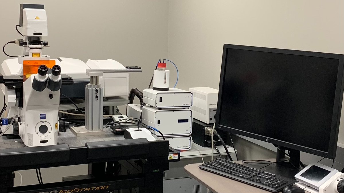
Let's explore the LCIF equipment inventory! Meet the popular ZEISS LSM 880 with super resolution Airyscan module capable of resolving structures up to 120nm. For further details or access to this scope, visit our website. prairieneuro.ca/lcif #Confocal #SuperResolution

Let's explore the LCIF equipment inventory! 
Meet the popular ZEISS LSM 880 with super resolution Airyscan module capable of resolving structures up to 120nm. For further details or access to this scope, visit our website. prairieneuro.ca/lcif 
#Confocal #SuperResolution
Live Cell Imaging Facility-LCIF (@lcif_uofm) 's Twitter Profile Photo

Next up is the Bruker Ultima Intravital 2-photon microscope, offering an expanded field of view with high resolution deep in scattering tissues. Find further info on this scope and how to gain access on our website. prairieneuro.ca/lcif #TwoPhotonMicroscopy #microscopy

Next up is the Bruker Ultima Intravital 2-photon microscope, offering an expanded field of view with high resolution deep in scattering tissues. Find further info on this scope and how to gain access on our website. prairieneuro.ca/lcif #TwoPhotonMicroscopy #microscopy
Live Cell Imaging Facility-LCIF (@lcif_uofm) 's Twitter Profile Photo

Next scope is ZEISS PALM IV laser dissection microscope! Enables uncontaminated isolation of fixed or live samples for downstream analysis like RNA extraction. For further details or access to this scope, visit our website. prairieneuro.ca/lcif #lasercapture #microdissection

Next scope is ZEISS PALM IV laser dissection microscope! Enables uncontaminated isolation of fixed or live samples for downstream analysis like RNA extraction. For further details or access to this scope, visit our website. prairieneuro.ca/lcif #lasercapture #microdissection
Live Cell Imaging Facility-LCIF (@lcif_uofm) 's Twitter Profile Photo

Lastly, the Bruker Ultima In-vitro 2-photon microscope allows you to visualize and record cell activities deep into slices and even photostimulate simultaneously! Visit our website to find out more about this amazing tool! prairieneuro.ca/lcif #twophotonmicroscopy

Lastly, the Bruker Ultima In-vitro 2-photon microscope allows you to visualize and record cell activities deep into slices and even photostimulate simultaneously! Visit our website to find out more about this amazing tool!
prairieneuro.ca/lcif #twophotonmicroscopy
Live Cell Imaging Facility-LCIF (@lcif_uofm) 's Twitter Profile Photo

Check out this sneak peek of our Intravital two-photon beam path in action! Aligning two lasers for dual laser imaging - Ti:Sapphire lasers are tuned to 720nm and 990nm to visualize the overlap. #microscopymonday #twophotonmicroscopy

Live Cell Imaging Facility-LCIF (@lcif_uofm) 's Twitter Profile Photo

For #FluorescentFriday, take a look at this image studying GluN1 expression in microglia. 🔴: GluN1 🟡: YFP-Neuron 🟢: GFP-Astrocyte 🔵: Iba1-Microglia. Image credits go to Meher Kantroo #Confocal #ZEISS #Microscopy

For #FluorescentFriday, take a look at this image studying GluN1 expression in microglia. 🔴: GluN1 🟡: YFP-Neuron 🟢: GFP-Astrocyte 🔵: Iba1-Microglia. 
Image credits go to <a href="/MeherKantroo/">Meher Kantroo</a>
#Confocal #ZEISS #Microscopy
Live Cell Imaging Facility-LCIF (@lcif_uofm) 's Twitter Profile Photo

Share that remarkable microscopic image that made you think, 'This is contest-worthy!' Submit your most captivating microscope images/videos taken on any LCIF microscope and stand a chance to win big prizes! Check out our website for more info! prairieneuro.ca/category/event… #microscopy

Share that remarkable microscopic image that made you think, 'This is contest-worthy!' Submit your most captivating microscope images/videos taken on any LCIF microscope and stand a chance to win big prizes! Check out our website for more info!
prairieneuro.ca/category/event…
#microscopy
Live Cell Imaging Facility-LCIF (@lcif_uofm) 's Twitter Profile Photo

🥇place: Farshid Gh The white matter (dark area in this image) within the cerebellum is called Tree of Life, and is covered by a three-layered cortex. Inner➡️outer: 1) Granular layer in green; 2) Purkinje cell layer; and 3) Molecular layer in red/purplish. #FluorescenceFriday

🥇place: <a href="/GhiyamihoorF/">Farshid Gh</a> The white matter (dark area in this image) within the cerebellum is called Tree of Life, and is covered by a three-layered cortex. Inner➡️outer: 1) Granular layer in green; 2) Purkinje cell layer; and 3) Molecular layer in red/purplish.
#FluorescenceFriday
Live Cell Imaging Facility-LCIF (@lcif_uofm) 's Twitter Profile Photo

🥈place: Christian Humphreys The image depicts HEK293T cells stained to label pannexin 2 (blue), wheat germ agglutinin (a plasma membrane marker, green) and calnexin (an ER marker, red), to visualize which cellular compartments pannexin 2 expresses. #FluorescenceFriday

🥈place: <a href="/christian_hump/">Christian Humphreys</a> 
The image depicts HEK293T cells stained to label pannexin 2 (blue), wheat germ agglutinin (a plasma membrane marker, green) and calnexin (an ER marker, red), to visualize which cellular compartments pannexin 2 expresses.
#FluorescenceFriday
Live Cell Imaging Facility-LCIF (@lcif_uofm) 's Twitter Profile Photo

🥉place: Rebecca Twilley Using magnified analysis of the proteome (MAP) an expansion microscopy technique which increases resolution 4x, this image shows pre- and post-synaptic proteins located in sub-synaptic domains of hippocampus neurons. #FluorescenceFriday

🥉place: Rebecca Twilley 
Using magnified analysis of the proteome (MAP) an expansion microscopy technique which increases resolution 4x, this image shows pre- and post-synaptic proteins located in sub-synaptic domains of hippocampus neurons. 
#FluorescenceFriday
Live Cell Imaging Facility-LCIF (@lcif_uofm) 's Twitter Profile Photo

Join us on August 27 to discover our latest service additions: Huygens and Imaris, two world-leading image processing/analysis software. Learn how these tools can improve your research. Register here or use the QR code below. #imageanalysis #Microscopy

Join us on August 27 to discover our latest service additions: Huygens and Imaris, two world-leading image processing/analysis software. Learn how these tools can improve your research. Register here or use the QR code below.
#imageanalysis #Microscopy
Live Cell Imaging Facility-LCIF (@lcif_uofm) 's Twitter Profile Photo

See the difference Huygens deconvolution can make! Reveal the hidden details in your images with just a few clicks, and efficiently process all your images in batches. Get in touch to try it out for free on your own data at LCIF! #Microscopy #Deconvolution #Huygens