Mireille Bitar, M.D. (@bitar_mireille) 's Twitter Profile
Mireille Bitar, M.D.

@bitar_mireille

Pediatric & Neuropathologist @UTSWMEDCENTER. Former faculty @NU_Pathology. Alumna @IcahnMountSinai @Columbiamed @ChildrensLA. My posts are not medical advice

ID: 1094074834135535616

calendar_today09-02-2019 03:25:42

1,1K Tweet

2,2K Followers

722 Following

Craig Horbinski (@craighorbinski) 's Twitter Profile Photo

Some ependymomas can really look papillary, like a choroid plexus papilloma. But if you look carefully, you'll see glial processes running from the tumor cells down to the vessels. #pathology #neuropath #pathtwitter

Some ependymomas can really look papillary, like a choroid plexus papilloma. But if you look carefully, you'll see glial processes running from the tumor cells down to the vessels. #pathology #neuropath #pathtwitter
Craig Horbinski (@craighorbinski) 's Twitter Profile Photo

Paraspinous mass in a patient with #nf1. What is the most likely diagnosis of this Mostly Problematic, Nasty, Spindly, and Terrifying lesion? #pathology #neuropath #PathTwitter

Paraspinous mass in a patient with #nf1. What is the most likely diagnosis of this Mostly Problematic, Nasty, Spindly, and Terrifying lesion? #pathology #neuropath #PathTwitter
Charles Herndon (@drcyclopath) 's Twitter Profile Photo

✅Schwannoma Elongated wavy bland nuclei with palisading ✅Hypercellular (Antoni A) and hypocellular (Antoni B Minimal pleomorphism, scant eosinophilic cytoplasm Hyalinized blood vessels, myxoid areas, collagen No clefts & deep dpa-dapa.com/public/display… #BSTPath #PathTwitter

✅Schwannoma
Elongated wavy bland nuclei with palisading
✅Hypercellular (Antoni A) and hypocellular (Antoni B
Minimal pleomorphism, scant eosinophilic cytoplasm
Hyalinized blood vessels, myxoid areas, collagen
No clefts & deep
dpa-dapa.com/public/display…
#BSTPath #PathTwitter
Mireille Bitar, M.D. (@bitar_mireille) 's Twitter Profile Photo

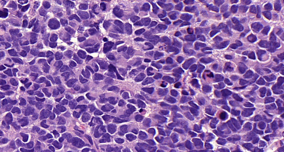
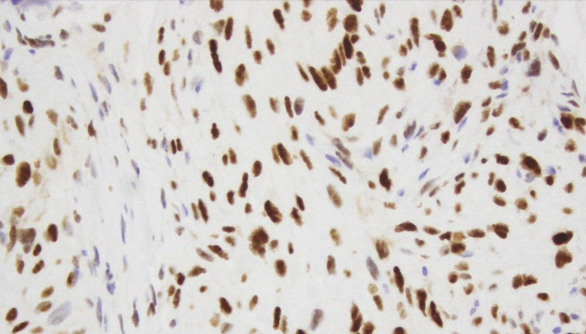
History of spine radiation as a child for a CNS neoplasm, few years later with a spinal bone lesion (H&E and p53 IHC).. no osteoid formation seen in the tumor, likely diagnosis? #neuropath #pedipath #molecpath #bonepath

History of spine radiation as a child for a CNS neoplasm, few years later with a spinal bone lesion (H&E and p53 IHC).. no osteoid formation seen in the tumor,  likely diagnosis? #neuropath #pedipath #molecpath #bonepath
Mireille Bitar, M.D. (@bitar_mireille) 's Twitter Profile Photo

One of the reasons I love hiking is because I’m reminded of the similarities between the human body and nature.. think of a neuron as a tree: dendrites are the branches, cell body is the trunk and the axon as roots...🧠🌲😊

One of the reasons I love hiking is because I’m reminded of the similarities between the human body and nature.. think of a neuron as a tree: dendrites are the branches, cell body is the trunk and the axon as roots...🧠🌲😊
Brain Pathology (@brainpathol) 's Twitter Profile Photo

Intraventricular mass in a 52-year-old woman WHAT IS YOUR DIAGNOSIS? WSI: isn-slidearchive.org/?col=ISN&fol=A… Case discussion: onlinelibrary.wiley.com/doi/10.1111/bp… #neuropath

Intraventricular mass in a 52-year-old woman
WHAT IS YOUR DIAGNOSIS?

WSI: isn-slidearchive.org/?col=ISN&fol=A…
Case discussion: onlinelibrary.wiley.com/doi/10.1111/bp…
#neuropath
Kenneth Aldape, MD (@kenneth_aldape) 's Twitter Profile Photo

Methylation Case of the Week: 5 year old female patient with a bithalamic tumor. what Might You Be incLined to call this? #pathology #PathTwitter #Neuropath

Methylation Case of the Week:

5 year old female patient with a bithalamic tumor. what Might You Be incLined to call this? #pathology #PathTwitter #Neuropath
Mireille Bitar, M.D. (@bitar_mireille) 's Twitter Profile Photo

Here is a KIAA1549::BRAF proven pilocytic #astrocytoma with a multinucleated tumor cell (pennies-on-a-plate pattern).. and large ganglionic-like cells… these tumors can also have entrapped neurons #pedipath #neuropath

Here is a KIAA1549::BRAF proven pilocytic #astrocytoma with a multinucleated tumor cell (pennies-on-a-plate pattern).. and large ganglionic-like cells… these tumors can also have entrapped neurons #pedipath #neuropath
Mireille Bitar, M.D. (@bitar_mireille) 's Twitter Profile Photo

I was fortunate to collaborate with an amazing group of veterinary pathologists and neuropathologists and glad to share our newly published research, aiming to facilitate the translation of knowledge between canine and human cancer #neuropath onlinelibrary.wiley.com/doi/10.1111/bp…

Brain Pathology (@brainpathol) 's Twitter Profile Photo

UNDER YOUR MICROSCOPE A pineal mass in a 39-year-old woman What is your diagnosis? #neuropath WSI: isn-slidearchive.org/?col=ISN&fol=A… Full case here: onlinelibrary.wiley.com/doi/full/10.11…
UNDER YOUR MICROSCOPE
A pineal mass in a 39-year-old woman
What is your diagnosis?
#neuropath
WSI: isn-slidearchive.org/?col=ISN&fol=A…
Full case here: onlinelibrary.wiley.com/doi/full/10.11…
Mireille Bitar, M.D. (@bitar_mireille) 's Twitter Profile Photo

On Mother's Day, sharing adapted work by Nizar Qabbani “Good morning my beautiful saint, it has been years since that boy sailed on his fairytale journey and hid in his pocket a damascene lilac, for no woman can comb my hair or lift me up like you do, oh mother, oh mother” ☺️

Mireille Bitar, M.D. (@bitar_mireille) 's Twitter Profile Photo

Here is a Wilms tumor (Nephroblastoma) with blastemal and epithelial components and a great resemblance to the CNS Embryonal tumor with multilayered rosettes (ETMR)! both are embryonal tumors that can be seen in DICER1 syndrome #pedipath #neuropath

Here is a Wilms tumor (Nephroblastoma) with blastemal and epithelial components and a great resemblance to the CNS Embryonal tumor with multilayered rosettes (ETMR)! both are embryonal tumors that can be seen in DICER1 syndrome #pedipath #neuropath
Mireille Bitar, M.D. (@bitar_mireille) 's Twitter Profile Photo

Look for the Line of Gennari along the banks of the calcarine sulcus of the occipital lobe, a visible band of myelinated axons formed by axonal projections from the thalamus to layer IV of the primary visual cortex "striate cortex” #neuroanatomy #neuropath #brainautopsy

Look for the Line of Gennari along the banks of the calcarine sulcus of the occipital lobe, a visible band of myelinated axons formed by axonal projections from the thalamus to layer IV of the primary visual cortex "striate cortex” #neuroanatomy #neuropath #brainautopsy