Dr. Basl Bernieh (@basldr) 's Twitter Profile
Dr. Basl Bernieh

@basldr

Cardiologist,
Sopron Hospital

ID: 1667646596

linkhttps://newmed.hu/kardiologia-sopron calendar_today13-08-2013 12:12:02

684 Tweet

158 Followers

174 Following

The Heart (@zheart11768530) 's Twitter Profile Photo

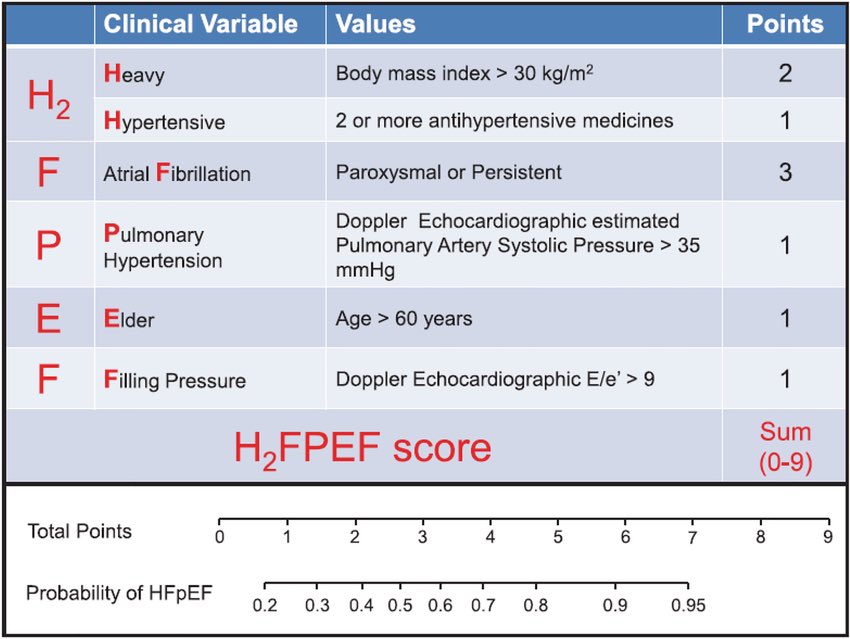
🔴A Simple, Evidence-Based Approach to Diagnosis of HFpEF⤵️ 🔹pmc.ncbi.nlm.nih.gov/articles/PMC62… #Cardiology #FOAMed #MedEd #medstudent #paramedic #Cardiology #CardioEd #medtwitter #meded #CardioTwitter #cardiotwiteros #MedX #cardiovascular #MedTwitter #medical #medEd #cardiotwitter

🔴A Simple, Evidence-Based Approach to Diagnosis of HFpEF⤵️ 

🔹pmc.ncbi.nlm.nih.gov/articles/PMC62…
 #Cardiology #FOAMed #MedEd #medstudent #paramedic #Cardiology #CardioEd #medtwitter #meded #CardioTwitter #cardiotwiteros  #MedX #cardiovascular #MedTwitter #medical #medEd #cardiotwitter
El Hamriti Mustapha (@melhamriti) 's Twitter Profile Photo

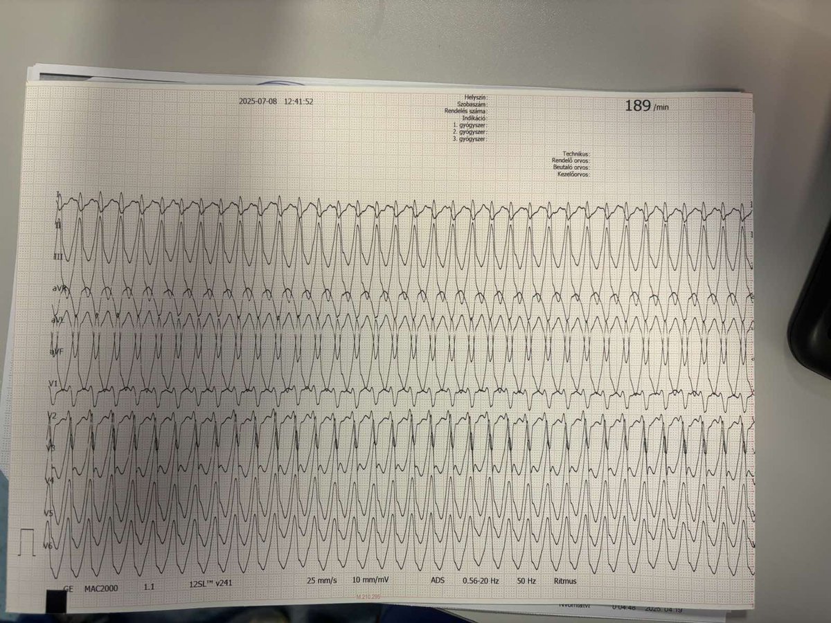
👇Fascicular VT (Part 1): Basic ECG Findings👇 •Left Posterior Fascicular VT: ECG pattern shows RBBB (right bundle branch block) and LAFB (left anterior fascicular block). •Left Anterior Fascicular VT: ECG pattern shows RBBB (right bundle branch block) and LPFB (left posterior

🇳🇴🇩🇰Asbjørn Støylen 🇬🇱🇵🇸🇺🇦 (@strain_rate) 's Twitter Profile Photo

IVR has constant volume, but shape change. All parts of the ventricle relax, but the apex relaxes most, and hence is most Energy demanding, the shortening of the base is not wasted work. The reconfiguration generates apical flow, giving momentum to the subsequent early filling

IVR has constant volume, but shape change. All parts of the ventricle relax, but the apex relaxes most, and hence is most Energy demanding, the shortening of the base is not wasted work. The reconfiguration generates apical flow, giving momentum to the subsequent early filling
Alexander Mladenow MD (@alex1708ander) 's Twitter Profile Photo

NeoCardioLab - Gabriel Altit Neonatal Heart Society Pediatric/congenital cardiology education Conquering CHD Jorge Faerron Oung Savly MD FACC FESC FASE FAAP FAPSC 🇰🇭 Great VSD tutorial 🤩 #echofirst VSD location ▶️ 👀 in ME RV in-out ▶️ important for the surgical approach 👉 3 types of VSD located at the base of the ❤️ ▶️ intraop 2D & 3DTEE 🪩2D & 3D images are horizontally and vertically mirrored🪩 #CHD American Society of Echocardiography #CVImaging

Dr. José Ángel Cabrera (@jacabreracardio) 's Twitter Profile Photo

Muscular arquitecture of the left atrium the 🔹subepicardial septopulmonary bundle overlaps with the 🔹subendocardial septoatrial bundle . Sharing with an enthusiastic group of young EP. Understanding anatomy is key to advancing the treatment of atrial fibrillation

Edgar Argulian (@argulian) 's Twitter Profile Photo

Spectral Doppler tracings provide important hemodynamic insights. This book is dedicated to interpretation of spectral Doppler recordings, similar to EKG books focused on EKG interpretation. Examples of some spectral Doppler patterns which may not be widely known: Notched aortic

Spectral Doppler tracings provide important hemodynamic insights. This book is dedicated to interpretation of spectral Doppler recordings, similar to EKG books focused on EKG interpretation. Examples of some spectral Doppler patterns which may not be widely known:

Notched aortic
CardiovascularCorner (@trackyourheart) 's Twitter Profile Photo

Cardiac Amyloidosis Diagnosis Cardiac amyloidosis is a restrictive cardiomyopathy caused by extracellular deposition of misfolded amyloid proteins in the myocardium, leading to stiffening and heart failure. Key Diagnostic Steps 1️⃣ Suspect in HFpEF with wall thickening, low

Cardiac Amyloidosis Diagnosis
Cardiac amyloidosis is a restrictive cardiomyopathy caused by extracellular deposition of misfolded amyloid proteins in the myocardium, leading to stiffening and heart failure.

Key Diagnostic Steps
1️⃣ Suspect in HFpEF with wall thickening, low
Alexander Mladenow MD (@alex1708ander) 's Twitter Profile Photo

Adam Christian Thebesius 1686-1732 To get the coronary sinus (CS) in TEE slowly advance the probe from the mid-esophageal 4Ch view till the MV disappears and instead of it the CS appears #echofirst By zooming in on the image the Thebesian valve can be occasionally seen

Carlos El-Tallawi, MD “HeartToProve” (@hearttoprove) 's Twitter Profile Photo

A coronary-cameral fistula: •Complication of septal myectomy •Septal-perforator ➡️ LV •Diastolic flow (vs. continuous in VSD) •AI physiology (“non-aortic valve AI”)