Alexandre Dumoulin @axonsalex.bsky.social
@axonsalex
Hello! find me on Bluesky: bsky.app/profile/axonsa… - I don't post on here anymore - Neurobiologist @UZH_Science
ID: 1294182584474374149
https://orcid.org/0000-0002-2420-6877 14-08-2020 08:02:50
382 Tweet
1,1K Followers
979 Following
 's Twitter Profile Photo")
To find out more about this work, we caught up with first author, Alexandre Dumoulin, and corresponding author, Esther Stoeckli, Professor University of Zurich: journals.biologists.com/dev/article/15…
 on Twitter photo To find out more about this work, we caught up with first author, Alexandre Dumoulin, and corresponding author, Esther Stoeckli, Professor <a href=\"/UZH_en/\">University of Zurich</a>:
journals.biologists.com/dev/article/15…")
 on Twitter photo Chicken commissural neurons stained for neurofilament-M (Cyan) and the primary cilium marker Arl13b (red). happy #FluoresenceFriday !")
 's Twitter Profile Photo")
 's Twitter Profile Photo")
 on Twitter photo AXON2025 is gearing up! If you're passionate about neural circuit development, this is the must-attend event of next year — set in an extraordinary, non-conventional location🚢that you won’t want to miss! #AXON2025 @JeroenPasterk")
 's Twitter Profile Photo")
 's Twitter Profile Photo")
The Vollum Institute is accepting applications for multiple faculty openings. We are interested in individuals whose research focuses on molecular and cellular neuroscience, genetics, neurodevelopment or signal transduction. #sciencejobs #neurotwitter academicjobsonline.org/ajo/jobs/28203
 on Twitter photo The Vollum Institute is accepting applications for multiple faculty openings. We are interested in individuals whose research focuses on molecular and cellular neuroscience, genetics, neurodevelopment or signal transduction. #sciencejobs #neurotwitter
academicjobsonline.org/ajo/jobs/28203")
 's Twitter Profile Photo")
 's Twitter Profile Photo")
Retinoic acid, an essential component of the roof plate organizer, promotes the spatiotemporal segregation of dorsal neural fates Read this #OpenAccess Research Article by Dina Rekler, Shai Ofek, Sarah Kagan, Gilgi Friedlander & Chaya Kalcheim Hebrew University: journals.biologists.com/dev/article/15…
 on Twitter photo Retinoic acid, an essential component of the roof plate organizer, promotes the spatiotemporal segregation of dorsal neural fates
Read this #OpenAccess Research Article by <a href=\"/DinaRekler/\">Dina Rekler</a>, Shai Ofek, Sarah Kagan, Gilgi Friedlander & <a href=\"/ChayaKalcheim/\">Chaya Kalcheim</a> <a href=\"/HebrewU/\">Hebrew University</a>:
journals.biologists.com/dev/article/15…")
 's Twitter Profile Photo")
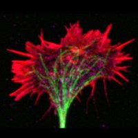
Baby you’re a fiiiiirework!!! Explanted Xenopus neural crest cell, microtubules (green) and actin (magenta). Imaging in frogs rocks- this is done room temp, no incubation, no problem. 🐸🔬✨ By Micaela Lasser, Helen Willsey Helen Willsey Lab ZEISS Microscopy LSM980 fast airyscan
 's Twitter Profile Photo")
 's Twitter Profile Photo")
 's Twitter Profile Photo")
Come and join the Williams lab The University of Manchester UoM Biology, Medicine and Health Medical Research Council We are using single-cell Multiomics and in vivo CRISPR approaches to understand the molecular mechanisms underlying lineage segregation from the neural plate border 🐣jobs.manchester.ac.uk/Job/JobDetail?…