André Forjaz (@andreforjaz) 's Twitter Profile
André Forjaz

@andreforjaz

PhD candidate @ Johns Hopkins Univ. | Wirtz Lab | Institute of Nano biotechnology (INBT) 🇵🇹

ID: 1464436845115482113

calendar_today27-11-2021 03:32:19

173 Tweet

133 Takipçi

218 Takip Edilen

Mina L Xu (@minaxu7) 's Twitter Profile Photo

Super happy to share this final manuscript in Cell. I am fortunate to have been mentored by Rong Fan, who dared me to learn things outside my comfort zone; helping forge a path forward within my own field for our future patients (1/2).nam12.safelinks.protection.outlook.com/?url=https%3A%…

Anirban Maitra (@aiims1742) 's Twitter Profile Photo

In this episode of MD Anderson Cancer Center Cancerwise Podcast, I chat with my colleague and friend Dr. Shubham Pant about clinical and research advances in #PancreaticCancer. We talk about clinical trials and what’s coming up on the horizon. Hope you enjoy! mdanderson.org/podcast.html

HOPTEC (@hoptec_) 's Twitter Profile Photo

We are beyond excited to announce the final panel for the 8th edition of the HOPTEC Webinar Series featuring guest speakers 𝗕𝗿𝗲𝗻𝗱𝗼𝗻 𝗗𝗮𝘃𝗶𝘀 and 𝗦𝗶𝗺ã𝗼 𝗥𝗼𝗰𝗵𝗮! 📅Date: 6th of May 2025 ⏰Time: 16H GMT - 11 AM EST 🔗Register here: forms.gle/cKMwGiRQJRLL7J…

We are beyond excited to announce the final panel for the 8th edition of the HOPTEC Webinar Series featuring guest speakers 𝗕𝗿𝗲𝗻𝗱𝗼𝗻 𝗗𝗮𝘃𝗶𝘀 and 𝗦𝗶𝗺ã𝗼 𝗥𝗼𝗰𝗵𝗮!

📅Date: 6th of May 2025
⏰Time: 16H GMT - 11 AM EST
🔗Register here: forms.gle/cKMwGiRQJRLL7J…
Rong Fan (@rongfan8) 's Twitter Profile Photo

Attending the 2nd Prescriptomics meeting in Caparica Portugal 🇵🇹. Thanks Jose jose luis capelo martinez for organizing such a stimulating and joyful conference to help scientists all over the world to join the forces and to advance precision health and personalized medicine.
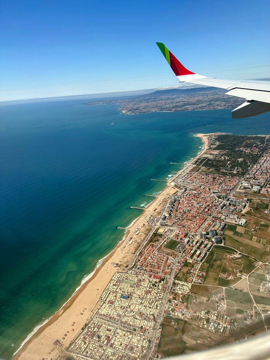
Attending the 2nd Prescriptomics meeting in Caparica Portugal 🇵🇹. Thanks Jose <a href="/CapeloMartinez/">jose luis capelo martinez</a> for organizing such a stimulating and joyful conference to help scientists all over the world to join the forces and to advance precision health and personalized medicine.

Denis Wirtz (@deniswirtz) 's Twitter Profile Photo

We introduce InterpolAI, an optical flow-based AI model to enhance 3D imaging (MRI, CODA, ssTEM, light sheet) Read about it here in our new NM paper: nature.com/articles/s4159…

Nature Methods (@naturemethods) 's Twitter Profile Photo

Can a deep learning model know your data well enough to predict missing parts? InterpolAI accurately interpolates synthetic images between pairs of authentic images to “repair” tissue damage and reduce stitching artifacts across modalities. Denis Wirtz bit.ly/453o1i1
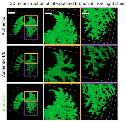
Can a deep learning model know your data well enough to predict missing parts? InterpolAI accurately interpolates synthetic images between pairs of authentic images to “repair” tissue damage and reduce stitching artifacts across modalities. <a href="/deniswirtz/">Denis Wirtz</a> bit.ly/453o1i1
Denis Wirtz (@deniswirtz) 's Twitter Profile Photo

To fix input images in a stack for 3D imaging, use InterpolAI. InterpolAI removes tissue tears and stitching + photobleaching artifacts in stacks of ssTEM, MRI, light-sheet, or histological images (CODA). Read it here: nature.com/articles/s4159… GitHub: github.com/sjoshi17jhu/In…

To fix input images in a stack for 3D imaging, use InterpolAI. 

InterpolAI removes tissue tears and stitching + photobleaching artifacts in stacks of ssTEM, MRI, light-sheet, or histological images (CODA).

Read it here: nature.com/articles/s4159…
GitHub: github.com/sjoshi17jhu/In…
André Forjaz (@andreforjaz) 's Twitter Profile Photo

Excited to share our work in Nature Methods! InterpolAI is a deep learning framework that uses optical flow to interpolate and restore missing or damaged biomedical images, allowing enhanced 3D maps across diverse imaging modalities. Read more: nature.com/articles/s4159…

Excited to share our work in Nature Methods!

InterpolAI is a deep learning framework that uses optical flow to interpolate and restore missing or damaged biomedical images, allowing enhanced 3D maps across diverse imaging modalities. 

Read more: nature.com/articles/s4159…
Denis Wirtz (@deniswirtz) 's Twitter Profile Photo

If your stack of images is full of defects, don't panic! InterpolaAI, a deep learning-based optical flow method, will fix issue tears, photobleaching and sticking artifacts for improved 3D imaging. Read our paper in Nature Methods: nature.com/articles/s4159…

If your stack of images is full of defects, don't panic!

InterpolaAI, a deep learning-based optical flow method, will fix issue tears, photobleaching and sticking artifacts for improved 3D imaging.

Read our paper in Nature Methods: nature.com/articles/s4159…
Arrate Munoz Barrutia (@arratemunoz) 's Twitter Profile Photo

This work is part of the results from my sabbatical stay at Prof. Denis Wirtz’s lab at Johns Hopkins during the 2022–2023 academic year. Very grateful to have been part of this exciting collaboration—congrats to the team on the Nature Methods publication!

Denis Wirtz (@deniswirtz) 's Twitter Profile Photo

It is very hard to avoid tissue tear when sectioning brain samples for ssTEM imaging. The majority of sections have such defects. No worry, we can fix these defects using InterpolAI, as shown in our recent Nature Methods paper: nature.com/articles/s4159…

It is very hard to avoid tissue tear when sectioning brain samples for ssTEM imaging.  The majority of sections have such defects.

No worry, we can fix these defects using InterpolAI, as shown in our recent Nature Methods paper: nature.com/articles/s4159…
Denis Wirtz (@deniswirtz) 's Twitter Profile Photo

3D spatial multi-omic assessment of tumors is not a luxury, it is necessary. 2D assessments of tumors are simply insufficient. The whole field of spatial omics needs to move to 3D. Read this work here: cell.com/cell-reports-m…

3D spatial multi-omic assessment of tumors is not a luxury, it is necessary.

2D assessments of tumors are simply insufficient.

The whole field of spatial omics needs to move to 3D.

Read this work here: cell.com/cell-reports-m…
Denis Wirtz (@deniswirtz) 's Twitter Profile Photo

How many whole slides or TMAs does it take to properly assess the 3D cellular heterogeneity of a single tumor? Many! Read about 2D vs. 3D multi-omic assessments of tissues and tumors here: cell.com/cell-reports-m…

How many whole slides or TMAs does it take to properly assess the 3D cellular heterogeneity of a single tumor?

Many!

Read about 2D vs. 3D multi-omic assessments of tissues and tumors here: cell.com/cell-reports-m…
Anirban Maitra (@aiims1742) 's Twitter Profile Photo

PIVOT is a very useful open source tool from Ashley Kiemen André Forjaz Denis Wirtz and colleagues at Johns Hopkins Kimmel Cancer Center that allows for co-registration and integration of multi-omic spatial data (eg, Visium, cyclicIF, imaging mass cytometry). Preprint: biorxiv.org/content/10.110…

PIVOT is a very useful open source tool from <a href="/AshleyKiemen/">Ashley Kiemen</a> <a href="/andreforjaz/">André Forjaz</a> <a href="/deniswirtz/">Denis Wirtz</a> and colleagues at <a href="/hopkinskimmel/">Johns Hopkins Kimmel Cancer Center</a> that allows for co-registration and integration of multi-omic spatial data (eg, Visium, cyclicIF, imaging mass cytometry). 
Preprint: 
biorxiv.org/content/10.110…
Denis Wirtz (@deniswirtz) 's Twitter Profile Photo

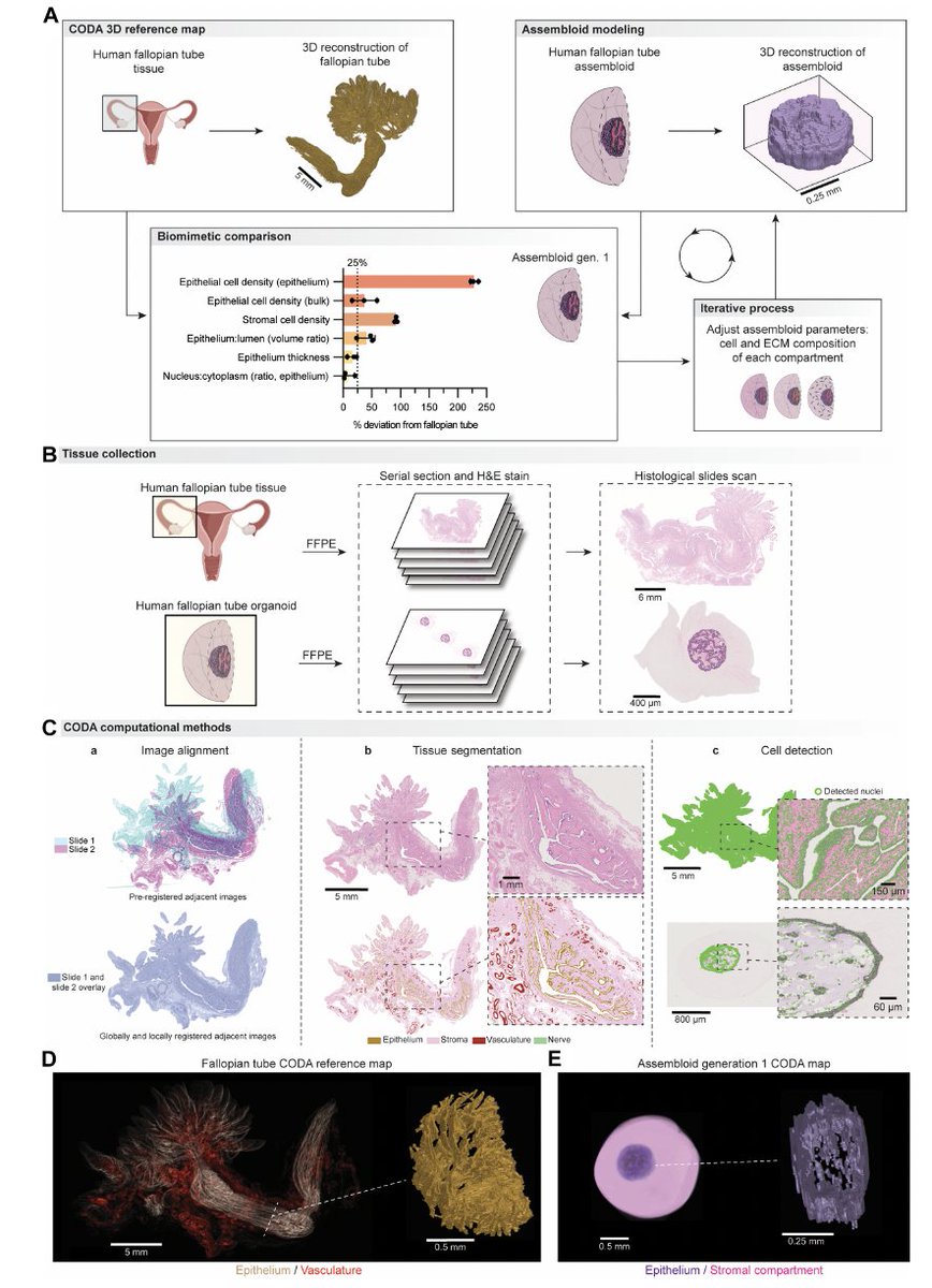
A fully functional organoid has to have the right form. We have developed a multi-compartment assembloid of a human organ, whose design converges iteratively towards the 3D map of that organ via CODA. Proof of principle for the human fallopian tube: science.org/doi/full/10.11…

A fully functional organoid has to have the right form.

We have developed a multi-compartment assembloid of a human organ, whose design converges iteratively towards the 3D map of that organ via CODA. 

Proof of principle for the human fallopian tube:
science.org/doi/full/10.11…
Denis Wirtz (@deniswirtz) 's Twitter Profile Photo

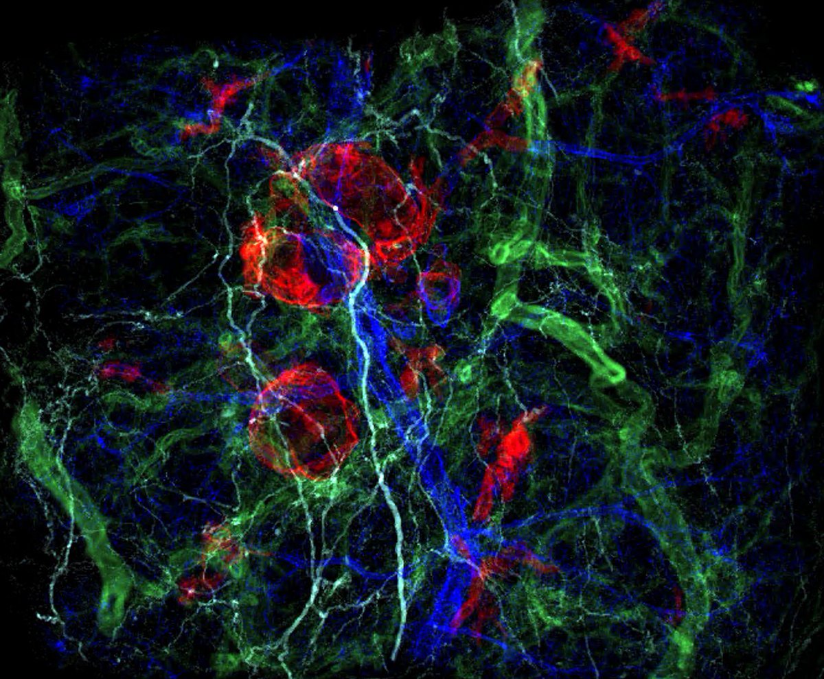
CODA reveals two different types of pre-cancer lesions in the human pancreas. CODA can map large volumes of tumors/tissues in 3D and at single-cell resolution. Red, PanIN lesions; navy blue: duct; light blue: nerve; green: blood vessel

CODA reveals two different types of pre-cancer lesions in the human pancreas.

CODA can map large volumes of tumors/tissues in 3D and at single-cell resolution. 

Red, PanIN lesions; navy blue: duct; light blue: nerve; green: blood vessel
Denis Wirtz (@deniswirtz) 's Twitter Profile Photo

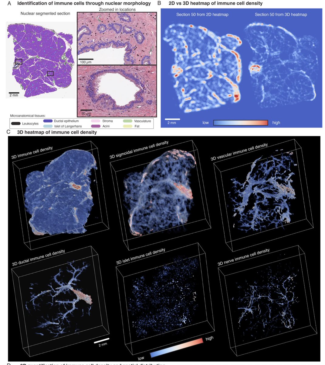
3D imaging and nuclear morphology. Through extension of our CODA platform, we integrate 3D imaging with nuclear segmentation to analyze nuclear morphological features in human tissue. More here: biorxiv.org/content/10.110…

3D imaging and nuclear morphology.

Through extension of our CODA platform, we integrate 3D imaging with nuclear segmentation to analyze nuclear morphological features in human tissue.

More here: biorxiv.org/content/10.110…