Mizar Imaging (@mizarimaging) 's Twitter Profile
Mizar Imaging

@mizarimaging

High Resolution Light Sheet Imaging. Get all the advantages of using a light sheet + now at high resolution

ID: 900353809796476928

linkhttp://mizarimaging.com calendar_today23-08-2017 13:47:30

764 Tweet

969 Takipçi

806 Takip Edilen

Elisabeth Kugler (@kuglerelisabeth) 's Twitter Profile Photo

Happy #FluorescenceFriday with some #sciArt Muller #glia cells of the #zebrafish retina. Here you see the depth-coded surface of Muller glia. #Science #womenWhoCode #womenInImageAnalysis #microscopy

Happy #FluorescenceFriday with some #sciArt Muller #glia cells of the #zebrafish retina. Here you see the depth-coded surface of Muller glia.

#Science #womenWhoCode #womenInImageAnalysis #microscopy
Marine Biological Laboratory (MBL) (@mblscience) 's Twitter Profile Photo

Calling all #StemCell and #Regeneration #Scientists! Don't miss your chance to apply to the Frontiers in Stem Cells and Regeneration (SCARE) course at the MBL! Course dates: Oct 2 - Oct 9, 2022 Applications due: July 5, 2022 mbl.edu/education/adva…

Calling all #StemCell and #Regeneration #Scientists! Don't miss your chance to apply to the Frontiers in Stem Cells and Regeneration (SCARE) course at the MBL! 

Course dates: Oct 2 - Oct 9, 2022
Applications due: July 5, 2022

mbl.edu/education/adva…
Stowers Institute (@sciencestowers) 's Twitter Profile Photo

This week's stunning #MicroscopyMonday shows the expression of ribosomal RNA within the neural tube of a mouse embryo. Image authors: Karla Terrazas-Falcon (Trainor Lab) and Yongfu Wang (Histology)

This week's stunning #MicroscopyMonday shows the expression of ribosomal RNA within the neural tube of a mouse embryo.

Image authors: Karla Terrazas-Falcon (Trainor Lab) and Yongfu Wang (Histology)
Lori O'Brien (@l_obrien_lab) 's Twitter Profile Photo

For this #FluorescenceFriday high mag images of kidney vasculature labeled with Evans Blue dye. Those striations are super cool to visualize with the EB.

For this #FluorescenceFriday high mag images of kidney vasculature labeled with Evans Blue dye. Those striations are super cool to visualize with the EB.
Stowers Institute (@sciencestowers) 's Twitter Profile Photo

This week's #MicroscopyMonday shows the head region of a sea anemone, with #neurons labeled with fluorescent protein. Despite its seemingly simple body plan, the sea anemone possesses a rather sophisticated cellular composition. Image author: Shuonan He (Gibson Lab) #innovation
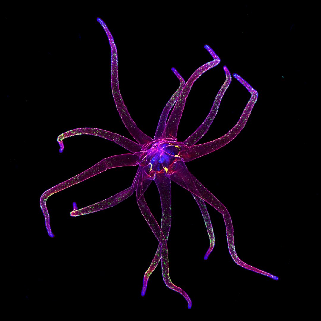

This week's #MicroscopyMonday shows the head region of a sea anemone, with #neurons labeled with fluorescent protein. Despite its seemingly simple body plan, the sea anemone possesses a rather sophisticated cellular composition. Image author: Shuonan He (<a href="/Gibson_Lab/">Gibson Lab</a>) #innovation
Marine Biological Laboratory (MBL) (@mblscience) 's Twitter Profile Photo

It's #MicroscopyMonday, so let's take a dive under the 🔬| This image of a live baby squid in transmitted light under polychromatic polarization microscope shows real colors, as it is seen by naked eye! Credit: Michael Shribak, MBL, in The University of Chicago 2022 "Science as Art" Competition

It's #MicroscopyMonday, so let's take a dive under the 🔬| This image of a live baby squid in transmitted light under polychromatic polarization microscope shows real colors, as it is seen by naked eye!

Credit: Michael Shribak, MBL, in <a href="/UChicago/">The University of Chicago</a> 2022 "Science as Art" Competition
Teledyne Photometrics (@photometrics) 's Twitter Profile Photo

Today the #scienceoffcamera time machine brings us to October 2020 and our wonderful interview with Dr. Paul Maddox, Founder and President of Mizar Imaging and Associate Professor at UNC-Chapel Hill We hope you take a listen! Find it where you get your podcasts and anchor.fm/science-off-ca…

Today the #scienceoffcamera time machine brings us to October 2020 and our wonderful interview with Dr. Paul Maddox, Founder and President of <a href="/MizarImaging/">Mizar Imaging</a> and Associate Professor at <a href="/UNC/">UNC-Chapel Hill</a> We hope you take a listen! Find it where you get your podcasts and anchor.fm/science-off-ca…
Marine Biological Laboratory (MBL) (@mblscience) 's Twitter Profile Photo

It's #MicroscopyMonday, so let's take a dive under the 🔬 This image shows multinucleated skeletal muscle cells form connections with multiple muscle precursor cells. Nuclei are in green and cell is in red. Credit: Helena Pinheiro (Helena Pinheiro)
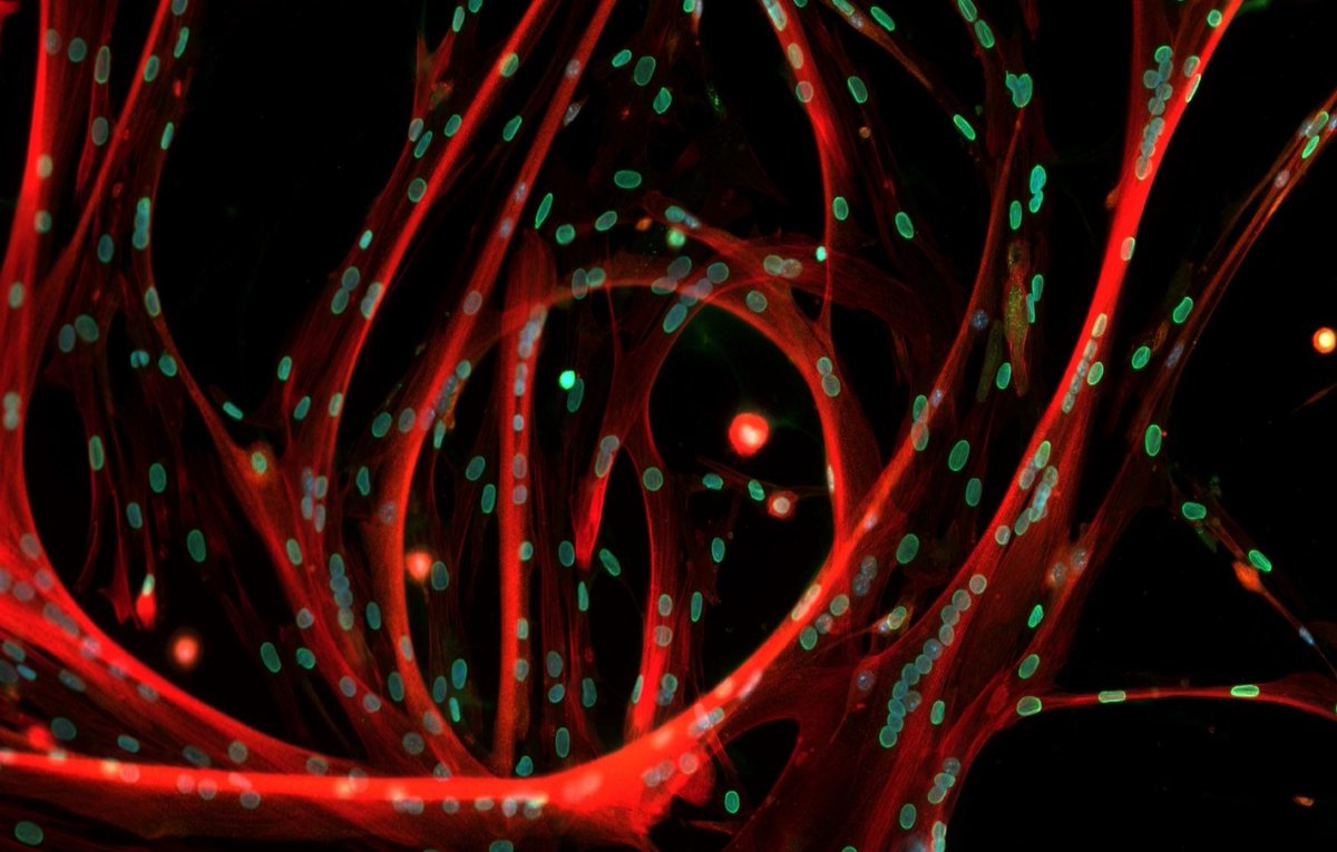
It's #MicroscopyMonday, so let's take a dive under the 🔬 

This image shows multinucleated skeletal muscle cells form connections with multiple muscle precursor cells. Nuclei are in green and cell is in red. 

Credit: Helena Pinheiro (<a href="/helenaapinheiro/">Helena Pinheiro</a>)
Marine Biological Laboratory (MBL) (@mblscience) 's Twitter Profile Photo

It's the last #MicroscopyMonday in #Squidtember, so let's dive under the 🔬! Check out this cluster of suckers at distal end of feeding tentacle in young longfin inshore squid (Doryteuthis pealeii)! Credit: Karen Crawford

It's the last #MicroscopyMonday in #Squidtember, so let's dive under the 🔬! Check out this cluster of suckers at distal end of feeding tentacle in young longfin inshore squid (Doryteuthis pealeii)!

Credit: Karen Crawford
Kristen Engevik (@thekengevik) 's Twitter Profile Photo

That one time I found a ghost in my cells 👻 Happy #MicroscopyMonday! #MicrosCreepy #boo #sciart #liveimaging #bioart #ghosted #microscopy

Marine Biological Laboratory (MBL) (@mblscience) 's Twitter Profile Photo

#MicroscopyMonday AND Halloween!? It's time to get spooky under the 🔬 ! Check out these amazing images of a short-tailed fruit bat (Carollia perspicillata) taken during an MBL Embryology course. Credit: Idoia Quintana-Urzainqui, Paola Bertucci, Peter Warth, Chi-Kuo Hu

#MicroscopyMonday AND Halloween!? It's time to get spooky under the 🔬 !

Check out these amazing images of a short-tailed fruit bat (Carollia perspicillata) taken during an MBL Embryology course. 

Credit: Idoia Quintana-Urzainqui, Paola Bertucci, Peter Warth, Chi-Kuo Hu
Okolab (@okolabgroup) 's Twitter Profile Photo

Always glad to support our partners! It was nice to get these images from a successful installation at Biozentrum, University of Basel. Nikon Microscope Solutions is running a demo of their Ti2 scope equipped with the Mizar Imaging TILT #LightSheet system and our Bold Line #TopStageIncubator. #CountOnOkolab
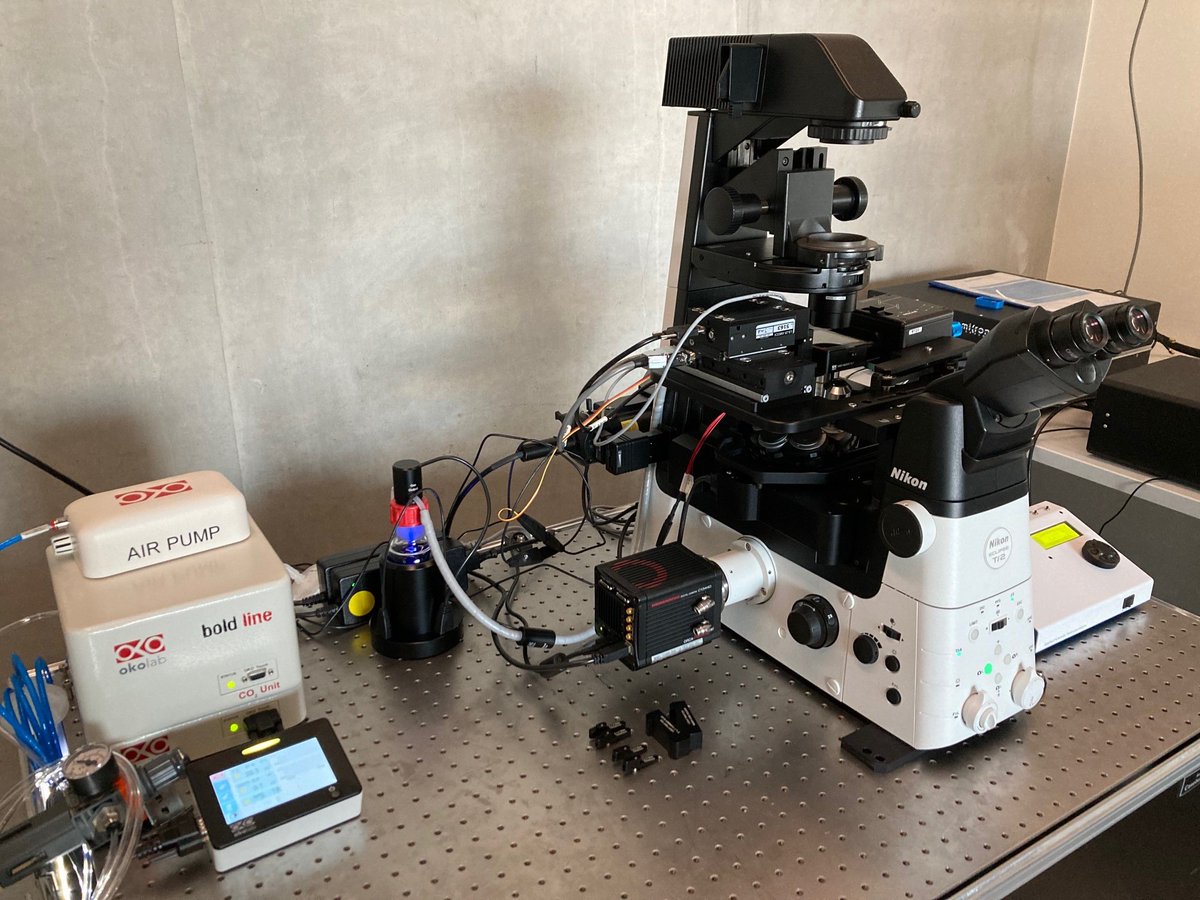
Always glad to support our partners! It was nice to get these images from a successful installation at <a href="/biozentrum/">Biozentrum, University of Basel</a>. <a href="/NikonInst/">Nikon Microscope Solutions</a> is running a demo of their Ti2 scope equipped with the <a href="/MizarImaging/">Mizar Imaging</a> TILT #LightSheet system and our Bold Line #TopStageIncubator. 
#CountOnOkolab