Joey Tidei (@j__today) 's Twitter Profile
Joey Tidei

@j__today

PhD candidate in the Beach (@myosincity) and Oakes (@pwoakes) Labs at Loyola University Chicago. Badger Alum. Former beer nerd. I like neurons. I like bread.

ID: 1445569521893326854

calendar_today06-10-2021 02:00:25

97 Tweet

204 Followers

292 Following

Joey Tidei (@j__today) 's Twitter Profile Photo

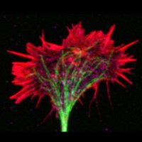
Extra spooky edition of #FluorescenceFriday. Primary rat hippocampal neurons at DIV3 stained for F-actin 🟠 and phosphorylated myosin 2 🟣.

Extra spooky edition of #FluorescenceFriday. Primary rat hippocampal neurons at DIV3 stained for F-actin 🟠 and phosphorylated myosin 2 🟣.
Alexandre Dumoulin @axonsalex.bsky.social (@axonsalex) 's Twitter Profile Photo

Wrapping up the week by visualizing the crazy axonal growth of dorsal root ganglia neurons in vitro using the SPY650-FastAct probe from Spirochrome! 🤩 Capturing one image every 10 minutes.

Joey Tidei (@j__today) 's Twitter Profile Photo

Since I missed #FluorescenceFriday this week I'll have to give it in person at #cellbio2023 ... Beach and Oakes Labs coming in hot.

PNASNews (@pnasnews) 's Twitter Profile Photo

Researchers grew rat neurons on a force sensor, which allowed them to discover that neurons develop tension after forming connections with other neurons, resulting in a contractile neural network. Without this contractility, neurons fail to fire. In PNAS: ow.ly/TQSj50QmtPO

Kevin Terretaz (@kwolbachia) 's Twitter Profile Photo

It was really cool to learn some jython programming for this tool. It's great for exploring all the LUTs, and once you have a set of favorite LUTs I recommend to use @biovoxxel's plugin "LUT channels tool" from the biovoxxel update site! x.com/biovoxxel/stat…

Jordan Beach (@myosincity) 's Twitter Profile Photo

The end of an era 😢 Melissa Quintanilla, the 1st PhD student to join the lab, has cleaned her desk and -20/-80 space, transferred her last data, and is ready to head to Paris to join the Piel Lab. What a pleasure it has been! Au revoir Dr. Quintanilla!!!
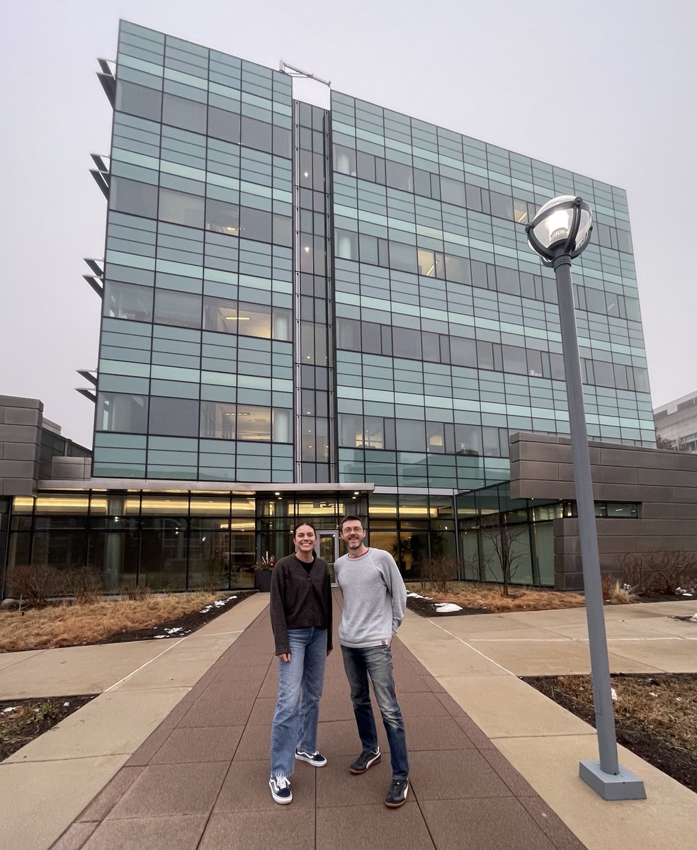

The end of an era 😢 
<a href="/fluoroforce/">Melissa Quintanilla</a>, the 1st PhD student to join the lab, has cleaned her desk and -20/-80 space, transferred her last data, and is ready to head to Paris to join the Piel Lab. 

What a pleasure it has been! 

Au revoir Dr. Quintanilla!!!
Journal of Cell Biology (@jcellbiol) 's Twitter Profile Photo

Quintanilla (Melissa Quintanilla), Oakes (Patrick Oakes), Beach (Jordan Beach) and colleagues Loyola University Chicago demonstrate subcellular biophysical mechanisms that enable myosin 2 filament assembly. hubs.ly/Q02l3klJ0 Matthew Akamatsu @Coronin @taraskalab #Biophysics #Cytoskeleton

Joey Tidei (@j__today) 's Twitter Profile Photo

Primary rat hippocampal neurons at DIV14 stained for Active-RhoA🔵and Phospho-Myosin Light Chain🟣. No AI required for this #FluorescenceFriday .

Primary rat hippocampal neurons at DIV14 stained for Active-RhoA🔵and Phospho-Myosin Light Chain🟣. No AI required for this #FluorescenceFriday .
Felipe Opazo (@felipeopaz_o) 's Twitter Profile Photo

Very happy to see the central Ph.D. topic (out of several) of Nikos Mougios in bioRxiv. He established a universal way to make all your antibody-based assays easily multiplexable by using erasable secondary #nanobodies. See it here: biorxiv.org/content/10.110…

Very happy to see the central Ph.D. topic (out of several) of <a href="/N_Mougios/">Nikos Mougios</a> in <a href="/biorxivpreprint/">bioRxiv</a>. He established a universal way to make all your antibody-based assays easily multiplexable by using erasable secondary #nanobodies. See it here: biorxiv.org/content/10.110…
Journal of Cell Biology (@jcellbiol) 's Twitter Profile Photo

Our April cover ➡️ hubs.la/Q02r6l130 shows overlayed temporal projection of actomyosin dynamics in the leading edge of migrating mouse embryonic fibroblast expressing EGFP-non-muscle myosin & mScarlet F-Tractin. From Melissa Quintanilla, Jordan Beach et al. hubs.la/Q02r6jGp0


Our April cover ➡️ hubs.la/Q02r6l130 shows overlayed temporal projection of actomyosin dynamics in the leading edge of migrating mouse embryonic fibroblast expressing EGFP-non-muscle myosin &amp; mScarlet F-Tractin. From <a href="/fluoroforce/">Melissa Quintanilla</a>, <a href="/myosincity/">Jordan Beach</a> et al. hubs.la/Q02r6jGp0
Pirta Hotulainen (@pirtahotulainen) 's Twitter Profile Photo

Our actin in AIS plasticity story, led by PhD student David Micinski, is now online in Frontiers in Molecular Neuroscience. We like to thank our excellent reviewers whose comments and requests clearly improved the paper. frontiersin.org/articles/10.33… Thread below👇

Our actin in AIS plasticity story, led by PhD student David Micinski, is now online in Frontiers in Molecular Neuroscience. We like to thank our excellent reviewers whose comments and requests clearly improved the paper.
frontiersin.org/articles/10.33…
Thread below👇
Joey Tidei (@j__today) 's Twitter Profile Photo

Life has been pulling me away from the scope (I had a baby named Henry!), but #FlourescenceFriday never takes a break. CAD cell with a dual tag myosin 2 construct - green on the motor heads and magenta on the tails, giving a🟢🟣🟢triplet. Can even see a few processive runs🏃‍♂️

Caillier Alexia (@alexiacaillier) 's Twitter Profile Photo

Really proud to see this paper finally out ! 🤩🎉🍾 Thank you to Journal of Cell Biology and the reviewers for their precious comments that definitely strengthened our work. See our previous thread for most of the details ! x.com/pwoakes/status…

Joey Tidei (@j__today) 's Twitter Profile Photo

Very fortunate to have access to an abundance of scopes to capture images like this. These are primary neurons treated with blebbistatin and stained for acetylated tubulin. Melissa Quintanilla iLUT again coming in HOT this #FluorescenceFriday

Very fortunate to have access to an abundance of scopes to capture images like this. These are primary neurons treated with blebbistatin and stained for acetylated tubulin. <a href="/fluoroforce/">Melissa Quintanilla</a>  iLUT again coming in HOT this #FluorescenceFriday
Journal of Cell Biology (@jcellbiol) 's Twitter Profile Photo

Our October issue is out! hubs.la/Q02R_lsP0 The cover shows a color-coded projection through time of confocal images of a mouse primary Th1 T cell expressing EGFP-Lifeact migrating in confinement. From Caillier Alexia, Patrick Oakes et al. (hubs.la/Q02R_xZC0)

Our October issue is out! hubs.la/Q02R_lsP0 The cover shows a color-coded projection through time of confocal images of a mouse primary Th1 T cell expressing EGFP-Lifeact migrating in confinement. From <a href="/AlexiaCaillier/">Caillier Alexia</a>, <a href="/pwoakes/">Patrick Oakes</a> et al. (hubs.la/Q02R_xZC0)