Hunterian Museum London(@HunterianLondon) 's Twitter Profileg
Hunterian Museum London

@HunterianLondon

The Hunterian Museum at the Royal College of Surgeons of England

Banner image - Concourse (2) by Barbara Hepworth, 1948, Barbara Hepworth © Bowness

ID:2414505733

linkhttp://www.hunterianmuseum.org calendar_today27-03-2014 14:53:55

1,8K Tweets

8,9K Followers

675 Following

Hunterian Museum London(@HunterianLondon) 's Twitter Profile Photo

Glass eyes made by Mollie Surman of Kingston, Surrey in the mid 1900s. Surman was from a family of glassblowers and made her first 'eye' when she was 12. The main eye would be blown from white glass before other colours of glass were added in to make the iris, pupil, and veins.

Glass eyes made by Mollie Surman of Kingston, Surrey in the mid 1900s. Surman was from a family of glassblowers and made her first 'eye' when she was 12. The main eye would be blown from white glass before other colours of glass were added in to make the iris, pupil, and veins.
account_circle
Hunterian Museum London(@HunterianLondon) 's Twitter Profile Photo

ANSWER: Wednesday might be hump day, but it's not a camel hump! It's the lining of one of the stomach chambers. Hunter described this as a ‘water stomach’ where the camel would store water. It is now thought it is probably involved in the absorption of water, rather than storage.

account_circle
Hunterian Museum London(@HunterianLondon) 's Twitter Profile Photo

Specimen in focus 🔍

The label on this incredible specimen reads ‘Camelus bactrianus’, telling us that it comes from a Bactrian Camel. But do you know which part of the animal it is from?

account_circle
Hunterian Museum London(@HunterianLondon) 's Twitter Profile Photo

Specimen in focus 🔍
These rabbit ears were part of an experiment carried out by John Hunter in 1777 to see how freezing affected living tissue. He hoped to discover if life could be extended through cryonics, freezing a living animal to bring it back to life in the future

Specimen in focus 🔍 These rabbit ears were part of an experiment carried out by John Hunter in 1777 to see how freezing affected living tissue. He hoped to discover if life could be extended through cryonics, freezing a living animal to bring it back to life in the future
account_circle
Hunterian Museum London(@HunterianLondon) 's Twitter Profile Photo

Great news! The Hunterian Museum has been shortlisted for 'Permanent Exhibition of the Year' at the Museum+ Heritage awards! 🤩
Enjoy this sneak peek of the galleries!

account_circle
Hunterian Museum London(@HunterianLondon) 's Twitter Profile Photo

Lithotomy – the patient's experience

In the 1700s lithotomy was dangerous and extremely painful, the operation had to be fast and the patient had to be held still. Some, grateful they had survived, kept the stones as mementos in inscribed boxes. The one shown is made of pewter.

Lithotomy – the patient's experience In the 1700s lithotomy was dangerous and extremely painful, the operation had to be fast and the patient had to be held still. Some, grateful they had survived, kept the stones as mementos in inscribed boxes. The one shown is made of pewter.
account_circle
Hunterian Museum London(@HunterianLondon) 's Twitter Profile Photo

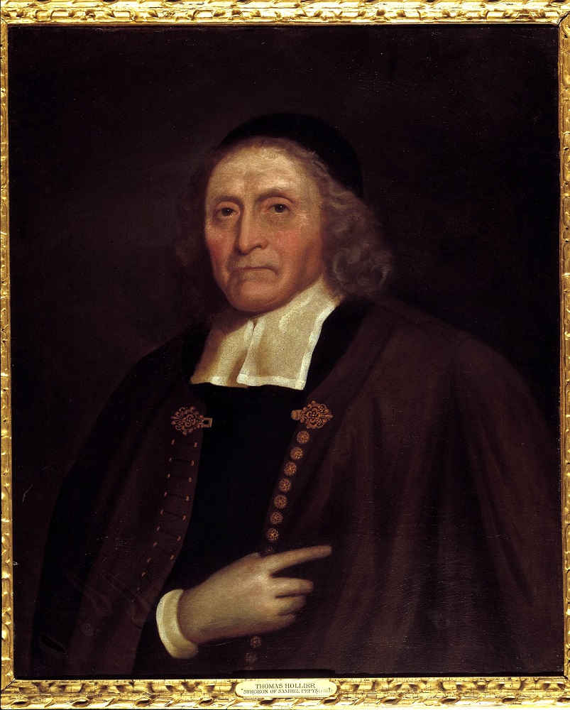
Thomas Hollier (1609 – 1690) was a London surgeon, skilled in lithotomy. His most famous patient the diarist, Samuel Pepys, underwent the operation on 26 March 1658 at the age of 25. Grateful to have survived, Pepys celebrated the anniversary of the operation every year after.

Thomas Hollier (1609 – 1690) was a London surgeon, skilled in lithotomy. His most famous patient the diarist, Samuel Pepys, underwent the operation on 26 March 1658 at the age of 25. Grateful to have survived, Pepys celebrated the anniversary of the operation every year after.
account_circle
Hunterian Museum London(@HunterianLondon) 's Twitter Profile Photo

The Art and Science of Surgery
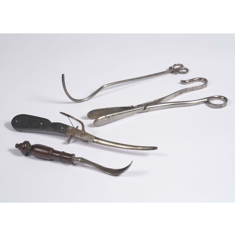
Lithotomy instruments, late 1700s, long and thin for insertion into the bladder or urethra. The hook or forceps were used to extract a whole stone through an incision in the bladder, while the scoop was used to remove fragments of crushed stone.

The Art and Science of Surgery Lithotomy instruments, late 1700s, long and thin for insertion into the bladder or urethra. The hook or forceps were used to extract a whole stone through an incision in the bladder, while the scoop was used to remove fragments of crushed stone.
account_circle
Hunterian Museum London(@HunterianLondon) 's Twitter Profile Photo

The Art and Science of Surgery
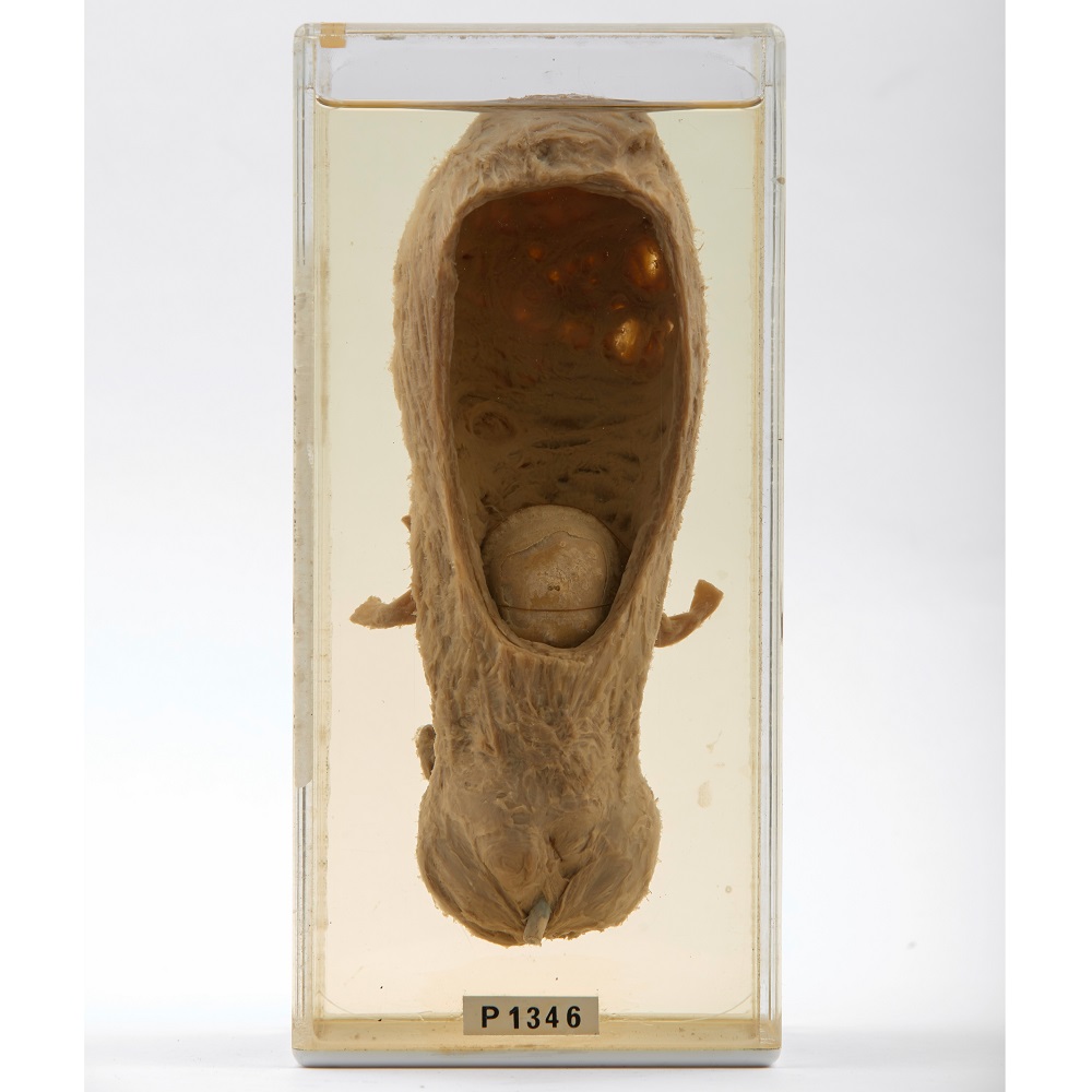
Cutting for the Stone. The specimen shows a human bladder with a large stone inside caused by the build up of mineral deposits. The surgical removal of bladder stones is called lithotomy, from the Greek litho - stone, and tomia - cutting.

The Art and Science of Surgery Cutting for the Stone. The specimen shows a human bladder with a large stone inside caused by the build up of mineral deposits. The surgical removal of bladder stones is called lithotomy, from the Greek litho - stone, and tomia - cutting.
account_circle
Hunterian Museum London(@HunterianLondon) 's Twitter Profile Photo

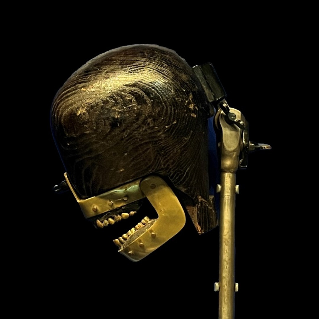
This dental phantom was made around 1900 for dental students to practice their skills before treating real patients. A hat-maker’s dummy has been added to represent the head of the patient. The jaws contain removable brass teeth allowing various dental procedures to be practiced.

This dental phantom was made around 1900 for dental students to practice their skills before treating real patients. A hat-maker’s dummy has been added to represent the head of the patient. The jaws contain removable brass teeth allowing various dental procedures to be practiced.
account_circle
Hunterian Museum London(@HunterianLondon) 's Twitter Profile Photo

Today is . Orthopaedic surgeon Dame Clare Marx PRCS (1954-2022) was the first woman to be President of the Royal College of Surgeons of England (2014 - 17). Photograph by Jane Brettle, bust by Consultant Plastic and Reconstructive Surgeon, Lisa Sacks FRCS.

Today is #InternationalWomensDay. Orthopaedic surgeon Dame Clare Marx PRCS (1954-2022) was the first woman to be President of the Royal College of Surgeons of England (2014 - 17). Photograph by Jane Brettle, bust by Consultant Plastic and Reconstructive Surgeon, Lisa Sacks FRCS.
account_circle
Hunterian Museum London(@HunterianLondon) 's Twitter Profile Photo

Today is . Fibrodysplasia Ossificans Progressiva (FOP) is a rare genetic condition in which the soft tissues are gradually replaced by bone, it was first described by St. Bart’s surgeon, John Freke, in 1736.
Image: Barts Health NHS Trust Archives Barts Health Archives


Today is #RareDiseaseDay. Fibrodysplasia Ossificans Progressiva (FOP) is a rare genetic condition in which the soft tissues are gradually replaced by bone, it was first described by St. Bart’s surgeon, John Freke, in 1736. Image: Barts Health NHS Trust Archives @BHAandM
account_circle
The Royal College of Surgeons of England(@RCSnews) 's Twitter Profile Photo

Participate in a brief survey to contribute to the development of our digital services and be entered into a prize draw to win £250. 💰

Responses are anonymous, and will help us provide you with the best possible digital experience. Complete by 10 March: ow.ly/AEoK50QEY22

account_circle
Hunterian Museum London(@HunterianLondon) 's Twitter Profile Photo

John Hunter used this specimen of a greater siren to show his idea that animals might be classed depending on the structure of their hearts. Although he didn’t think one feature could be used to classify an animal, he thought it could help when new species were discovered.

John Hunter used this specimen of a greater siren to show his idea that animals might be classed depending on the structure of their hearts. Although he didn’t think one feature could be used to classify an animal, he thought it could help when new species were discovered.
account_circle
Hunterian Museum London(@HunterianLondon) 's Twitter Profile Photo

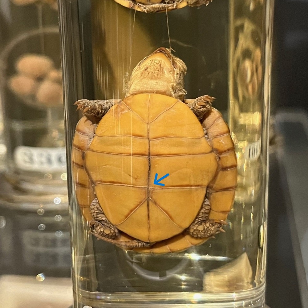
These three young tortoises were prepared by Hunter to show the closing of the umbilical aperture. This is where the yolk sac would be attached to the underside of the tortoise (plastron) when in the egg. The tortoises are in age order with the youngest at the top.

These three young tortoises were prepared by Hunter to show the closing of the umbilical aperture. This is where the yolk sac would be attached to the underside of the tortoise (plastron) when in the egg. The tortoises are in age order with the youngest at the top.
account_circle